
Abstract
Podoplanin (PDPN) obtained from various animal species has been characterized using specific anti-PDPN monoclonal antibodies (mAbs), namely, PMab-1, PMab-2, PMab-32, PMab-38, PMab-44, and PMab-52 against mouse, rat, rabbit, dog, bovine, and cat PDPN, respectively. PDPN is expressed in type I alveolar cells in lungs, lymphatic endothelial cells, and kidney podocytes. In this study, we investigated possible cross-reactions between anti-PDPN mAbs and sheep PDPN. Type I alveolar cells from sheep lung were strongly detected by PMab-44 using immunohistochemical analyses. These results indicate that PMab-44 may be useful for the detection of sheep PDPN.
Introduction
P
PDPN is a specific marker for type I alveolar cells of the lung.(2,8) Therefore, anti-PDPN monoclonal antibodies (mAbs) are useful in distinguishing type I alveolar cells of the lung from the type II cells. We characterized the PDPNs of various animal species using specific anti-PDPN mAbs. We established antimouse (PMab-1),(9) antirat (PMab-2),(10) antirabbit (PMab-32),(11) antidog (PMab-38(12) and PMab-48(13)), antibovine (PMab-44),(14) and anticat (PMab-52)(15) PDPN antibodies. PMab-38 was found to be cancer cell specific since it did not react with lymphatic endothelial cells and type I alveolar cells,(12) but reacted with squamous cell carcinomas(16) and melanomas.(17) We developed several antihuman PDPN mAbs, including NZ-1.2,(9) LpMab-7,(18,19) LpMab-10,(20) LpMab-13,(21) and LpMab-17.(22) We also produced antiglycopeptide mAbs (GpMabs) against human PDPN, namely, LpMab-3,(18) LpMab-9,(18) LpMab-12,(23) LpMab-19,(24) and LpMab-21.(25,26) Cancer-specific mAbs (CasMabs) against human PDPN, such as LpMab-2(18,27) and LpMab-23,(28,29) were also established. Because PDPN is expressed in several normal tissues, CasMabs against human PDPN should be utilized for clinical applications.
Antisheep PDPN mAbs have not been reported until date. In this study, we used immunohistochemical analyses to investigate the potential cross-reactions between anti-PDPN mAbs established for several species, particularly for sheep PDPN.
Materials and Methods
Immunohistochemical analyses
Normal sheep lungs were collected from two sheep after autopsy at Hokkaido University, fixed in 10% neutral-buffered formalin, and processed routinely to make paraffin-embedded tissue sections. Histological sections of thickness 4 μm were directly autoclaved in EnVision FLEX Target Retrieval Solution, high pH (Agilent Technologies, Inc., Santa Clara, CA) for 20 minutes. After blocking with SuperBlock T20 (PBS) Blocking Buffer (Thermo Fisher Scientific, Inc. Waltham, MA), sections were incubated with PMab-44 (10 μg/mL) for 1 hour at room temperature and treated using Envision+ Kit (Agilent Technologies, Inc.) for 30 minutes. Then color was developed using 3,3′-diaminobenzidine tetrahydrochloride (DAB; Agilent Technologies, Inc.) for 2 minutes, and counterstaining was performed with hematoxylin (FUJIFILM Wako Pure Chemical Corporation, Osaka, Japan).
Results and Discussion
Previously, we have developed a mouse antibovine PDPN mAb, PMab-44 (IgG1, kappa), which specifically detects bovine PDPN in immunohistochemical analyses.(14) Furthermore, a series of bovine PDPN deletion or point mutants were used for investigating the binding epitopes of PMab-44 using flow cytometry and Western blotting.(30) The PLAG3 of bovine PDPN was identified as the PMab-44 critical epitope. A comparison of amino acid sequences revealed an 85% homology between bovine and sheep PDPN (Supplementary Fig. S1). Therefore, we further investigated whether PMab-44 cross-reacts with sheep PDPN.
Flow cytometry indicated that PMab-44 reacted with CHO-K1 cells that overexpressed sheep PDPN (CHO/sPDPN) (Supplementary Fig. S2). Other anti-PDPN mAbs, such as antimouse (PMab-1),(9) antirat (PMab-2),(10) antirabbit (PMab-32),(11) antidog PMab-38(12) and PMab-48(13), and anticat (PMab-52(15)), did not react with CHO/sPDPN (data not shown). These results indicate that only PMab-44 is useful for the detection of sheep PDPN.
Furthermore, we investigated the expression of sheep PDPN in sheep lungs using immunohistochemical analyses. For this purpose, we used lungs obtained from two sheep (No. 1 and No. 2). Both lungs were stained using PMab-44 (10 μg/mL) when EnVision FLEX Target Retrieval Solution, high pH, was used for an antigen retrieval procedure. Representative results (No. 2) are depicted in Figure 1. Type I alveolar cells, but not the bronchial cells, were strongly stained with PMab-44 (Fig. 1A, B). Staining was not observed without PMab-44 (Fig. 1C, D). The identical tissue (No. 2) was stained moderately using PMab-44 (1 μg/mL) when citrate buffer (pH 6) was used for antigen retrieval (Supplementary Fig. S3), indicating that a specific staining by PMab-44 was observed at a low concentration of PMab-44 (1 μg/mL) or under universal antigen retrieval conditions (i.e., when citrate buffer was used). Unfortunately, the sheep kidney and colon were not stained by PMab-44 in this study (data not shown). This may be attributed to the varying expression levels of sheep PDPN among different tissues.
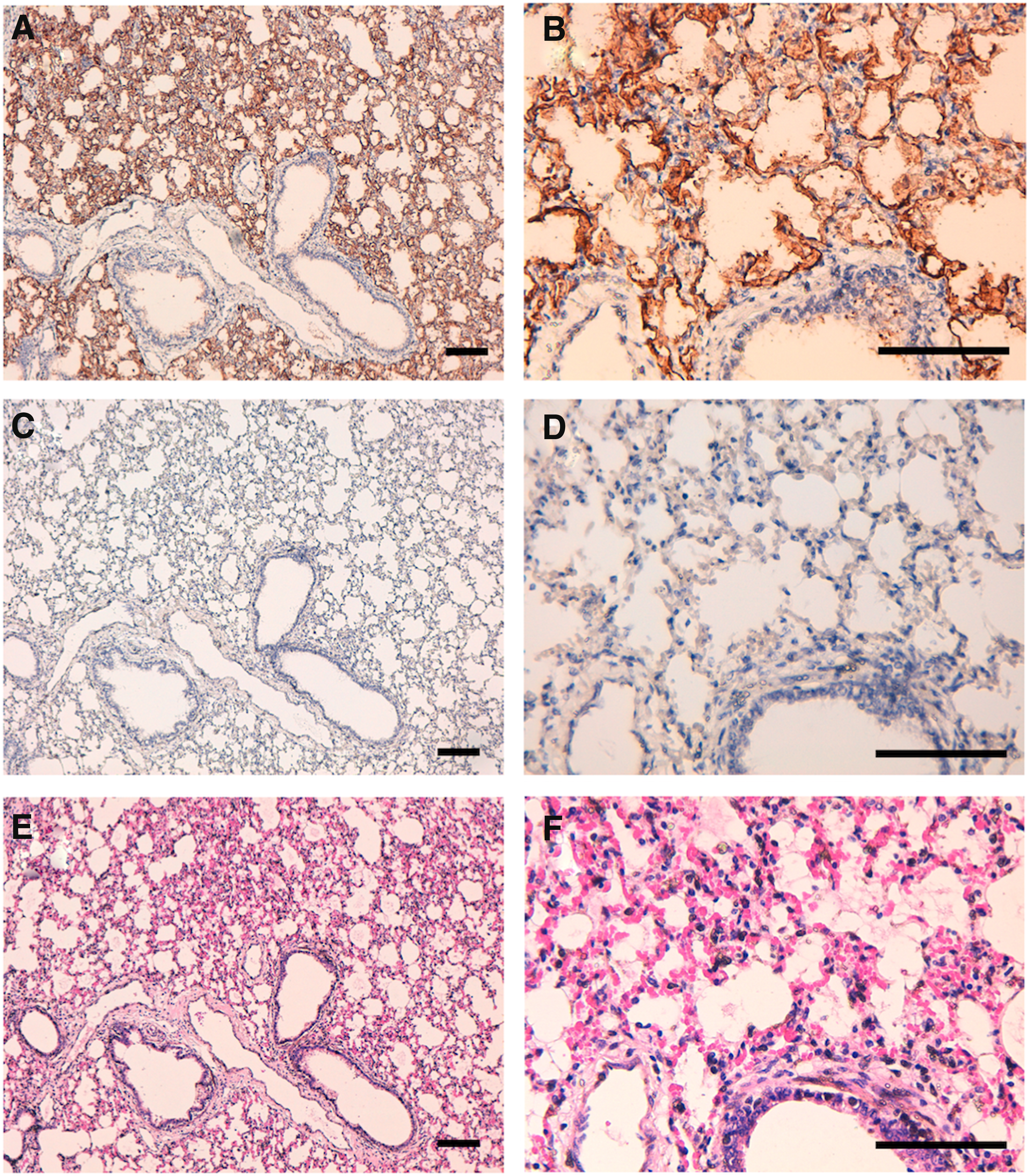
Immunohistochemical analyses using sheep tissues. Histological sections of the sheep tissues were directly autoclaved in EnVision FLEX Target Retrieval Solution, high pH, for 20 minutes. After blocking, the sections were incubated with 10 μg/mL of PMab-44
In conclusion, the antibovine PDPN mAb PMab-44 is useful for the detection of sheep PDPN in sheep lungs using immunohistochemical analyses. Further studies are necessary to demonstrate the capacity of PMab-44 for detection of sheep PDPN in other tissues.
Footnotes
Acknowledgments
This research was supported, in part, by AMED under grant numbers: JP18am0101078 (Y.K.), JP18am0301010 (Y.K.), and JP18ae0101028 (Y.K.), and by JSPS KAKENHI grant number 17K07299 (M.K.K.) and grant number 16K10748 (Y.K.).
Author Disclosure Statement
No competing financial interests exist.
References
Supplementary Material
Please find the following supplemental material available below.
For Open Access articles published under a Creative Commons License, all supplemental material carries the same license as the article it is associated with.
For non-Open Access articles published, all supplemental material carries a non-exclusive license, and permission requests for re-use of supplemental material or any part of supplemental material shall be sent directly to the copyright owner as specified in the copyright notice associated with the article.