
Abstract
von Willebrand factor (VWF) is a glycoprotein that plays a central role in the initiation of blood coagulation. VWF performs two important functions: it acts as a molecular bridge between platelets and as a carrier for coagulation factor VIII (FVIII). von Willebrand disease (VWD) and acquired von Willebrand syndrome (AVWS) are caused by the absence of and/or abnormality in VWF. The pathophysiology of VWD and AVWS is complex and, therefore, it is difficult to diagnose them by conducting the same laboratory tests in all patients. To develop useful monoclonal antibodies (mAbs) for the diagnosis of VWD and AVWS, rat mAbs against human VWF were generated. Immunoblotting analysis revealed that mAbs recognized the reduced and nonreduced VWF protein and endogenous VWF in normal human plasma. Furthermore, we developed a highly sensitive monoclonal antibody-based sandwich enzyme-linked immunosorbent assay technique. In conclusion, the development of VWF-specific mAbs would be useful in the diagnosis of VWD and AVWS.
Introduction
Von Willebrand factor (VWF) is a large glycoprotein that circulates in plasma at a concentration of 5–10 μg/mL and plays a central role in the initiation of blood coagulation.(1,2) VWF mediates platelet adhesion to the subendothelium during vascular injury. It is synthesized as a monomer of ∼220 kDa containing multiple domains and secreted by endothelial cells and megakaryocytes.(3–6) VWF translation product contains 2813 amino acids that consist of a 22-amino acid signal peptide, a 741-amino acid propeptide, and a mature subunit of 2050 amino acids.(7) VWF is composed of multiple domains; some domains contribute to dimerization, whereas others mediate binding to various proteins. The C-terminal cysteine knot domain dimerizes VWF.(8,9) Many interactions with VWF binding partners, including binding to platelet GPIb and collagen, are localized in the A domain.(10) D′ and D3 domains bind coagulation factor VIII (FVIII).(11) These interactions depend upon its location, the local microenvironment, and physiological or pathological cues.
Any deficiency and/or abnormality of VWF leads to increased bleeding tendency, and this disorder was named von Willebrand disease (VWD).(12) VWD is the most common bleeding disorder, with a prevalence in up to 1% of the population, and caused by inherited defects in the concentration, structure, or function of VWF.(13,14) Mutations in VWF have been described that affect platelet binding, collagen binding, secretion, and synthesis; however, these do not necessarily lead to VWD. Therefore, knowing personal and family medical history is sufficient to make a diagnosis of VWD by laboratory testing.(15) In addition, acquired von Willebrand syndrome (AVWS) is another disorder related to diseases, such as malignancies, hypothyroidism, cardiovascular diseases, and cardiac replacement devices,(16,17) and occurs predominantly in older patients. The mechanism of an AVWS subtype involves the development of an anti-VWF antibody that can induce both a loss of function and increased clearance of VWF.(18) However, the pathophysiology of VWD and AVWS is complex, and it is difficult to diagnose them by performing the same laboratory tests in all patients.
Therefore, the first aim of this investigation was to generate useful monoclonal antibodies (mAbs) for the diagnosis of VWD and AVWS. In this study, we report the generation and application of a series of three mAbs. These mAbs would be useful in immunoblotting, enzyme-linked immunosorbent assay (ELISA), and sandwich ELISA. These experimental tools will allow us to detect VWF in patient's plasma.
Materials and Methods
Production of rat mAbs
Antihuman VWF rat mAbs were generated based on the rat lymph node method established by Sado and colleagues.(19) An 8-week-old female WKY/Izm rat (SLC, Shizuoka, Japan) was injected in the hind footpads with 100 μg of human Factor VIII-free VWF protein (Fitzgerald Industries International, Concord, MA) and Freund's complete adjuvant (Becton Dickinson, Sparks, MD). After 2 weeks, the cells from the medial iliac lymph nodes of the rat immunized with an antigen were fused with mouse myeloma SP2 cells at a ratio of 5:1 in 50% polyethylene glycol (PEG1500; Roche, Indianapolis, IN) solution. The resulting hybridoma cells were plated onto 96-well plates and cultured in HAT selection medium (Hybridoma-SFM [Life Technologies, Grand Island, NY], 10% fetal bovine serum, 5% BM Condimed H1 [Roche], 100 μM hypoxanthine, 0.4 μM aminopterin, and 16 μM thymidine). After 10 days postfusion, the hybridoma supernatants were screened by means of an ELISA against VWF protein. Positive clones were subcloned and rescreened by ELISA and immunoblotting.
The specific immunoglobulin class of three mAbs was determined using a rat isotyping kit (BD Biosciences, San Diego, CA). The analysis indicated that mAbs 3B4, 3E7, and 7A12 were rat IgG2a (κ).
Labeling of mAbs with peroxidase (POD) was performed using POD Labeling Kit (Dojindo, Kumamoto, Japan) in accordance with the manufacturer's protocol.
Enzyme-linked immunosorbent assay
For the first screening of generated mAbs, VWF protein (5 μg/mL) in Tris-buffered saline (TBS, 20 mM Tris-HCl, pH 7.5 containing 150 mM NaCl) was adsorbed on the surface of Costar Serocluster 96-well U-bottomed plates (Corning, Kennebunk, ME) by overnight incubation at 4°C. To avoid nonspecific binding, the plates were blocked with 1% bovine serum albumin (BSA; Sigma, St. Louis, MO) in TBS-T (20 mM Tris-HCl, pH 7.5 containing 150 mM NaCl, and 0.1% Tween-20). Hybridoma supernatants were incubated for 1 hour at room temperature and then washed three times with TBS-T. The plates were incubated for 30 minutes at room temperature with alkaline phosphatase (AP)-conjugated antirat IgG antibody (Sigma) at a dilution of 1:5000. After washing three times with TBS-T, immunoreactivity was visualized by means of p-nitrophenyl phosphate substrate system (p-NPP; Sigma). The reaction was quenched with 3 N NaOH and the absorbance was measured at 405 nm.
Immunoblotting analysis
VWF protein was added to sodium dodecyl sulfate-polyacrylamide gel electrophoresis (SDS-PAGE) sample buffer with or without beta-mercaptoethanol (2-ME; Wako, Osaka, Japan). This sample was resolved by SDS-PAGE using 4% polyacrylamide/1% agarose mixed gel and subsequently transferred to Immobilon-P polyvinylidene fluoride transfer membranes (PVDF; Millipore, Bedford, MA). Human plasma (Coagtrol N; Sysmex, Kobe, Japan) was added to SDS-PAGE sample buffer with 2-ME and was resolved by SDS-PAGE using 6% polyacrylamide gel and subsequently transferred to PVDF membranes. These membranes were blocked overnight at 4°C with 3% skim milk in TBS-T and incubated for 1 hour at room temperature with mAbs. After washing three times with TBS-T, the membranes were incubated for 30 minutes at room temperature with AP-conjugated antirat IgG antibody. Next the membranes were again washed three times with TBS-T and developed by treatment with p-nitro blue tetrazolium chloride (NBT; Wako) and 5-bromo-4-chloro-3-indolyl phosphate (BCIP; Wako).
Sandwich ELISA
The wells of 96-well plates were first coated with 10 μg/mL of 7A12 as a capture antibody in ELISA buffer (10 mM sodium phosphate [pH 7.0]) overnight at 4°C. To avoid nonspecific binding, the plates were blocked with 2% BSA in phosphate-buffered saline (PBS) and incubated overnight at 4°C. VWF protein (125–2000 ng/mL) was incubated for 1 hour at room temperature and then washed three times with PBS. The plates were then incubated for 1 hour at room temperature with 2 μg/mL of POD-labeled 3E7 as a detection antibody. After washing three times with PBS, immunoreactivity was visualized using 3,3′,5,5′-tetramethylbenzidine (TMB; Dojindo) system. The reaction was quenched with 7% phosphoric acid. Absorbance was measured at 450 nm and a standard curve for the concentration of human VWF was plotted.
Results
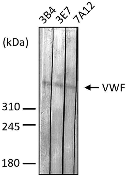
mAbs were produced against human VWF using the rat medial iliac lymph node method. Human VWF protein was used as an antigen to immunize rat with a single injection in hind footpads. Two weeks postimmunization, the lymphocytes were collected from the enlarged lymph nodes of the rat. Hybridoma obtained after fusing lymphocytes with mouse myeloma SP2 cells was tested for the production of mAbs that react with VWF protein in ELISA (data not shown). Twenty-six hybridoma supernatants that were positive for ELISA were examined by immunoblotting against the reduced and nonreduced human VWF protein. The results revealed that three mAbs designated 3B4, 3E7, and 7A12, showed a strong signal against the reduced and nonreduced human VWF protein (Fig. 1). These mAbs were selected for further studies, and examined by immunoblotting for specificity toward normal human plasma. As shown in Figure 2, the immunoblotting analysis of the normal human plasma showed that three mAbs detected only one band. To specifically quantify human VWF protein, we first determined which combination of capture and detection antibodies showed high sensitivity and linearity. We found that a combination of capture antibody 7A12 and detection antibody 3E7 exhibited higher sensitivity and linearity than that exhibited by each of them individually (data not shown). The standard curve using human VWF protein showed high linearity in the range 125–2000 ng/mL (Fig. 3).

Anti-VWF rat mAbs 3B4, 3E7, and 7A12 react with both human reduced and nonreduced VWF protein. Reduced or nonreduced VWF proteins with or without 2-ME were separated on 4% polyacrylamide/1% agarose mix gel.
Reactivity of anti-VWF rat mAbs 3B4, 3E7, and 7A12 in human normal plasma. Reduced human normal plasma was separated on 6% SDS-PAGE gel, transferred to a PVDF membrane, and immunoblotted with three mAbs. Molecular mass markers in kDa are indicated on the left.
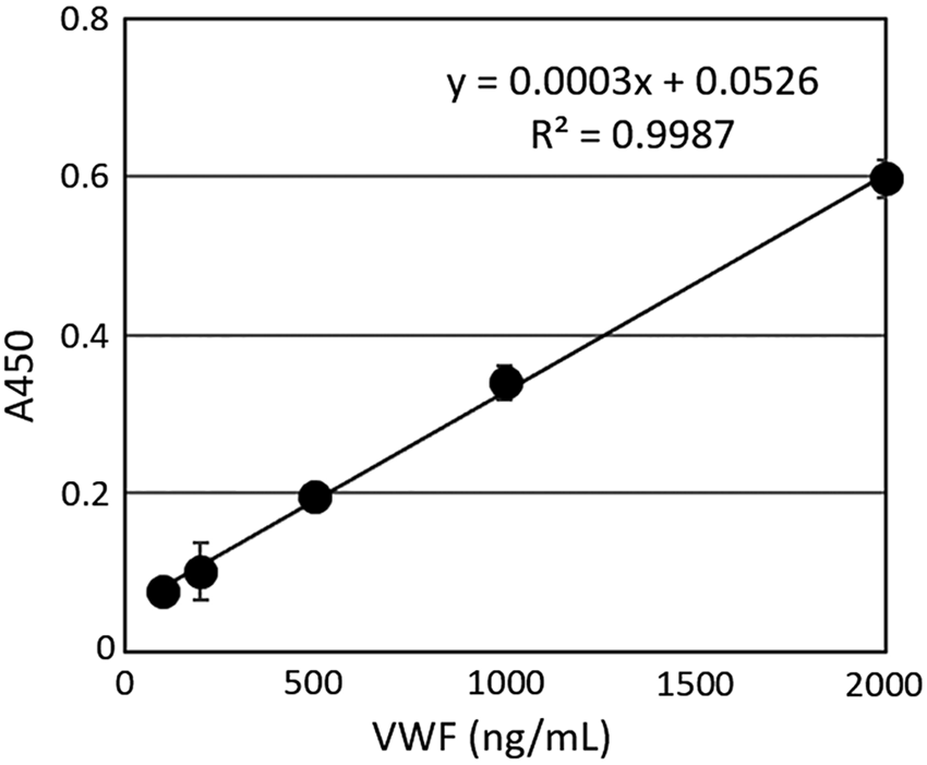
The standard curve for the concentration of human VWF was plotted using a sandwich ELISA system using 7A12 as a capture antibody and POD-labeled 3E7 as a detection antibody. ELISA, enzyme-linked immunosorbent assay; POD, peroxidase.
Discussion
In this study, for the diagnosis of VWD and AVWS, we generated rat mAbs against human VWF. mAbs 3B4, 3E7, and 7A12 were used for immunoblotting, ELISA, and sandwich ELISA. VWF is a large glycoprotein and forms a multimer in plasma. These mAbs reacted with reduced and nonreduced forms of VWF, and also with endogenous VWF in normal human plasma. We predict that these mAbs could be used in understanding the physiological significance of VWF in the human plasma. Furthermore, VWF interacts with many binding partners. Thus, these mAbs may be used as tools in the analysis of these interactions by inhibiting them.
VWD and AVWS lead to increased bleeding tendency. Measurement of VWF protein in patient's plasma is important for the diagnosis. In addition, the mechanism of AVWS involves the development of an anti-VWF antibody that can induce both a loss of function and increased clearance of VWF. Therefore, it is essential to measure anti-VWF antibody levels in patient's plasma. In general, patients with autoimmune VWD (AiVWD) show a very low VWF activity. However, unexpectedly high levels of VWF antigen are often detected by ELISA.(20) In addition, immunoblotting analysis of their plasma shows low levels of high-molecular weight VWF multimer. The presence of autoantibodies against VWF is also detected by ELISA.(20,21) Since AiVWD may be exacerbated with steroid tapering,(21) patient's VWF antigen and anti-VWF autoantibody levels must be monitored by ELISA for a long period of time. Incidentally, AiVWD has been declared as one of the designated intractable diseases by the Japanese Ministry of Health, Labour and Welfare in 2017. Our rat mAbs will be useful for the ELISA of VWF antigen and anti-VWF autoantibody as well as immunoblotting for VWF multimer analysis.
In summary, we generated rat mAbs against human VWF and developed sandwich ELISA system using these mAbs. The sandwich ELISA technique can be used to quantitatively determine protein levels in fluids, such as plasma. Our study indicates that mAbs may be useful to quantitatively determine human VWF protein and anti-VWF antibody levels in patient's plasma using a sensitive sandwich ELISA. We believe that our sandwich ELISA system is useful for the diagnosis of VWD, AVWS, and AiVWD.
Footnotes
Acknowledgment
This study was supported by a research grant from the Japanese Ministry of Health, Labour and Welfare.
Author Disclosure Statement
No competing financial interests exist.