
Abstract
Podoplanin (PDPN) is utilized as a specific marker of type I alveolar cells of lung and lymphatic endothelial cells of every tissue. Therefore, sensitive and specific monoclonal antibodies detecting PDPN are necessary for immunohistochemical analyses, especially using formalin-fixed paraffin-embedded tissues. Recently, we developed an anti-bear PDPN (bPDPN) mAb, PMab-247, which is useful for Western blot, flow cytometry, and immunohistochemical analyses. In this study, immunohistochemical analyses showed that PMab-247 strongly detected bPDPN, which is expressed in type I alveolar cells and lymphatic endothelial cells of bear lung and podocytes of bear kidney. These findings suggest that PMab-247 could be useful for pathophysiological analyses using immunohistochemistry.
Introduction
Podoplanin (PDPN) is a type I transmembrane sialoglycoprotein.(1) PDPN is also known as gp44 (2)/Aggrus,(3) which induces platelet aggregation through C-type lectin-like receptor-2 (CLEC-2).(4,5) The sialic acid of platelet aggregation-stimulating domain of PDPN is known to be important for PDPN-induced platelet aggregation.(6,7)
PDPN is expressed in several normal cells, including pulmonary type I alveolar cells, renal epithelial cells, and lymphatic endothelial cells.(1) PDPN is used to distinguish lymphatic endothelial cells from vascular endothelial cells in pathophysiological studies.(8) The PDPN–CLEC-2 interaction was reported to facilitate the separation of embryonic blood and lymphatic vessels.(9)
The expression of human PDPN (hPDPN) has been reported in many malignant cancers, such as lung cancers,(10) oral cancers,(11) and esophageal cancers.(12) The expression of hPDPN is associated with cancer metastasis.(3,13) We previously developed monoclonal antibodies (mAbs) against human,(14) mouse,(15) rat,(16) rabbit,(17) bovine,(18) dog,(19) cat,(20) pig,(21) horse,(22) goat,(23) tiger,(24) alpaca,(25) whale,(26) and Tasmanian devil(27) PDPNs. Furthermore, we developed an anti-bear PDPN (bPDPN) mAb, PMab-247.(28) In this study, we performed immunohistochemical analyses using PMab-247 against bPDPN.
Materials and Methods
Immunohistochemical analyses
Brown bear (Ursus arctos) tissues were collected after autopsy at Hokkaido University, fixed in 10% neutral-buffered formalin, and routinely processed to make paraffin-embedded tissue sections. Histological sections (4-μm thick) were directly autoclaved in citrate buffer (pH 6.0; Nichirei Biosciences, Inc., Tokyo, Japan) for 20 minutes. Then, sections were blocked using the SuperBlock T20 (phosphate buffered saline) Blocking Buffer (Thermo Fisher Scientific, Inc., Waltham, MA), incubated with PMab-247 (5 μg/mL) for 1 hour at room temperature, and treated with the EnVision+ Kit for mouse (Agilent Technologies, Inc., Santa Clara, CA) for 30 minutes. Color was developed using 3,3′-diaminobenzidine tetrahydrochloride (Agilent Technologies, Inc.) for 2 minutes, and counterstaining was performed using hematoxylin (FUJIFILM Wako Pure Chemical Corporation, Osaka, Japan).
Results and Discussion
Although we have established many anti-PDPN mAbs, such as anti-human,(14) anti-mouse,(15) anti-rat,(16) anti-rabbit,(17) anti-bovine,(18) anti-dog,(19) anti-cat,(20) anti-pig,(21) anti-horse,(22) anti-goat,(23) anti-tiger,(24) anti-alpaca,(25) anti-whale,(26) and anti-Tasmanian devil(27) PDPNs, sensitive and specific anti-bPDPN mAbs, which are useful for immunohistochemical analyses, had not been developed. Recently, two mice were immunized with Chinese hamster ovary (CHO)/bPDPN cells.(28) The developed hybridomas were seeded into 96-well plates and cultivated for 9 days. Wells positive for CHO/bPDPN and negative for CHO-K1 were selected by flow cytometry. After several additional screenings, PMab-247 (IgG1, kappa) was finally selected. PMab-247 specifically detected CHO/bPDPN cells by Western blot and immunohistochemistry, suggesting that the potential usefulness of PMab-247 for the functional analyses of bPDPN.
In this study, we performed immunohistochemical analyses against the brown bear. PDPN has been reported to be expressed in several tissues, including lung and kidney(1); we selected bear lung (Fig. 1) and bear kidney (Fig. 2) for immunohistochemical analyses. Expectedly, PMab-247 stained type I alveolar cells of bear lung (Fig. 1B), lymphatic endothelial cells of bear lung (Fig. 1C), and podocytes of bear kidney (Fig. 2B). These staining patterns are consistent with our previous immunohistochemical studies using several anti-PDPN mAbs.(21) No staining was observed without primary antibodies against type I alveolar cells of bear lung (Fig. 1E), lymphatic endothelial cells of bear lung (Fig. 1F), and podocytes of bear kidney (Fig. 2D).
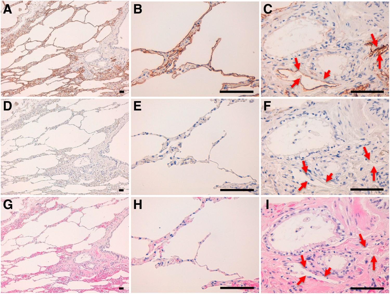
Immunohistochemical analyses for bear lung. Bear lung sections were incubated with 5 μg/mL of PMab-247
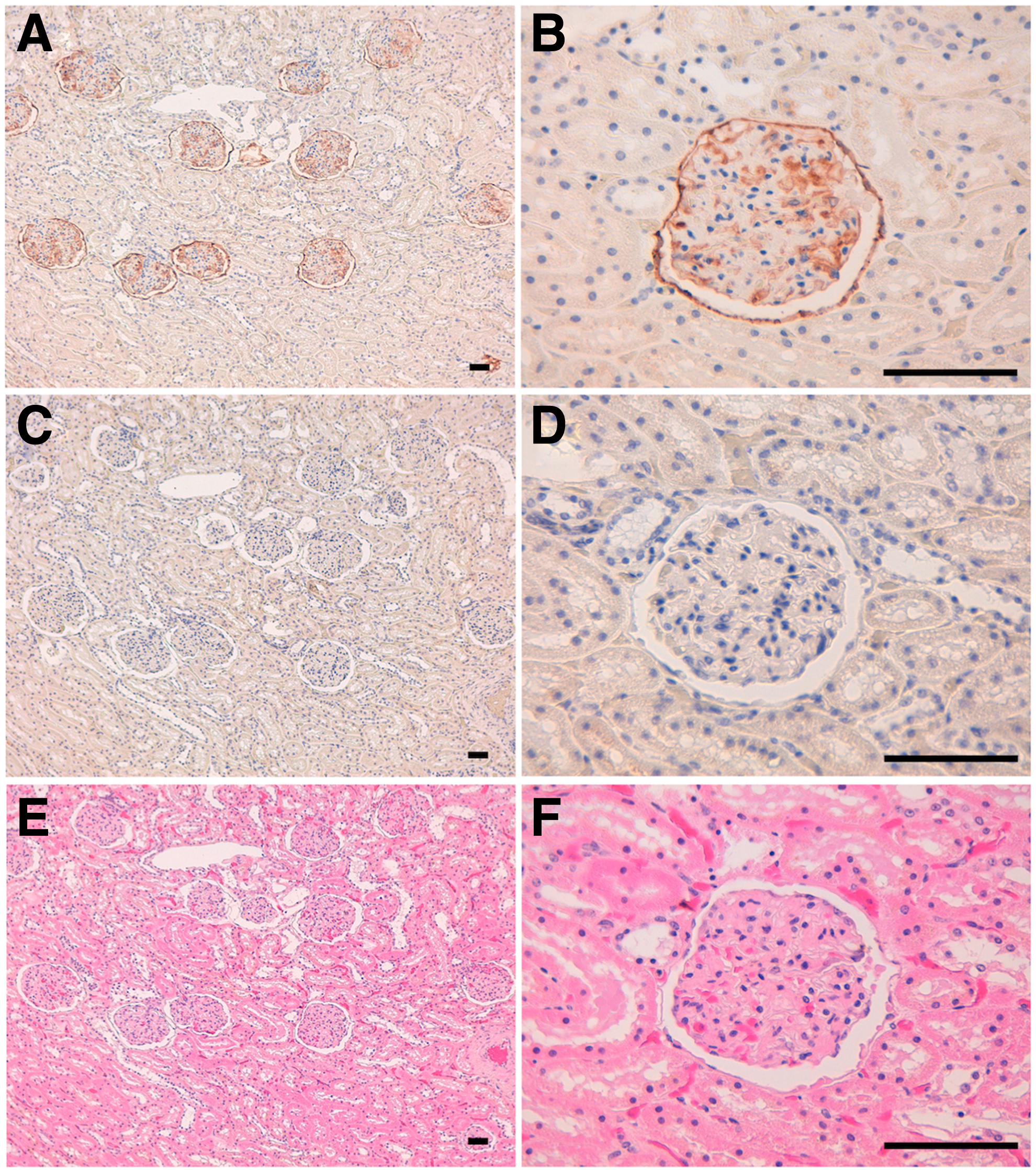
Immunohistochemical analyses for bear kidney. Bear kidney sections were incubated with 5 μg/mL of PMab-247
In conclusion, PMab-247 is useful for immunohistochemical analyses against the bPDPN. The epitope of PMab-247 needs further investigation to clarify the sensitivity and specificity of PMab-247 against bPDPN. PMab-247 could be useful in elucidating the pathophysiological functions of bPDPN in future studies.
Footnotes
Acknowledgments
This research was supported in part by AMED under Grant Numbers JP19am0101078 (Y.K.), JP18am0301010 (Y.K.), and JP19ae0101028 (Y.K.), and by JSPS KAKENHI Grant Numbers 17K07299 (M.K.K.), 16K10748 (Y.K.), and 19K07705 (Y.K.).
Author Disclosure Statement
No competing financial interests exist.