
Abstract
The diacylglycerol kinases (DGKs) catalyze the phosphorylation of the cell membrane lipid diacylglycerol (DG), which is important in lipid biochemistry and signal transduction into phosphatidic acid. DG-mediated signal transduction downstream of the T cell receptor has been reported to be terminated by DGKζ, 1 of 10 DGK isoforms in most cases. We previously established an anti-DGKζ monoclonal antibody (mAb) DzMab-1 (rat IgG1, kappa), which reacts with both mouse DGKζ and human DGKζ (hDGKζ). In this study, we characterized the binding epitope of DzMab-1 using Western blotting, and found that Met1 and Pro3 residues of hDGKζ are important for facilitating DzMab-1 binding to hDGKζ. Furthermore, DzMab-1 was shown to be useful for immunohistochemical analyses for formalin-fixed paraffin-embedded HeLa cells. These findings could be applied for the production of more functional anti-hDGKζ mAbs.
Introduction
Diacylglycerol kinases (DGKs) phosphorylate the cell membrane lipid diacylglycerol (DG) into phosphatidic acid.(1–3) DG functions as an important second messenger in T cells.(4) DG-mediated signal transduction downstream of T cell receptors (TCR) was shown to be terminated by 2 of the 10 DGK isoforms, DGKα and DGKζ.(5) Especially, DGKζ is known to be the dominant isoform.(6) T cells deficient in either DGKα or DGKζ are hyper-responsive, leading to enhanced proliferation and secretion of cytokines in response to TCR activation.(7–9) CD8+ T cells deficient in DGKs exhibit enhanced activity against xenografts after adoptive transfer of T cells when expressing TCRs or chimeric antigen receptors specific for tumor antigens.(10) Furthermore, targeting DGKζ may increase the efficacy of adoptive T cell and immune checkpoint therapies in the treatment of leukemia.(11) In this study, we characterized the binding epitope of an anti-DGKζ monoclonal antibody (mAb), DzMab-1 using Western blotting.
Materials and Methods
Western blotting
Lysates were boiled in sodium dodecyl sulfate (SDS) sample buffer (Nacalai Tesque, Inc., Kyoto, Japan). Otherwise, lysates were immunoprecipitated using amylose resin (New England Biolabs, Inc., Beverly, MA) and boiled in SDS sample buffer (Nacalai Tesque, Inc.). The samples were electrophoresed on 5%–20% polyacrylamide gels (Nacalai Tesque, Inc. or FUJIFILM Wako Pure Chemical Corporation, Osaka, Japan) and transferred onto a polyvinylidene difluoride membrane (Merck KGaA, Darmstadt, Germany). After blocking with 4% skim milk (Nacalai Tesque, Inc.) for 1 hour, the membrane was incubated with DzMab-1 or NZ-1 (anti-PA tag)(12) for 1 hour, followed by biotin-conjugated anti-rat IgG (1:1000 dilution; Agilent Technologies, Inc., Santa Clara, CA) for 30 minutes, and further incubated with VECTASTAIN Elite ABC HRP Kit (Vector Laboratories, Inc., Burlingame, CA) for 30 minutes. The membrane was finally developed with the ImmunoStar LD Chemiluminescence Reagent (FUJIFILM Wako Pure Chemical Corporation) using the Sayaca-Imager (DRC Co., Ltd., Tokyo, Japan). All procedures of Western blotting were performed at room temperature.
Immunohistochemical analyses
Cell blocks of HeLa cells (American Type Culture Collection, Manassas, VA) were produced using iPGell (Genostaff Co., Ltd., Tokyo, Japan). Histological sections of 4-μm thickness were deparaffinized in xylene, then rehydrated, and were autoclaved in EnVision FLEX Target Retrieval Solution High pH (Agilent Technologies, Inc.) for 20 minutes and then blocked using SuperBlock T20 (PBS) Blocking Buffer (Thermo Fisher Scientific, Inc., Waltham, MA). Samples were incubated with DzMab-1 (5 μg/mL) for 1 hour at room temperature, followed by biotin-conjugated anti-rat IgG (1:1000 dilution; Agilent Technologies, Inc.) for 1 hour, and further incubated with VECTASTAIN Elite ABC HRP Kit (Vector Laboratories, Inc.) for 1 hour. The tissue sections were stained using 3,3′-diaminobenzidine tetrahydrochloride (DAB; Agilent Technologies Inc.) for 5 minutes and counterstained using hematoxylin (FUJIFILM Wako Pure Chemical Corporation).
Results and Discussion
Previously, DzMab-1 (rat IgG1, kappa), which can react with both mouse and human DGKζ, was established.(13) Western blot analysis demonstrated that DzMab-1 did not react with DGKα or DGKγ, indicating that DzMab-1 specifically reacts with human DGKζ (hDGKζ).
We herein characterized the binding epitope of DzMab-1 using Western blotting. As shown in Figure 1, we produced seven C-terminal deletion mutants of hDGKζ (i.e., dC700, dC600, dC500, dC400, dC300, dC200, and dC100). Western blotting demonstrated that DzMab-1 detected all deletion mutants, which were also detected by an anti-PA tag mAb, NZ-1 (Fig. 1A), indicating that the N-terminus of the DzMab-1-epitope exists between 1st amino acid (aa) and 100th aa (Fig. 1B).
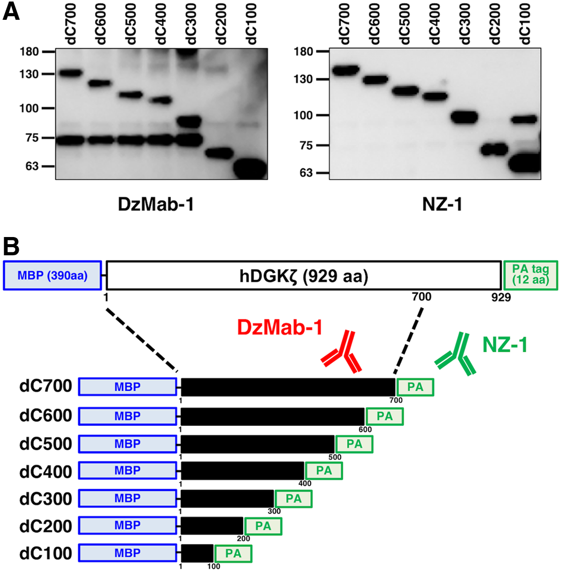
Epitope mapping of DzMab-1 using C-terminal deletion mutants of hDGKζ.
We next produced nine N-terminal deletion mutants of hDGKζ-dC200 (i.e., dN10, dN20, dN30, dN40, dN50, dN60, dN70, dN80, and dN90). Western blotting demonstrated that DzMab-1 did not react with those deletion mutants; in contrast, anti-PA tag mAb, NZ-1, detected them (Fig. 2A, B), indicating that the N-terminus of the DzMab-1-epitope exists between 1st aa and 10th aa (Fig. 2C).

Epitope mapping of DzMab-1 using N-terminal deletion mutants of hDGKζ. Immunoprecipitates
We produced the following 10 point mutants using hDGKζ-dC200: M1A, E2A, P3A, R4A, D5A, G6A, S7A, P8A, E9A, and A10G. Western blotting demonstrated that the anti-PA tag mAb, NZ-1, detected all point mutants (Fig. 3A). In contrast, DzMab-1 did not detect M1A and P3A. These results are summarized in Figure 3B.
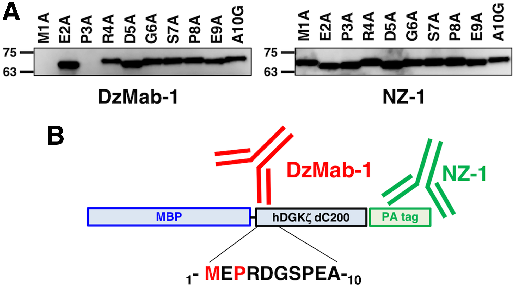
Epitope mapping of DzMab-1 using point mutants of hDGKζ.
In our previous reports, DzMab-1 was shown to be useful for immunocytochemical analyses.(13) In this study, we investigated whether DzMab-1 is also applicable for immunohistochemical analyses. As depicted in Figure 4, DzMab-1 reacted with HeLa cells in immunohistochemical analyses using formalin-fixed paraffin-embedded (FFPE) cells, indicating that DzMab-1 could also be useful for FFPE samples of patient tissues.

Immunohistochemical analyses of DzMab-1 for FFPE HeLa cells. Sections were incubated with 5 μg/mL of DzMab-1
In conclusion, Met1 and Pro3 residues of hDGKζ are important for facilitating DzMab-1 binding to hDGKζ. These findings could be applied for the production of more functional anti-hDGKζ mAbs.
Footnotes
Acknowledgments
This research was supported in part by AMED under grant numbers JP18am0101078 (Y.K.), JP18am0301010 (Y.K.), and JP18ae0101028 (Y.K.), and by JSPS KAKENHI grant numbers 17K07299 (M.K.K.), 16K10748 (Y.K.), and 19K07705 (Y.K.).
Author Disclosure Statement
Y.K. received research funding from Ono Pharmaceutical Co., Ltd. The other authors have no conflicts of interest.