
Abstract
Podoplanin (PDPN)/T1alpha is a type I transmembrane sialoglycoprotein, which is expressed on podocytes of the kidneys and type I alveolar cells of the lungs. PDPN is also known as Aggrus, a platelet aggregation-inducing factor, which comprises three platelet aggregation-stimulating (PLAG) domains (PLAG1, PLAG2, and PLAG3) in the N-terminus and PLAG-like domains (PLDs) in the middle of the PDPN protein. We have previously established a mouse anti-bear PDPN (bPDPN) monoclonal antibody (mAb) clone, PMab-247 using the Cell-Based Immunization and Screening (CBIS) method. PMab-247 is very useful in flow cytometry, Western blotting, and immunohistochemical (IHC) analyses; however, the binding epitope of PMab-247 has not been elucidated. In this study, we aimed to investigate the epitope of PMab-247 using enzyme-linked immunosorbent assay and IHC analyses. The results revealed that the critical epitopes of PMab-247 are Asp76, Arg78, Glu80, and Arg82 of bPDPN. The Glu80 and Arg82 are included in PLD of bPDPN. The findings of our study can be applied to the production of more functional anti-bPDPN mAbs.
Introduction
Podoplanin (PDPN)/T1
In our previous study, we used the Cell-Based Immunization and Screening (CBIS) method to develop sensitive and specific monoclonal antibodies (mAbs) against bear PDPN (bPDPN) to facilitate the immunohistochemical (IHC) analysis of paraffin-embedded tissue sections.(10) Flow cytometry revealed that PMab-247 (mouse IgG1, kappa), one of the established clones, recognized CHO/bPDPN but did not react with CHO-K1. Western blotting demonstrated that PMab-247 could detect bPDPN as a 48-kDa band in CHO/bPDPN cells. IHC analyses revealed that PMab-247 strongly stained pulmonary type I alveolar cells, lymphatic endothelial cells, and podocytes of the kidney.(11) This study aimed to determine the binding epitope of PMab-247 to bPDPN using enzyme-linked immunosorbent assay (ELISA) and IHC analyses.
Materials and Methods
Enzyme-linked immunosorbent assay
Synthesized bPDPN peptides using PEPScreen (Sigma-Aldrich Corp., St. Louis, MO) were immobilized on Nunc Maxisorp 96-well immunoplates (Thermo Fisher Scientific, Inc., Waltham, MA) at 10 μg/mL for 30 minutes at 37°C. After blocking with SuperBlock T20 (PBS) Blocking Buffer (Thermo Fisher Scientific, Inc.), the plates were incubated with purified PMab-247 (1 μg/mL), followed by a peroxidase-conjugated anti-mouse IgG (dilution of 1:2000; Agilent Technologies Inc., Santa Clara, CA). Enzymatic reactions were performed using 1-Step Ultra TMB-ELISA (Thermo Fisher Scientific, Inc.). Optical density was measured at 655 nm using an iMark microplate reader (Bio-Rad Laboratories, Inc., Berkeley, CA). These reactions were performed at 37°C using a total sample volume of 50–100 μL.
Immunohistochemical analyses
Malayan Sun Bear (Helarctos malayanus) tissues were collected after autopsy at Hokkaido University, fixed in 10% neutral-buffered formalin, and routinely processed to make paraffin-embedded tissue sections. Histological sections (4-μm thick) were directly autoclaved in citrate buffer (pH 6.0; Nichirei Biosciences, Inc., Tokyo, Japan) for 20 minutes. These tissue sections were then blocked with the SuperBlock T20 (PBS) Blocking Buffer (Thermo Fisher Scientific, Inc.), incubated with PMab-247 (5 μg/mL) or PMab-247 (5 μg/mL) + peptides (50 μg/mL) for 1 hour at room temperature, and treated using an Envision + Kit (Agilent Technologies, Inc.) for 30 minutes. Color was developed with 3,3′-diaminobenzidine tetrahydrochloride (Agilent Technologies, Inc.) for 2 minutes, and counterstaining was performed using hematoxylin (FUJIFILM Wako Pure Chemical Corporation, Osaka, Japan).
Results and Discussion
We first synthesized a series of bPDPN peptides, which are summarized in Table 1. Using ELISA, PMab-247 detected the 68–87 peptide corresponding to the 68th–87th amino acids (AAs) of bPDPN. We then synthesized point mutants of 68–87 peptides (Table 2). Using ELISA, PMab-247 strongly detected P68A, I69A, N70A, R71A, E72A, G73A, V74A, T75A, L77A, L79A, D81A, T83A, T84A, E85A, S86A, and T87A, and weakly detected R82A. Conversely, it did not detect D76A, R78A, and E80A, indicating that Asp76, Arg78, Glu80, and Arg82 are the critical epitopes of PMab-247.
Determination of PMab-247 Epitope by Enzyme-Linked Immunosorbent Assay
+++, OD655 ≥ 0.6; −, OD655 < 0.1.
Determination of PMab-247 Epitope by Enzyme-Linked Immunosorbent Assay Using Point Mutants
+++, OD655 ≥ 0.6; +, 0.1 ≤ OD655 < 0.4; −, OD655 < 0.1.
We then performed a blocking assay using IHC to confirm the PMab-247 epitope. PMab-247 reacted with lung type I alveolar cells (Fig. 1A, B). These reactions were completely neutralized by P68A (Fig. 1C, D). Conversely, R78A did not block these reactions of PMab-247 (Fig. 1E, F). Likewise, D76A, E80A, and R82A did not inhibit the PMab-247 reaction (Supplementary Fig. S1). Negative control (blocking buffer) did not show any staining (Fig. 1G, H). As shown in Figure 2A and B, PMab-247 reacted with the podocytes and Bowman's capsules of the kidneys. The reactions of PMab-247 in the kidneys were completely neutralized by P68A (Fig. 2C, D) although R78A did not block these reactions of PMab-247 (Fig. 2E, F). Negative control (blocking buffer) did not show any staining (Fig. 2G, H). These IHC results indicate that Asp76, Arg78, Glu80, and Arg82 of bPDPN are essential for PMab-247 binding to bPDPN.
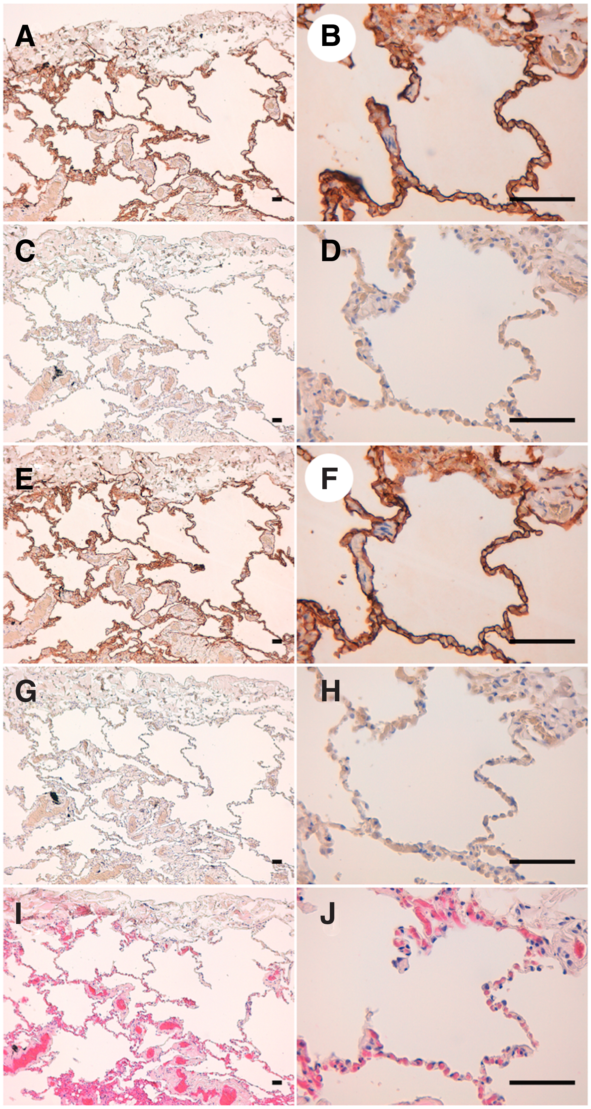
IHC analyses of bear lungs using PMab-247 and point mutants of bPDPN. Histological sections of the bear lungs were directly autoclaved in citrate buffer for 20 minutes. After blocking with SuperBlock T20 (PBS) Blocking Buffer, the sections were incubated with

IHC analyses of bear kidneys using PMab-247 and point mutants of bPDPN. Histological sections of the bear kidneys were directly autoclaved in citrate buffer for 20 minutes. After blocking with SuperBlock T20 (PBS) Blocking Buffer, the sections were incubated with
In summary, the critical epitopes of PMab-247 are Asp76, Arg78, Glu80, and Arg82 of bPDPN. Among these AAs, Glu80 and Arg82 are included in PLAG-like domains of bPDPN, which is important for PDPN/CLEC-2 interaction (Fig. 3). These findings can be applied to the production of more functional anti-bPDPN mAbs.

Schematic illustration of the epitope recognized by PMab-247. PLAG and PLD. Red AAs indicate the critical epitope of PMab-247. AAs, amino acids; PLAG, platelet aggregation-stimulating; PLD, PLAG-like domain.
Footnotes
Acknowledgment
We thank Yoshikazu Satoh for excellent technical assistance.
Author Disclosure Statement
No competing financial interests exist.
Funding Information
This research was supported in part by AMED under grant numbers JP19am0101078 (Y.K.), JP18am0301010 (Y.K.), and JP19ae0101028 (Y.K.), and by JSPS KAKENHI grant numbers 17K07299 (M.K.K.) and 19K07705 (Y.K.).
References
Supplementary Material
Please find the following supplemental material available below.
For Open Access articles published under a Creative Commons License, all supplemental material carries the same license as the article it is associated with.
For non-Open Access articles published, all supplemental material carries a non-exclusive license, and permission requests for re-use of supplemental material or any part of supplemental material shall be sent directly to the copyright owner as specified in the copyright notice associated with the article.