




Abstract
Killer cell lectin-like receptor subfamily G member 1 (KLRG1), a type II transmembrane protein, was identified as an inhibitory receptor expressed on natural killer (NK) cells and certain T cells. The protein regulates effector functions and developmental processes in these cells. In this study, we established a specific and sensitive monoclonal antibody (mAb) for human KLRG1 (hKLRG1), which is useful for flow cytometry, using a Cell-Based Immunization and Screening (CBIS) method. The established anti-hKLRG1 mAb, KLMab-1 (mouse IgG1, kappa), reacted with overexpressed hKLRG1 in Chinese hamster ovary-K1 (CHO/hKLRG1) and human NK cells, which also expressed endogenous hKLRG1 as confirmed by flow cytometry. KLMab-1, which was established by the CBIS method, could be useful for elucidating the hKLRG1-related biological response by flow cytometry.
Introduction
Natural killer (NK) cells generate rapid responses to virus-infected cells, without prior activation of the innate immunity.(1) NK cells are characterized by the expression of CD16 and CD56, and the lack of CD3. The proportion of NK cells in peripheral blood lymphocytes is ∼7%.(2) Several activating and inhibitory receptors are also expressed on NK cell surfaces.(3,4)
Killer cell lectin-like receptor subfamily G member 1 (KLRG1) is a type II transmembrane protein and is an inhibitory receptor expressed on NK and some T cells.(5–7) The protein is also expressed on group 2 innate lymphoid cells in mice and humans.(8,9) It is reported that the percentage of KLRG1-expressing NK cells is increased by viral infection.(6) IL-2 receptor signaling is essential for the development of KLRG1+ terminally differentiated T regulatory cells.(10) KLRG1 also binds to E-, N-, and R-cadherin. Of these proteins, E-cadherin is known to inhibit T cell proliferation, NK cell cytotoxicity, and NK or T cell activation by binding to KLRG1; therefore, KLRG1 regulates effector functions and developmental processes in NK or T cells.(11–15) Moreover, the interactive inhibition between E-cadherin and KLRG1 prevents metastasis in the 4T1 metastatic breast cancer model.(16)
Programed cell death 1 (PD-1) and cytotoxic T lymphocyte-associated protein 4 (CTLA-4) are inhibitory receptors similar to KLRG1 and are putative therapeutic targets for immune checkpoints.(17–20) Anti-PD-1 monoclonal antibodies (mAbs) and anti-CTLA-4 mAbs are reported as potential anticancer drugs.(21,22) By contrast, mAbs against KLRG1 have not been developed as potential drugs for cancer therapies.
In this study, we developed a novel anti-KLRG1 mAb using a Cell-Based Immunization and Screening (CBIS) method(23,24) and investigated its application in flow cytometry.
Materials and Methods
Cell lines
Chinese hamster ovary-K1 (CHO-K1) and P3X63Ag8U.1 (P3U1) were obtained from the American Type Culture Collection (Manassas, VA). For flow cytometry, NK cells (donor lot 4022602 NK1602) were purchased from Takara Bio, Inc. (Shiga, Japan). Synthesized DNA (Eurofins Genomics KK, Tokyo, Japan) encoding human KLRG1 (hKLRG1; accession no: NP_001316028.1) plus N-terminal PA tag(25) was subcloned into a pCAG-Ble vector (FUJIFILM Wako Pure Chemical Corporation, Osaka, Japan). The hKLRG1 plasmid was transfected into CHO-K1 cells using Lipofectamine LTX with Plus Reagent (Thermo Fisher Scientific Inc., Waltham, MA). Stable transfectants were identified using a cell sorter (SH800; Sony Corp., Tokyo, Japan) and cultivated in a medium containing 0.5 mg/mL zeocin (InvivoGen, San Diego, CA) to generate CHO/PA-hKLRG1 cells.
CHO-K1, P3U1, and CHO/PA-hKLRG1 cells were cultured in Roswell Park Memorial Institute (RPMI) 1640 medium (Nacalai Tesque, Inc., Kyoto, Japan), supplemented with 10% heat-inactivated fetal bovine serum (Thermo Fisher Scientific Inc.), 100 U/mL penicillin, 100 μg/mL streptomycin, and 0.25 μg/mL amphotericin B (Nacalai Tesque, Inc.). Cells were grown in a humidified incubator at 37°C with 5% CO2.
Antibodies
An anti-hKLRG1 mAb (clone 14C2A07; mouse IgG2a, kappa) was purchased from BioLegend (San Diego, CA). Secondary Alexa Fluor 488-conjugated anti-mouse IgG was purchased from Cell Signaling Technology, Inc. (Danvers, MA).
Hybridoma production
Female BALB/c mice (6 weeks old) were purchased from CLEA Japan (Tokyo, Japan). Animals were housed under specific pathogen-free conditions. Animal studies were performed according to the relevant guidelines and regulations to minimize animal suffering and distress. Studies were approved by the Animal Care and Use Committee of Tohoku University (permit: 2019NiA-001). Mice were monitored daily during the 4-week study. A reduction of >25% of total body weight was defined as a humane endpoint. Mice were euthanized by cervical dislocation, and death was verified by respiratory and cardiac arrest.
We used CBIS method to develop mAbs against hKLRG1.(24) In brief, two BALB/c mice were intraperitoneally immunized with CHO/PA-hKLRG1 cells (1 × 10⁸) together with Imject Alum (Thermo Fisher Scientific Inc.). The procedure required three additional immunizations followed by a final booster intraperitoneal injection administered 2 days before spleen cells were harvested. Once harvested, cells were fused with P3U1 cells using polyethylene glycol 1500 (Roche Diagnostics, Indianapolis, IN). For selection, resultant hybridomas were grown in RPMI medium supplemented with hypoxanthine, aminopterin, and thymidine (Thermo Fisher Scientific Inc.). Culture supernatants were screened by flow cytometry.
Flow cytometry
Cells were harvested after a brief exposure to 0.25% trypsin and 1 mM ethylenediaminetetraacetic acid (Nacalai Tesque, Inc.). They were washed in 0.1% bovine serum albumin in phosphate-buffered saline, treated with primary mAbs for 30 minutes at 4°C, and then treated with Alexa Fluor 488-conjugated anti-mouse IgG (1:2000; Cell Signaling Technology, Inc.). Fluorescence data were collected using the SA3800 Cell Analyzer (Sony Corp., Tokyo, Japan).
Determination of binding affinity by flow cytometry
CHO/PA-hKLRG1 cells were suspended in 100 μL serially diluted anti-hKLRG1 mAbs, and Alexa Fluor 488-conjugated anti-mouse IgG was added (1:200; Cell Signaling Technology, Inc.). Fluorescence data were collected using the BD FACSLyric instrument (BD Biosciences, Franklin Lakes, NJ). The dissociation constant (KD) was calculated by fitting binding isotherms to built-in one-site binding models in GraphPad PRISM 8 (GraphPad Software, Inc., La Jolla, CA).
Results
Establishment of anti-hKLRG1 mAbs
CBIS method was used to establish anti-hKLRG1 mAbs (Fig. 1). First, two mice were immunized with CHO/PA-hKLRG1 cells, and hybridomas were produced by fusing splenocytes with P3U1 myeloma cells. Next, we selected hybridomas whose supernatants were positive for CHO/PA-hKLRG1 but negative for CHO-K1 cells, using flow cytometry. The first screening identified strong signals from CHO/PA-hKLRG1 cells and weak or no signals from CHO-K1 cells in 22 of 960 wells (2.3%). After limiting dilutions and several additional screenings, KLMab-1 (IgG1, kappa), which reacted most strongly with CHO/PA-hKLRG1, was established.

Production of anti-hKLRG1 mAbs. Schematic representation of CBIS.
Flow cytometric analyses
KLMab-1 recognized CHO/PA-hKLRG1 but not CHO-K1 cells (Fig. 2A). Also, KLMab-1 reacted with endogenous KLRG1 in NK cells (Fig. 2A). A 0.01 μg/mL concentration was adequate for KLMab-1 recognition of CHO/PA-hKLRG1, but the KLMab-1 recognition of NK cells was weak (Fig. 2A). As a positive control, clone 14C2A07 (BioLegend) reacted similarly with all cell types (Fig. 2B).
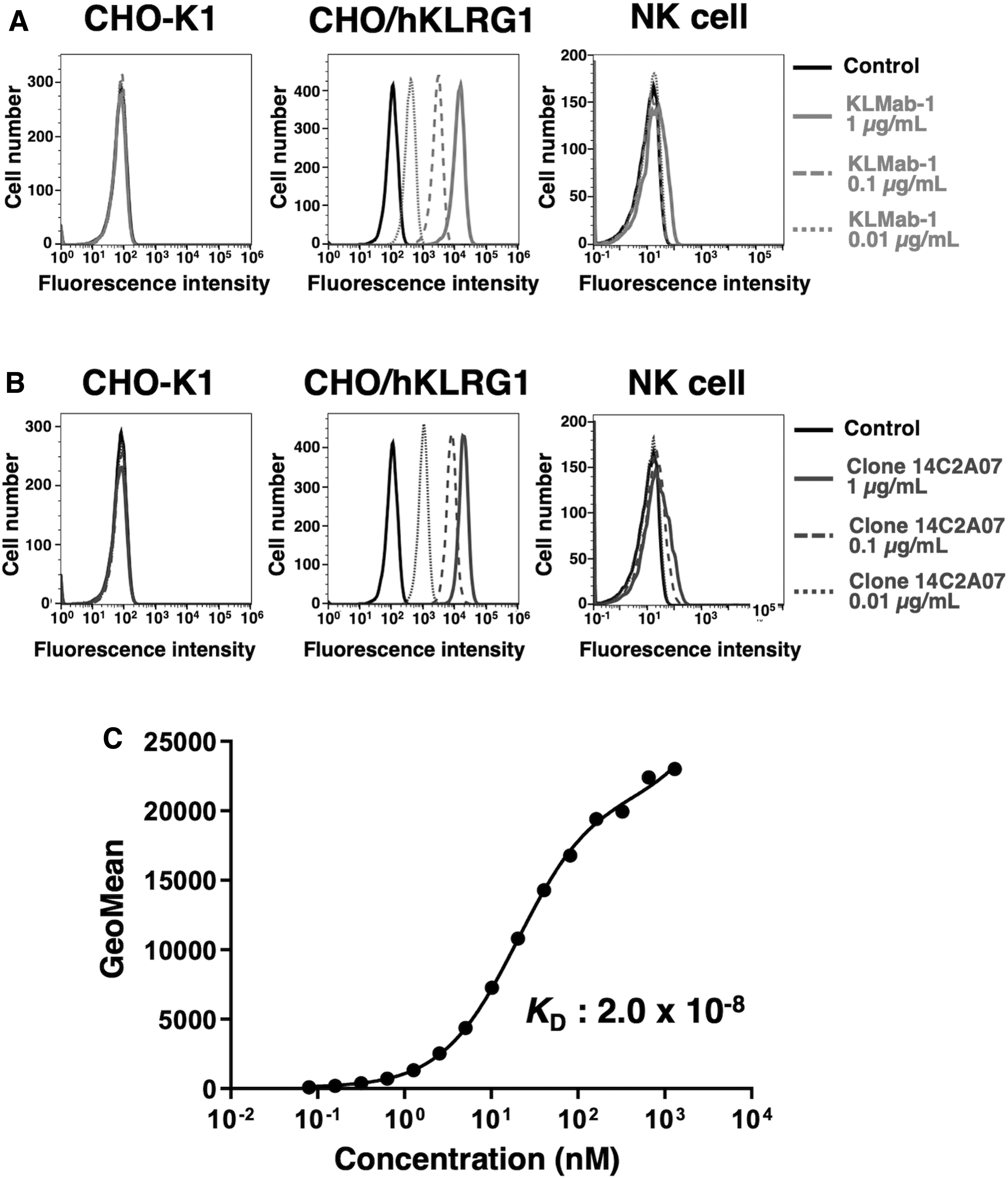
Flow cytometry using anti-hKLRG1 mAbs.
Determination of binding affinity using KLMab-1 in hKLRG1-expressing cells
We conducted a kinetic analysis of the interaction between KLMab-1 and CHO/PA-hKLRG1 cells by flow cytometry to assess the binding affinity of KLMab-1 with CHO/PA-hKLRG1 cells. The geometric mean of fluorescence intensity was plotted against different KLMab-1 concentrations and fitted by one-site binding models. The KD of KLMab-1 for CHO/PA-hKLRG1 cells was determined to be 2.0 × 10⁻⁸ M (Fig. 2C), suggesting that KLMab-1 possessed a moderate binding affinity for these cells.
Discussion
By using CBIS method, the biological structure and modification of proteins, such as glycosylation and folding, could be retained. In commercial products, two mAbs for flow cytometry, including clone 14C2A07 from BioLegend (Fig. 2B), have been developed using cell-based immunization (Supplementary Table S1). It generates a more natural and functional antigen than do synthetic peptides as immunogens. Furthermore, it is easier to acquire mAbs against multipass transmembrane proteins, because purified proteins are not required as immunogens and for screening purposes. Previously, we developed mAbs against several membrane proteins, such as CD20,(26) CD44,(27) CD133,(24) EpCAM,(28) and TROP2,(29) using CBIS method. In this study, we successfully developed a sensitive anti-hKLRG1 mAb, KLMab-1. KLMab-1 recognized overexpressed and endogenous hKLRG1 in our cell lines (Fig. 2). Because hKLRG1 is considered the next pivotal immune checkpoint molecule,(30) KLMab-1 and in vivo mouse models may be used to investigate molecular mechanisms behind hKLRG1-related diseases.
Footnotes
Authors' Contributions
T.A., R.N., and T.N. performed experiments; M.K.K. designed the experiments; T.A. and Y.K. wrote the article.
Author Disclosure Statement
The authors have no conflict of interest.
Funding Information
This research was supported in part by Japan Agency for Medical Research and Development (AMED) under Grant Numbers: JP20am0401013 (Y.K.) and JP20am0101078 (Y.K.).

References
Supplementary Material
Please find the following supplemental material available below.
For Open Access articles published under a Creative Commons License, all supplemental material carries the same license as the article it is associated with.
For non-Open Access articles published, all supplemental material carries a non-exclusive license, and permission requests for re-use of supplemental material or any part of supplemental material shall be sent directly to the copyright owner as specified in the copyright notice associated with the article.
