




Abstract
The development of specific antibodies is essential to understand a wide variety of biological phenomena and pathophysiological analyses. Podoplanin (PDPN), a type I transmembrane glycoprotein, is known as a diagnostic marker. Anti-PDPN monoclonal antibodies (mAbs) against many species, such as human, mouse, rat, rabbit, dog, bovine, cat, tiger, horse, pig, goat, alpaca, Tasmanian devil, bear, whale, and sheep, have been established in recent studies. However, sensitive and specific mAbs against elephant PDPN (elePDPN) have not been established. Thus, this study established a novel mAb against African savanna elephant (Loxodonta africana) PDPN using the Cell-Based Immunization and Screening method. elePDPN-overexpressed Chinese hamster ovary-K1 (CHO/elePDPN) cells were immunized, and mAbs were screened against elePDPN using flow cytometry. One of the mAbs, PMab-265 (IgM, κ), specifically detected CHO/elePDPN cells by flow cytometry. These findings suggested the potential usefulness of PMab-265 for the functional analyses of elePDPN.
Introduction
Elephants are known to be extremely cancer resistant; however, about 3% of elephants develop cancer.(1,2) The analysis of elephant cancers will help in the elucidation of the mechanism of cancer development. For pathophysiological analysis, specific and sensitive antibodies against elephant cells are essential.
Podoplanin (PDPN) is a type I transmembrane glycoprotein expressed in many cell types, including lymphatic endothelial cells.(3) Therefore, PDPN has been used as a lymphatic endothelial cell marker to distinguish lymphatic endothelial cells from vascular endothelial cells in pathophysiological studies.(4) In humans, PDPN expression has been reported in several tumors, including malignant brain tumors,(5–8) oral squamous cell carcinomas,(9) lung cancers,(10) esophageal cancers,(11) malignant mesotheliomas,(12,13) osteosarcomas,(14–16) chondrosarcomas,(15) and testicular tumors.(17) Moreover, PDPN expression is associated with malignant progression and cancer metastasis.(5,18,19) Therefore, anti-PDPN monoclonal antibodies (mAbs) are useful for investigating PDPN expression in various cancers.
Previously, we have developed anti-PDPN mAbs for cat,(20) tiger,(21) horse,(22) pig,(23) goat,(24) alpaca,(25) Tasmanian devil,(26) bear,(27) whale,(28) and sheep(29) using the Cell-Based Immunization and Screening (CBIS) method.(30–32) These mAbs have all been proven useful in flow cytometry, Western blotting, and immunohistochemical analyses. However, sensitive and specific mAbs for elephant PDPN (elePDPN) have not yet been developed. In this study, we immunized mice with MAP16-tagged elePDPN-overexpressed Chinese hamster ovary-K1 (CHO/MAP16-elePDPN) cells, and established anti-elePDPN mAbs, which can be used for flow cytometry.
Materials and Methods
Cell lines
CHO-K1 and P3X63Ag8U.1 (P3U1) cells were obtained from the American Type Culture Collection (Manassas, VA). African savanna elephant fibroblast-like cells (LACF-NaNaI) were obtained from the RIKEN BioResource Center (Ibaraki, Japan).(33) The cDNA encoding the full-length open reading frame (ORF) of elePDPN (accession no. XM_010593105.2) was obtained by polymerase chain reaction using cDNA derived from LACF-NaNaI cells. The elePDPN ORF with N-terminal MAP16 tag (MAP16-elePDPN) or RIEDL tag (RIEDL-elePDPN) was subcloned into a pCAG-Ble vector (FUJIFILM Wako Pure Chemical Corporation, Osaka, Japan). Plasmids were transfected using Lipofectamine LTX with Plus Reagent (Thermo Fisher Scientific, Inc., Waltham, MA). Stable transfectants were selected by limiting dilution and cultivation in a medium containing 0.5 mg/mL Zeocin (InvivoGen, San Diego, CA). CHO/MAP16-elePDPN cells were subsequently used as an immunogen, whereas CHO/RIEDL-elePDPN cells were used for further analyses. P3U1, CHO-K1, CHO/MAP16-elePDPN, and CHO/RIEDL-elePDPN cells were cultured in RPMI-1640 (Nacalai Tesque, Inc., Kyoto, Japan) supplemented with 10% heat-inactivated fetal bovine serum (FBS; Thermo Fisher Scientific, Inc.), 100 U/mL penicillin, 100 μg/mL streptomycin, and 0.25 μg/mL amphotericin B (Nacalai Tesque, Inc.), and incubated at 37°C in a humidified atmosphere containing 5% CO2. LACF-NaNaI cells were cultured in minimum essential medium (Nacalai Tesque, Inc.) supplemented with 10% heat-inactivated FBS (Thermo Fisher Scientific, Inc.), 100 U/mL penicillin, 100 μg/mL streptomycin, and 0.25 μg/mL amphotericin B (Nacalai Tesque, Inc.), 5 μg/mL plasmocin (InvivoGen), and 1 mM L-sodium pyruvate (Nacalai Tesque, Inc.) and incubated at 37°C in a humidified atmosphere containing 5% CO2.
Hybridoma production
Female BALB/c mice (6 weeks old) were purchased from CLEA Japan (Tokyo, Japan). The animals were housed under specific pathogen-free conditions. The Animal Care and Use Committee of Tohoku University approved all animal experiments. The CBIS method was used to develop mAbs against elePDPN. Two BALB/c mice were immunized with CHO/MAP16-elePDPN cells (1 × 10⁸) intraperitoneally with Imject Alum (Thermo Fisher Scientific, Inc.). The procedure included three additional immunizations with CHO/MAP16-elePDPN cells (1 × 10⁸), followed by a final booster injection of CHO/MAP16-elePDPN cells (1 × 10⁸) 2 days before harvest of splenic cells. Subsequently, splenocytes were fused with P3U1 cells using polyethylene glycol 1500 (Roche Diagnostics, Indianapolis, IN). The hybridomas were then grown in RPMI-1640 supplemented with 10% heat-inactivated FBS (Thermo Fisher Scientific, Inc.), 100 U/mL of penicillin, 100 μg/mL streptomycin, and 0.25 μg/mL amphotericin B (Nacalai Tesque, Inc.), 5 μg/mL plasmocin (InvivoGen), and hypoxanthine/aminopterin/thymidine (Thermo Fisher Scientific, Inc.). The culture supernatants were screened for anti-elePDPN antibody production using flow cytometry.
Flow cytometry
CHO/RIEDL-elePDPN and parental CHO-K1 cells were harvested after a brief exposure to 0.25% trypsin in 1 mM ethylenediaminetetraacetic acid (Nacalai Tesque, Inc.). After washing with 0.1% bovine serum albumin (BSA; Nacalai Tesque, Inc.) in phosphate-buffered saline (PBS; Nacalai Tesque, Inc.), cells were treated with anti-elePDPN mAb (1 μg/mL) for 30 minutes at 4°C, followed by Alexa Fluor 488-conjugated anti-mouse IgG (1:1000; product no. 4408; Cell Signaling Technology, Inc., Danvers, MA). Fluorescence data were collected using BD FACSLyric (Becton, Dickinson and Company, Franklin Lakes, NJ).
Results and Discussion
To develop anti-elePDPN mAbs, the CBIS method was employed using stable transfectants for immunization and flow cytometry (Fig. 1). Two mice were immunized with CHO/MAP16-elePDPN cells that overexpressed elePDPN with an N-terminal MAP16 tag. Hybridomas were seeded into 96-well plates, and CHO/RIEDL-elePDPN-positive and CHO-K1-negative wells were identified. The first screening identified strong signals from CHO/RIEDL-elePDPN cells and weak or no signals from CHO-K1 cells in 4 of 1054 wells (0.38%). After limiting dilution and several additional screenings, PMab-265 (IgM, κ), which reacted most strongly with CHO/RIEDL-elePDPN, was finally established.

Production of anti-elePDPN mAbs. Schematic representation of the CBIS method. (1) CHO/MAP16-elePDPN cells were immunized into BALB/c mice by intraperitoneal injection. (2) Splenocytes were fused with myelomas for hybridoma production. (3) Screening of anti-elePDPN mAb-producing hybridomas was performed by flow cytometry using CHO/RIEDL-elePDPN and parental CHO-K1 cells. (4) Anti-elePDPN mAb-producing hybridomas were cloned using the limiting dilution method. CBIS, Cell-Based Immunization and Screening.
Next, flow cytometric analyses were performed using PMab-265 with CHO/RIEDL-elePDPN, CHO-K1, and LACF-NaNaI cells. LACF-NaNaI cells are fibroblast-like cells derived from African savanna elephants.(33) Cells were treated with PMab-265 (Fig. 2, black line) or without PMab-265 (Fig. 2, gray shading). PMab-265 recognized CHO/RIEDL-elePDPN cells (Fig. 2A) but not CHO-K1 cells (Fig. 2B). PMab-265 also recognized endogenous elePDPN in LACF-NaNaI cells (Fig. 2C). These results demonstrated that PMab-265 is applicable for detecting both overexpressed elePDPN and endogenous elePDPN using flow cytometry. Although elephants are extremely cancer-resistant, about 3% of them develop cancers.(1,2) Since PMab-265 is an elePDPN-specific mAb, it may be useful for the pathophysiological analysis of lymphangiogenesis in elephant cancers.
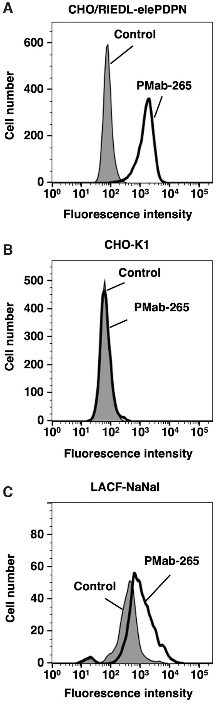
Detection of elePDPN by flow cytometry using PMab-265. CHO/RIEDL-elePDPN
This study succeeded in developing anti-elePDPN using the CBIS method. The CBIS method was previously successful in developing several mAbs against membrane proteins, such as CD20,(34) CD44,(32) and TROP2.(35) This method helps establish mAbs against membrane proteins because purified proteins are not required as immunogens and for screening purposes. Moreover, the biological structure and modification of proteins, such as glycosylation and folding, could be retained; therefore, mAbs established using the CBIS method could detect endogenous proteins.
This study showed that PMab-265 is useful for detecting endogenous elePDPN by flow cytometric analysis; however, it is not clear whether PMab-265 is applicable for the other analyses, such as immunohistochemistry and Western blotting. Future studies need to assess the usability of PMab-265 in immunohistochemistry and Western blotting analyses of elephant tissues.
Footnotes
Author Disclosure Statement
No competing financial interests exist.
Funding Information
This research was supported in part by Japan Agency for Medical Research and Development (AMED) under Grant Numbers: JP21am0401013 (to Y.K.) and JP21am0101078 (to Y.K.), and by the Japan Society for the Promotion of Science (JSPS) Grants-in-Aid for Scientific Research (KAKENHI) grant nos. 21K15523 (to T.A.), 21K07168 (to M.K.K.), 20K16322 (to M.S.), and 19K07705 (to Y.K.).