
Abstract
CC chemokine receptor type-2 (CCR2) is a member of the G protein-coupled receptors, and is mainly expressed on cell surface of immune cells. CCR2 binds to its ligand, C-C motif chemokine 2 (also named as monocyte chemoattractant protein-1), which involves in the tumor progression by modulating the tumor microenvironment. Therefore, the monoclonal antibody (mAb) targeting CCR2 could be one of the strategies for cancer treatment. In this study, we investigated the critical epitope of C2Mab-6, an anti-mouse CCR2 (mCCR2) mAb developed by N-terminal peptides immunization. We first performed enzyme-linked immunosorbent assay (ELISA) using N-terminal peptides of mCCR2 and demonstrated that C2Mab-6 recognizes 1–19 amino acids of mCCR2. We further performed ELISA using 20 alanine-substituted peptides of mCCR2. C2Mab-6 lost the reaction to the alanine-substituted peptides of D3A, N4A, M6A, P8A, Q9A, and F10A. These results indicate that the binding epitope of C2Mab-6 includes Asp3, Asn4, Met6, Pro8, Gln9, and Phe10 of mCCR2.
Introduction
Chemokine receptors are G protein-coupled receptor (GPCR) with seven transmembrane regions, which are localized on the plasma membrane. Chemokines are divided into four different subfamilies of XC, CC, CXC, and CX3C, according to the number and position of conserved N-terminus cysteine residues. 1 Chemokines orchestrate the cellular function of immune cells through their interaction with their receptors, as represented by cell migration. 2
CC chemokine receptor type-2 (CCR2) is expressed in various immune cells including T lymphocytes, natural killer cells, macrophages, dendritic cells, and monocytes. CCR2 is primarily involved in the regulation of migration and positioning of immune cells.3,4 CCR2 is the major receptor of C-C motif chemokine 2 (CCL2)/monocyte chemoattractant protein-1. CCL2–CCR2 axis plays pivotal roles in the regulation of immune system. 5
At the inflammation site, CCR2-expressing cells are associated with tissue damage. During influenza A virus infection, interferon-γ-stimulated CCR2+ monocytes infiltration has been reported as a driver of lung damage. 6 High levels of CCR2 in peripheral blood have been detected in severe COVID-19 patients. 7 Genome-wide studies suggest that 3p21.31 is involved in the risk of severe COVID-19 aggravation and is involved in upregulation of chemokine receptors, including CCR2 in monocytes and macrophages. 8
CCL2 expression has been reported to be upregulated in several tumors, including breast, bladder, and bone cancers.9–11 High CCR2 expression in tumor-infiltrating immune and stromal cells has been confirmed,12–14 and associated with poor prognosis.15,16 Therefore, the CCL2–CCR2 axis is thought to be an important target for cancer therapy.
We have developed monoclonal antibodies (mAbs) against chemokine receptors, including mouse CCR2 (mCCR2), 17 mouse CCR3, 18 mouse CCR4, 19 mouse CCR8, 20 human CCR9, 21 and mouse CXCR6 22 by the Cell-Based Immunization and Screening method or the N-terminal peptide immunization method. In this study, we investigated the epitope of an anti-mCCR2 mAb, C2Mab-6, 17 by using enzyme-linked immunosorbent assay (ELISA).
Materials and Methods
Enzyme-linked immunosorbent assay
The mCCR2 peptides (accession no.: NM_009915), including three N-terminal peptides (Table 1) and 20 point mutants (Table 2), were synthesized by utilizing PEPscreen (Sigma-Aldrich Corp., St. Louis, MO). Each peptide was immobilized on Nunc Maxisorp 96-well immunoplates (Thermo Fisher Scientific, Inc., Waltham) at a concentration of 10 μg/mL for 30 minutes at 37℃. After washing with phosphate-buffered saline containing 0.05% Tween20 (PBST), wells were blocked with 1% bovine serum albumin-containing PBST for 30 minutes at 37℃.
Identification of the C2Mab-6 Epitope Using N-Terminal Mouse CCR2 Peptides
+++, OD655 ≧ 0.3; −, OD655 < 0.1.
Identification of the C2Mab-6 Epitope Using Alanine-Substituted Mouse CCR2 Peptides
+++, OD655 ≧ 0.3; ++, 0.2 ≦ OD655 < 0.3; +, 0.1 ≦ OD655 < 0.2; −, OD655 < 0.1.
The plates were then incubated with C2Mab-6 (1 μg/mL), followed by a 1:2000 (1:20000) dilution of peroxidase-conjugated anti-rat immunoglobulins (Sigma-Aldrich Corp.). Enzymatic reactions were performed using the ELISA POD Substrate TMB Kit (Nacalai Tesque, Inc., Japan). Optical density was detected at 655 nm using an iMark microplate reader (Bio-Rad Laboratories, Inc., Berkeley, CA).
Results
Epitope identification using N-terminal mCCR2 peptides
An anti-mCCR2 mAb, C2Mab-6 (rat IgG1, kappa), was established by immunization with the mixture of three keyhole limpet hemocyanin-conjugated mCCR2 N-terminal peptides. 17 The sequences are listed in Table 1. To characterize the binding epitope of C2Mab-6, we first investigated the reactivity of C2Mab-6 to the N-terminal synthetic peptides: 1–19 amino acids (aa), 11–29 aa, and 21–39 aa of mCCR2 (Table 1). The results of ELISA demonstrated that C2Mab-6 recognized 1–19 aa of mCCR2 (Fig. 1A). These results are summarized in Figure 1B.
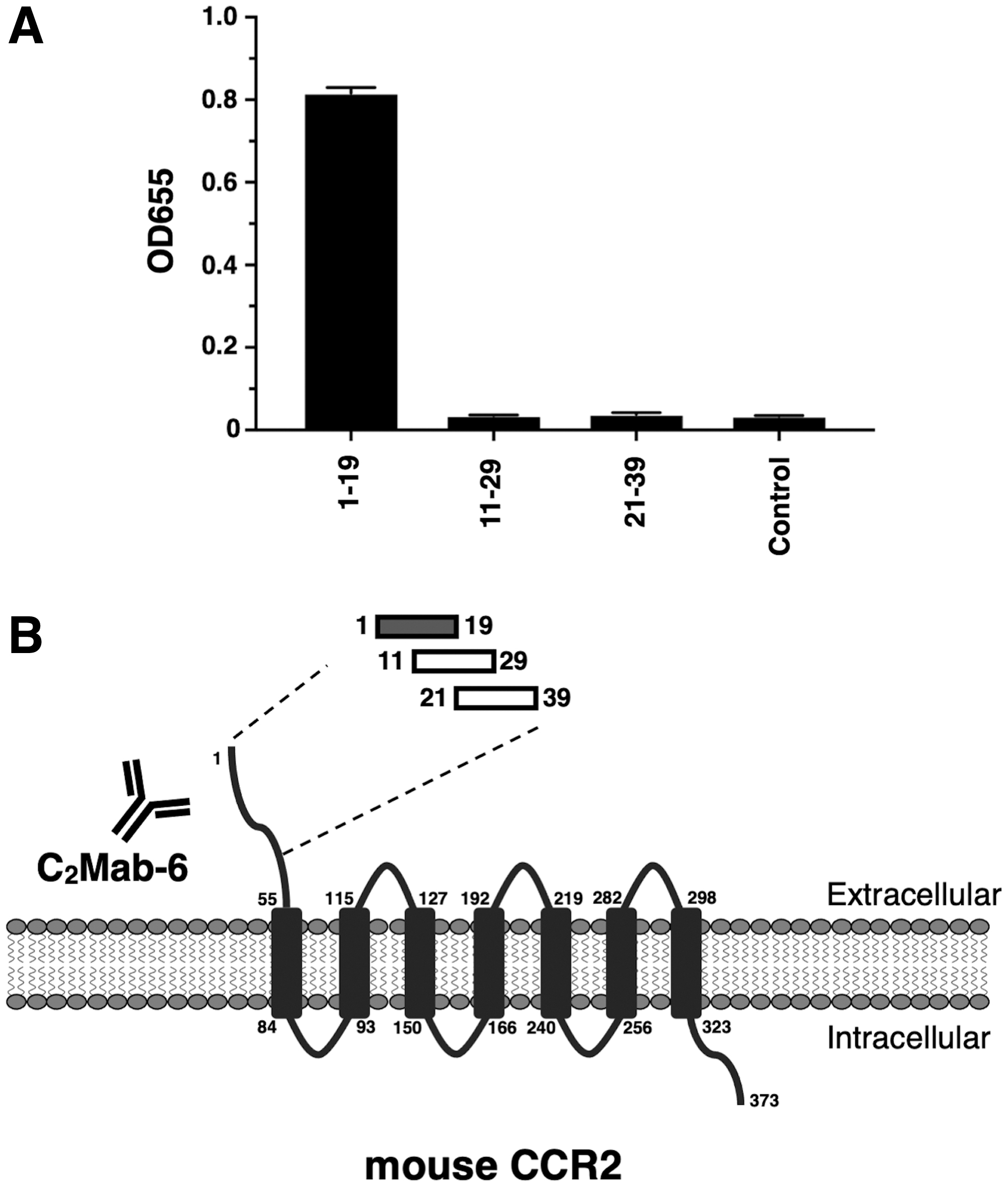
Identification of the C2Mab-6 epitope for mCCR2 by ELISA using N-terminal peptides.
Epitope identification using alanine-substituted mCCR2 peptides
We further synthesized 20 different alanine-substituted mCCR2 peptides (Table 2). The results of ELISA demonstrated that C2Mab-6 bound to point mutants, such as M1A, E2A, N5A, L7A, I11A, H12A, G13A, I14A, L15A, S16A, T17A, S18A, H19A, and S20A as well as the 1–20 aa wild type (WT) sequence (positive control) (Fig. 2A). In contrast, C2Mab-6 did not bind to point mutants, such as the D3A, N4A, M6A, P8A, Q9A, and F10A (Fig. 2A), indicating that Asp3, Asn4, Met6, Pro8, Gln9, and Phe10 were determined to be the critical aa, included in the C2Mab-6 epitope. The results are summarized in Figure 2B.
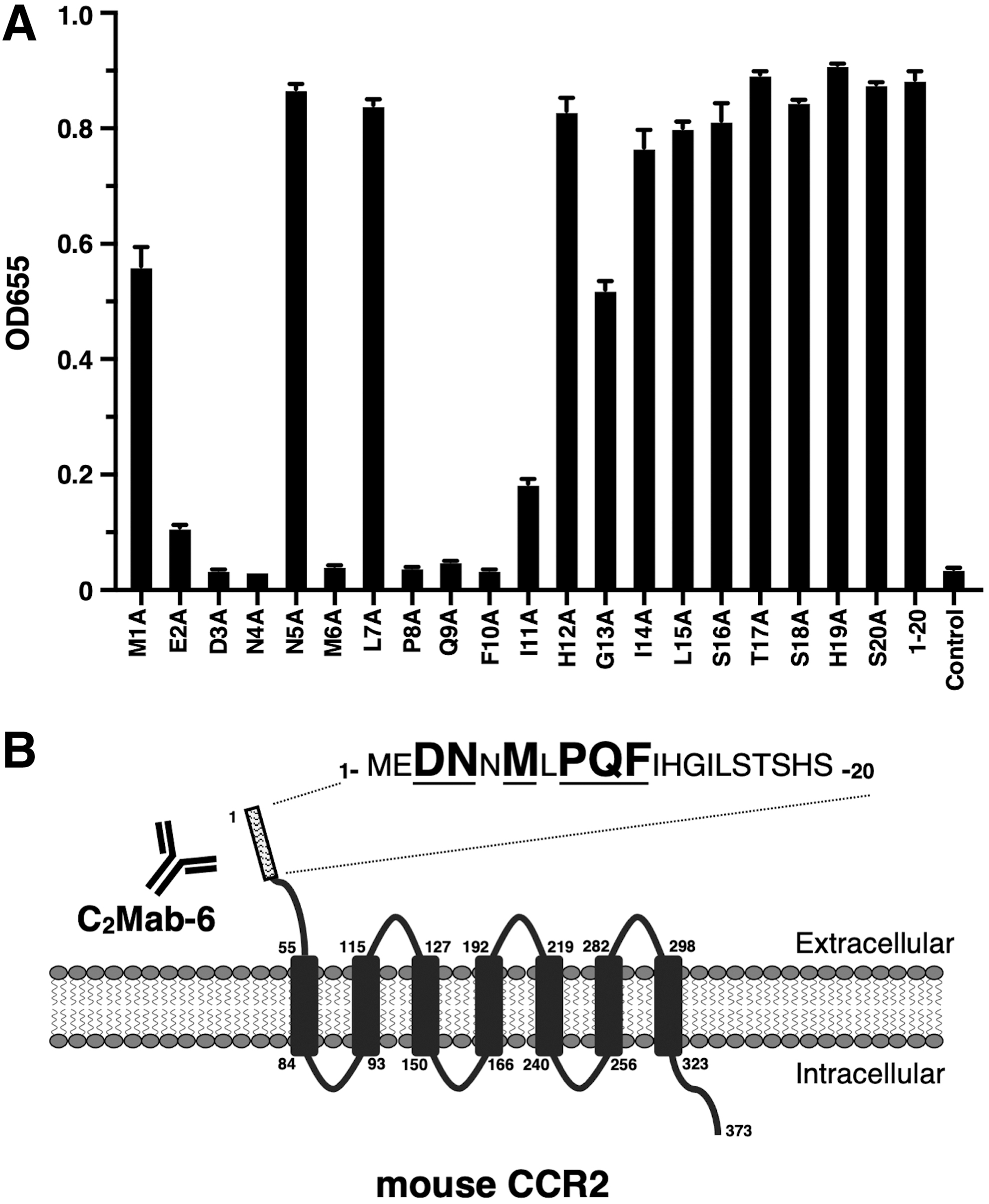
Identification of the C2Mab-6 epitope for mCCR2 by ELISA using alanine-substituted peptides.
Discussion
We previously developed an anti-human CCR2 mAb (clone C2Mab-9) by using N-terminal peptide immunization method and determined the epitope of C2Mab-9 as Phe23, Phe24, Asp25, and Tyr26. 23 In this study, we successfully determined that the epitope of an anti-mCCR2 mAb (clone C2Mab-6) 17 as Asp3, Asn4, Met6, Pro8, Gln9, and Phe10 (Fig. 2). The N-terminal regions of some GPCRs, including CCR2, CCR3, CCR5, and CXCR1, have been identified as their ligand-binding sites. 1 Recently, cryoelectron microscopy structure of human CCL2 bound to CCR2-G-protein complex was reported.
The structural analysis reveals that N-terminal CCL2 deeply inserts into the extracellular half of the CCR2 transmembrane domain, and forms substantial interactions with CCR2 (Cys113, Thr117, Tyr120, His121, Cys190, Gly191, and P192) through the most N-terminal glutamine of CCL2. Furthermore, N-terminal Gly29, Ala30, Cys32, and His33 of CCR2, closed to the C2Mab-9 epitope, are also involved in the interaction with CCL2. 24 Therefore, there is a possibility that C2Mab-9 affects the CCL2–CCR2 interaction. The 29-GAPCH-33 sequence in human CCR2 is conserved as 42-GEPCH-46 in mCCR2. Since the C2Mab-6 epitope is apart from the CCL2-binding sequence in mouse, further studies are essential to evaluate the biological activity of C2Mab-6.
The inhibition of CCR2 functions has been reported to enhance the antitumor immunity of immune checkpoint blockades including anti-programmed cell death-1 mAb in mouse model. 11 C2Mab-6 could be useful for the investigation of mCCR2 expression in immune cells, and the combination therapy with immune checkpoint blockade in mice tumor models.
Footnotes
Author Disclosure Statement
No competing financial interests exist.
Funding Information
This research was supported in part by Japan Agency for Medical Research and Development (AMED) under Grant Nos. JP22ama121008 (to Y.K.), JP22am0401013 (to Y.K.), JP22bm1004001 (to Y.K.), JP22ck0106730 (to Y.K.), and JP21am0101078 (to Y.K.).