
Abstract
Cross-resistance to macrolide, lincosamide, and streptogramin B (MLSB) antibiotics is mainly mediated by the erm (erythromycin ribosome methylation) genes that encode 23S rRNA methylases in enterococi, and various mechanisms are involved in the streptogramin B resistance. Prevalence of MLSB resistance and its genetic mechanisms were analyzed for a total of 159 strains of Enterococcus faecium isolated from clinical specimens in a university hospital in Japan from 1997 to 2006. Resistance to erythromycin (EM) and clindamycin was detected in 88.1% and 89.9% of all the strains examined, respectively, and expression of resistance was totally constitutive. Although none of the strain was resistant to quinupristin/dalfopristin (Q/D), 28 strains (17.6%) showed intermediate resistance to Q/D (MIC: 2 μg/ml). The erm(B) gene was detected in 139 strains (87.4%), and msrC was found in all the strains examined, whereas no other known MLSB resistance genes were identified. The erm(B) regulator region (RR) containing a coding region of the leader peptide was classified into 13 genetic variations (L1–L3, M, S1–S7, D, and R genotypes) in 56 strains. However, no relatedness was identified between the erm(B) RR genotype and EM resistance, or reduced susceptibility to Q/D, although most of Q/D-intermediate strains were assigned to the L1, L2, and S1 genotypes. Q/D-intermediate strains were classified into five multiple-locus variable-number tandem-repeat analysis (MLVA) types, including four types of clonal complex (CC)-C1, five sequence types (STs), including four STs of CC-17, and several resistance gene/virulence factor profiles. The present study revealed the occurrence of Q/D-intermediate E. faecium, which are composed of heterogeneous strains in Japan, and more genetic diversity in the erm(B) RRs than those reported previously.
Introduction
Quinupristin/dalfopristin (Q/D), a mixture of semisynthetic streptogramin B/A compounds, is used as an efficient therapeutic option for infections caused by multiresistant gram-positive bacteria, including vancomycin-resistant E. faecium. However, resistance to Q/D in E. faecium has been found in clinical isolates from humans in several countries, and an intrahospital dissemination of Q/D-resistant E. faecium was also reported.6,7,20,31,48 Although the detection rate is still low in humans, increase and spread of Q/D-resistant E. faecium are considered as concerns for successful therapy for VRE infections.
Both resistance mechanisms to streptogramin A and to streptogramin B are necessary for occurrence of Q/D resistance. 49 The resistance mechanism against streptogramin B (quinupristin) is related to that against macrolide and lincosamide. Resistance to macrolides in enterococci is generally due to methylation of 23S rRNA by a methylase encoded by the erm (erythromycin ribosome methylation) gene, 29 which confers cross-resistance to macrolide, lincosamide, and streptogramin B (MLSB) antibiotics. Efflux of macrolides and streptogramin B is mediated by an ABC porter encoded by msrC in E. faecium.33,40,46 Despite still a low frequency in E. faecium, streptogramin B resistance is also conferred by Vgb lactonase via destruction of the ring molecule, causing inactivation of this compound.2,17 Resistance to streptogramin A in E. faecium is mediated by streptogramin acetyltransferases VatD and VatE.34,51
MLSB resistance mediated by erm-encoding methylase is inducible or constitutive, which has been shown to be related to sequence variation in the regulator region (RR) located upstream of the erm genes. 8 Deletion, insertion, and point mutations in a leader peptide-coding region in the RR are implicated in constitutive expression of erm(A) and erm(C) in the staphylococcal and streptococcal species.8,13 Similarly, a large deletion in the RR was identified in enterococci and streptococci, exhibiting constitutive resistance to MLSB.24,36 However, in enterococci, the relation of the genetic diversity in the RR to the expression mode of the erm gene is still ambiguous23,26; therefore, further investigation is necessary in this regard.
In Japan, the frequency of VRE isolation from clinical specimens is still low (120 cases in 2010), 15 and the Q/D-resistant enterococcus has not yet been reported. However, a total reported case of VRE infection has been gradually increasing recently, and hospital outbreaks are also documented. 14 On the other hand, high resistance rates against macrolides were observed for E. faecium and E. faecalis in a cross-sectional study, 53 while their resistance mechanism has not been well characterized. MLSB resistance due to erm(B)-encoding methylase is revealed to decrease the bactericidal activity of Q/D in vitro as well as in experimental endocarditis in rabbits.4,11 Therefore, it is of significance to survey the resistance to macrolide and its resistance mechanisms to alert a reduced susceptibility to Q/D in E. faecium. In the present study, the prevalence of macrolides and Q/D resistances and genetic variations in the RR of the erm(B) genes was investigated for E. faecium strains isolated in a Japanese hospital.
Materials and Methods
Bacterial strains
A total of 159 clinical isolates of E. faecium were analyzed. All the strains were isolated from the specimens of patients with clinical symptoms of infection (single strain per patient), in the Sapporo Medical University Hospital, Sapporo, Japan, for four periods (January 1997–December 1998, 46 strains; January–June 2001, 11 strains; October 2002–September 2004, 83 strains; January–June 2006, 19 strains). E. faecium was identified in our previous study. 45 All the E. faecium isolates showed ampicillin resistance (MIC ≥16 μg/ml), but no strain was resistant to vancomycin. Individual bacterial strains had been stored at −80°C in Microbank™ (PRO-LAB Diagnostics), and were recovered when they were analyzed.
Antimicrobial susceptibility testing
Minimum inhibitory concentrations (MICs) of erythromycin (EM) and Q/D were measured by a broth microdilution test and interpreted as sensitive or resistant based on the National Clinical and Laboratory Standards Institute (CLSI) guidelines. 5 For the present study, Q/D (Synercid) was kindly provided by Astellas Pharma, Inc. Susceptibility to josamycin (JM) and clindamycin (CLDM) was examined by a disk-diffusion test. All the isolates were tested for inducible resistance using D-test as described previously.26,54 The EM disk (15 microgram) and CLDM disk (2 microgram) and EM disk and JM disk (30 microgram) were placed 15 mm and 5 mm apart on an agar plate. After incubation for 18 hr at 37°C, D-zone formation, that is, blunting of the inhibition zone of JM or CLDM proximal to EM disk, was observed. MIC and susceptibility of EM and Q/D were determined also by E-test (BioMerieux). For the selected 21 strains, the MICs were determined for the representative antimicrobial agents (azithromycin, clarithromycin, JM, leucomycin, CLDM, ampicillin, cefoxitin, vancomycin, gentamycin, fosfomycin, tetracycline, and ciprofloxacin) by the broth microdilution test.
Detection of antimicrobial resistance genes and virulence factors by PCR
For all the strains, presence of bacterial genes that are associated with resistance to MLSB antibiotics and reported for enterococcus, streptococcus, and staphylococcus was examined by PCR with the use of the primers published previously. Genes examined and literatures of primers were as follows: erm(A), erm(C), 25 erm(T), 9 vatA-vatE, vgbA, vgbB, 41 vgaA, vgaB, lnu(A), lnu(B), mef(A/E), 6 msrAB, 40 and msrC.46,50 Primers to detect erm(B) (ermBN1: CGAGTGAAAAAGTACTCAACCA, ermBN2: CGGTGAATATCCAAGGTACG) were designed in this study. For the representative strains, presence of the beta-lactamase gene, tetracycline resistance genes, and aminoglycoside resistance genes was examined by PCR as described previously.25,30,45 Bacterial DNA was extracted with achromopeptidase as described previously. 22 In each PCR tube, a reaction mixture (50 μl) containing 1 μl bacterial DNA, 5 units of Ex-Taq™ DNA polymerase (TaKaRa), 200 μM each of dNTP, 30 pM each of primer, 10 mM Tris–HCl (pH8.3), 50 mM KCl, and 1.5 mM MgCl2 was prepared. The reaction mixture in the tube was subjected to 30 PCR cycles of denaturation at 94°C (30 sec), annealing at 55°C or other temperature depending on the primers (30 sec), and primer extension at 72°C (2 min) in a thermal cycler. PCR products with specific sizes of individual resistance genes were detected by agarose gel electrophoresis and staining with ethidium bromide.
The presence of the virulence factors reported for E. faecium (agg, gelE, cylA, esp, efa-Afm, hyl, acm, cpd, cob, and ccf) in the representative strains was examined by PCR using the primers as described previously.3,10
Sequence analysis of erm(B) and the RR of erm(B) and 23S rRNA
Full-length gene sequences of erm(B) were determined directly from PCR products with primers ermB-F1 (CAGATAACTAAAATTACAAACAAATCG) and ermB-R1 (TCTAATAATTTATCTCCATTCCCT). The RR of erm(B), which includes the coding region of the leader peptide, was amplified by PCR with the primer pairs, ermB-N2 and ermB-RR1 (CTTAGAAGCAAACTTAAGAG), or ermB-N2 and ermB-P1 (GTATAATAGGAATTGAAGTTA), for all the erm(B)-positive strains. Depending on the size of the PCR product with these primers, the strains were classified into some groups, from which representative strains were selected for sequence determination. Sequencing reaction was performed with fluorescent dideoxy-chain-termination chemistry using the BigDye Terminator version 3.1 cycle sequencing kit (Applied Biosystems). The DNA sequence was determined by the use of the ABI Prism 3100 genetic analyzer (Applied Biosystems). The secondary structures of the erm(B) RR were predicted by two Web-based programs, the mfold Web server (http://mfold.rna.albany.edu/?q=mfold) and GeneBee-Molecular Biology server (www.genebee.msu.su/genebee.html). The partial sequence of 23S rRNA domain V was determined to check the presence of mutation, by using the PCR product with the primers previously described for S. pneumoniae. 42
Genotyping
For selected strains, the genotype was determined by the multiple-locus variable-number tandem-repeat (VNTR) analysis (MLVA) and multilocus sequence typing (MLST), as described previously.12,43 The MLVA Website (www.umcutrecht.nl/subsite/MLVA/) was used for MLVA type assignment. An allelic profile (allele number) was obtained from the MLST Website (www.mlst.net), and the sequence type (ST) was further analyzed to determine the clonal complex (CC) by eBURST version.3 software.
GenBank accession numbers
Sequences of erm(B) and its RR for representative 13 strains determined in the present study were deposited in the GenBank database under the accession numbers JN899582–JN899594.
Results
Resistance phenotypes and resistance genes
Resistance to EM, CLDM, and JM was detected in 140 (88.1%), 143 (89.9%), and 139 (87.4%) strains, respectively, of all the 159 E. faecium strains examined (Table 1). D-zone was not detected around the JM, CLDM, or EM disks in any strains exhibiting resistance to them, indicating that expression of resistance to these antibiotics was totally constitutive. Most of EM-resistant strains were also resistant to CLDM and JM, except for nine EM-resistant strains susceptible to CLDM and/or JM. Although none of the strain was resistant to Q/D, 28 strains (17.6%) showed intermediate resistance with an MIC of 2 μg/ml by a broth microdilution test, which was confirmed by E-test (1.5–3 μg/ml).
S, susceptible; I, intermediately resistant; R, resistant.
Among the 18 genes related to MLSB resistance in gram-positive cocci, only erm(B) and msrC were detected by PCR in the E. faecium strains in the present study. The erm(B) gene was detected in 139 strains (87.4%), all of which showed resistance to EM with an MIC of ≥128 micrograms/ml. In contrast, among 20 erm(B)-negative strains, six and two strains were judged as resistant to CLDM and JM, respectively. Among the 28 Q/D-intermediate strains, 26 strains possessed erm(B). The msrC was detected in most of the strains (151 strains) by PCR with a primer pair msrC3 and msrC4 described by Werner et al. 46 However, the remaining eight negative strains were PCR-positive with another primer pair, 52 yielding PCR products with an expected size (405 bp). When PCR products from four of the eight strains were determined, the sequences were found to be 95%–99% identical to the msrC gene reported for the E. faecium strains (e.g., DO, TX1330, TX2465, and FAIR-E349) by a BLAST analysis (data not shown). Therefore, all the E. faecium strains were regarded as msrC-positive.
On the basis of the above results, a subsequent analysis was focused on the genetic diversity of erm(B) (and its regulatory region) and the reduced susceptibility to Q/D.
Sequence analysis of erm(B) and the erm(B) RR
A total of 56 strains were randomly selected from the strains showing different sizes of the PCR products from the erm(B) RR, and from different isolation periods, and different susceptibility to Q/D (susceptible or intermediate resistant). The RR sequence located upstream of erm(B) determined for the 56 strains was classified into 13 genotypes designated as L1–L3, M, S1–S7, D, R, and the alignment of individual sequences is shown in Fig. 1. Among these RR genotypes, the difference in the nucleotide length between −10 box and the erm(B) initiation codon was detected (Table 2). In the coding region of the leader peptide, partial duplication of 12 nucleotides (CAAATGCGTTAT) of S1–S7 was found in the L1–L3 types. Compared with the S1–S7 types, type M had two additional nucleotides in this region. Compared with the coding region of the leader peptide of L2 and L3 and S1–S7 (96 and 84 bp in length, respectively), considerably shorter regions were noted in the RR genotypes L1 and M due to nucleotide substitution to cause a termination codon or frameshift alteration by nucleotide insertion, respectively (Table 2 and Fig. 1). The RR genotype D was a deleted form of the genotype S1–S7 sequences, lacking a 62–bp sequence containing an initiation codon of their leader peptide regions, and has no coding region of the leader peptide. In contrast, in the genotype R, an 81-bp sequence containing a 3′-end portion of the leader peptide region was repeated tandemly (Fig. 1).
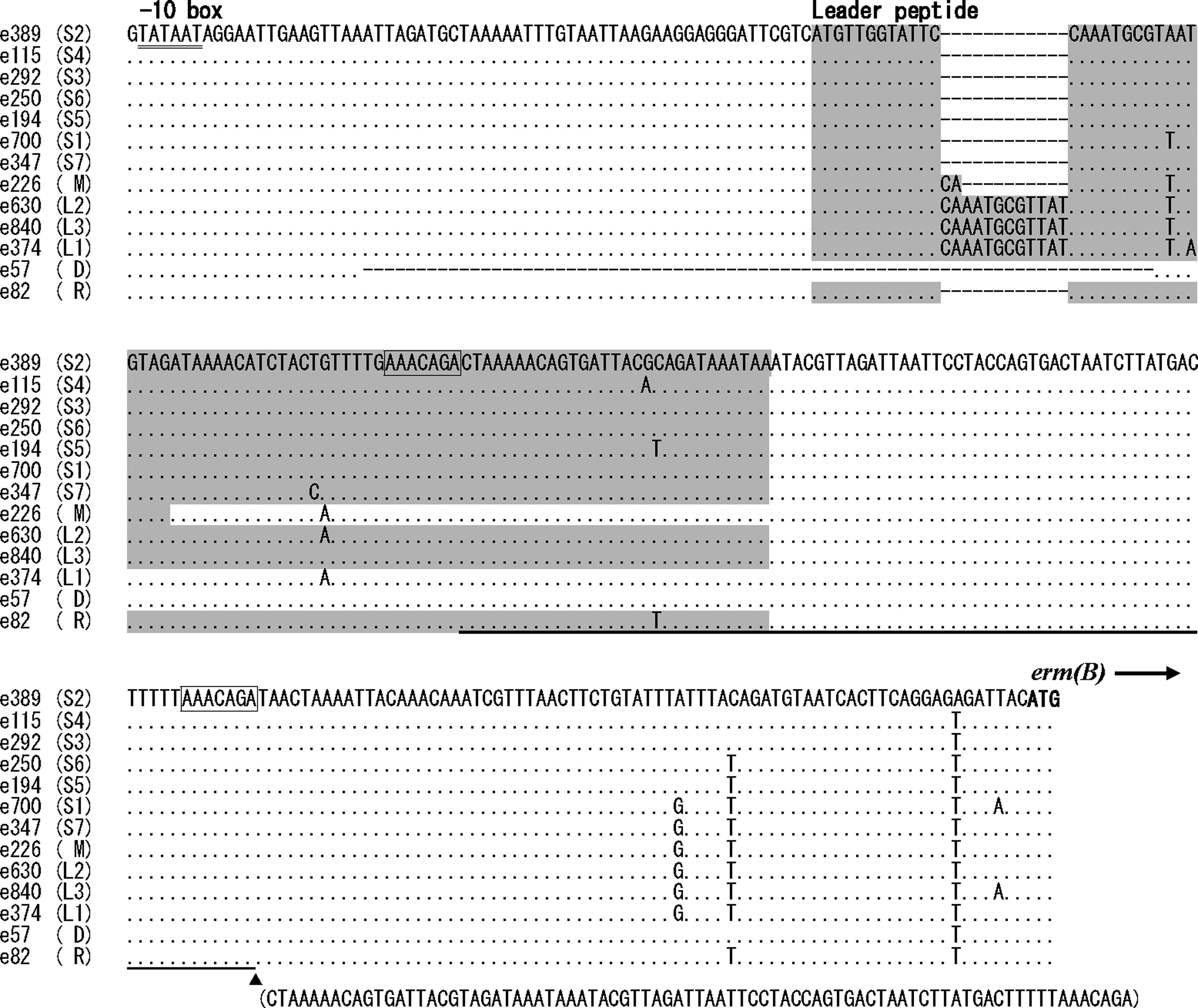
Alignment of the erm(B) regulator region (RR) sequences between −10 box (underlined) and initiation codon of erm(B) representing 13 genotypes (S1–S7, M, L1–3, D, and R). Strain names and RR genotypes are indicated on the left. The identical nucleotide to that in the sequence on the top (genotype S2) is shown by dot. Dash denotes gap. Putative amino-acid-coding region in the leader peptide is shaded. In the genotype R, a sequence with underline (an 81-bp portion that is identical to that in the parenthesis at the bottom) is inserted just after this sequence, at the site of closed triangle, forming a tandem repeat. Direct repeat sequences detected just before the repeat region and 3′-end of the repeating sequence are boxed.
Incidence of the L (L1–L3), M, and S (S1–S7) types among the 56 strains was 18, 9, and 25 strains, respectively, and the RR genotypes L1, L2, M, S1, S2, and S5 were more frequently observed (Table 2). Most of the Q/D-intermediate strains were assigned to the L1, L2, and M genotypes, although these strains belonged to seven different RR genotypes.
Although the erm(B) RR of 13 genotypes was classified into three different secondary structures that were predicted by two programs, the SD sequence of erm(B) was predicted to be located commonly within the loop of the stem–loop structures, at the proximal side to erm(B) (Supplementary Fig. S1; Supplementary Data are available online at www.liebertpub.com/mdr).
The sequence of the erm(B) gene open reading frame (ORF) was determined for 17 representative strains, including those with all the 13 RR genotypes, and Q/D-susceptible and Q/D-intermediate strains. Among these strains, the nucleotide difference was detected in six sites, whereas the amino acid difference in three sites (Table 3). Based on these nucleotide variations, erm(B) was divided into seven subtypes [erm(B)1–erm(B)7].
Codons containing nucleotides that are different from those of strain e374 [erm(B) 1] are underlined and shown in boldface.
MLVA, multiple-locus variable-number tandem-repeat analysis; ST, sequence type; RR, regulator region; EM, erythromycin; Q/D, quinupristin–dalfopristin.
Analysis of 23S rRNA
The nucleotide sequence of 23S rRNA domain V was determined for 15 strains, including 10 Q/D-intermediate strains (eight erm(B)-positive and two erm(B)-negative strains) and five Q/D-susceptible strains (three erm(B)-positive and two erm(B)-negative strains). At three sites in the domain V where mutations associated with macrolide and streptogramin resistance had been reported for S. pneumoniae, 42 all the 15 strains had identical nucleotides to those of the MLS-susceptible strains (Supplementary Fig. S2).
Genotyping, susceptibility, resistance gene, and virulence factors
Clonal heterogeneity and genetic traits of E. faecium strains were analyzed for 21 representative strains. Among them, erm(B)-positive 17 strains were selected from individual erm(B)-RR genotypes (one or two strains per genotype), and from strains with or without Q/D-intermediate resistance and of different origin of specimens. Four erm(B)-negative strains, two Q/D-intermediate strains, and the other two Q/D-susceptible strains derived from different specimens were also selected for this analysis.
Among the 17 erm(B)-positive strains, 10 MLVA types (MTs), including eight types grouped into CC-C1, were identified (Table 4 and Supplementary Table S1). While five types were found in the Q/D-intermediate strains, MT25, MT293, and MT385 were detected in both Q/D-susceptible and Q/D-intermediate strains. STs determined for nine Q/D-intermediate strains belonged to five types, among which four STs (ST78, ST203, ST204, and ST234) were classified into CC17. However, STs belonging to CC17 (ST17, ST78, and ST426) were also found in the Q/D-susceptible strains.
Clonal complex. CC of MT was assigned according to literature (43,51).
Intermediate resistance to Q/D.
blaZ, ant(3'')-Ia, ant(4')-Ia, ant(9)-Ia, ant(9)-Ib, aph(2'')-Ib, aph(2'')-Ic, and aac(6')-Im were not detected in any strains.
agg, gelE, cylA, cpd, and cob were not detected in any strains.
n.t., not typed; n.a., not assigned.
The representative 17 strains with erm(B) showed high MICs (>256 micrograms/ml) to the three macrolides (clarithromycin, azithromycin, and leucomycin), and were mostly resistant to beta-lactams, tetracycline, and ciprofloxacin (Table 4). Most of the strains possessed tet(M), ant(6)-Ia, and aph(3′′)-IIIa, as well as aac(6′)-Ii. erm(B)-positive strains were discriminated into seven resistance gene profiles, among which five profiles were found in the Q/D-intermediate strains. Most of the Q/D-intermediate strains were susceptible to gentamycin and/or fosfomycin. However, a Q/D-intermediate strain e823 had tet(L), tet(M), and aac(6′)-Ie-aph(2′′)-Ia, showing resistance to all the antimicrobials tested, except for vancomycin and Q/D.
The representative 17 erm(B)-positive strains and 4 erm(B)-negative strains commonly possessed the efaAfm and acm genes (Table 4). Both esp and hyl were detected in strains with the erm(B) RR genotypes L1, L2, L3, and M (one strain). erm(B)-positive strains were discriminated into five virulence factor profiles, and all the five profiles were found in the Q/D-intermediate strains.
Discussion
Clinical isolates of E. faecium have been documented to be highly resistant to macrolides,31,35,38,39,53 associated with a high prevalence of erm(B).35,37 Similarly, in the present study, a high rate of EM resistance (88.1%) was observed, and most of them were erm(B)-positive (139/140, 99.3%). Although Q/D resistance in E. faecium was not detected in the report from Europe in 1997–1998, 39 thereafter, 23% of clinical isolates in 1999–2002 were described to be resistant to Q/D in Belgium. 6 Isolation of Q/D-resistant E. faecium was reported in the United States and Korea,7,20,31 whereas a reduced susceptibility to Q/D was observed in Brazil and Greece.19,32 On the other hand, high detection rates of resistance to streptogramins were documented for E. faecium strains from animals in the United States and European countries.16,18,47 In Japan, although Q/D-resistant E. faecium has not yet been isolated, strains showing reduced susceptibility to Q/D were first reported in the present study, with a detection rate of 17.6%. The Q/D-intermediate strains were classified into 5 types each of the MLVA type, ST, resistance gene, and virulence factor profiles, some of which were found in the Q/D-susceptible strains. Q/D-intermediate resistance was found in the strains with the erm(B) subtypes 1, 2, 4, and 6, among which subtypes 1, 2, and 4 were also seen in Q/D-susceptible strains. These findings indicate that the Q/D-intermediate strains are composed of heterogeneous strains that are not distinguishable from Q/D-susceptible strains. Most of MLVA types identified in erm(B)-positive strains were grouped into CC-C1 in the present study. On the other hand, the Q/D-intermediate strains were mostly classified into STs belonging to CC17, and were simultaneously assigned to MLVA types of the CC-C1. The CC17 of MLST is recognized as an emerging subpopulation of E. faecium causing hospital-associated infections worldwide, 44 and a combination of MLST CC17 and MLVA CC-C1 has been reported as an epidemic, virulent, hospital-adapted lineage. 50 The present study suggested that the resistance to MLSB antibiotics is prevalent in E. faecium strains from this lineage.
In the present study, except for erm(B) and msrC, no known gene associated with MLSB resistance was detected in the Q/D-intermediate strains, and no mutation relevant to MLSB resistance was identified in domain V of 23S rRNA. Similarly to our results, Karanika and coworkers reported Q/D-intermediate-resistant E. faecium (28.9% in clinical isolates) in Greece, which did not harbor known streptogramin resistance genes, except for erm(B), suggesting the presence of an undetermined mechanism responsible for a low-level Q/D resistance. 19 Therefore, the causing mechanism of the Q/D-intermediate resistance in E. faecium was not evident. However, the fact that almost all the Q/D-resistant or Q/D-intermediate strains possess erm(B)7,19 suggests the involvement of erm(B) in an altered susceptibility to Q/D. Lopez and coworkers described that the presence of erm(B) was associated with a reduced susceptibility of Q/D in E. faecium clinical isolates. 23 Although erm(B) alone does not confer resistance to Q/D, erm(B)-encoding methylase is revealed to decrease the bactericidal activity of Q/D in vitro, 4 suggesting that excess expression of erm(B) may affect the antibacterial function of Q/D. Secondly, the chromosomal gene msrC was shown to confer a low-level reduction in susceptibility to macrolide and streptogramin B. 40 Thus, the synergistic activity of Q/D is suggested to be inhibited by the excretion of quinupristin from bacterial cells mediated by increased expression of msrC. Although it was not investigated in detail in the present study, msrC in E. faecium appears to be genetically divergent, which was supported by determination of partial sequences, and also suggested by a failure of PCR detection of this gene in some strains using a certain primer pair. 46 It is also conceivable that the functional difference in MsrC, that is, the efficacy in efflux of macrolides and streptogramin B, may be caused by such divergent nature of msrC.
The RR of erm genes is considered to be associated with its inducible expression, and constitutive expression of erm(C) in staphylococcal plasmid pE194 was attributed to a large deletion in this region.8,13 In erm(B) RR from several streptococcal strains and an E. faecalis strain, point mutations in the control peptide, large deletion, or DNA duplication was identified and implicated in change to constitutive expression of macrolide resistance. 36 Two studies23,26 reported the sequence diversity of the erm(B) RR in many clinical isolates of enterocooci. Min and coworkers detected four sequence variations [erm(Bv), erm(Bv1), erm(Bv2), and erm(Bv3)] among 36 isolates exhibiting inducible macrolide resistance, 26 while two genetic variations (A and B) were found in 19 E. faecium strains with various susceptibilities to Q/D by López et al. 23 In the present study, more genetic variations in erm(B) (13 RR genotypes), including those reported previously, were identified. The RR genotype S6 was identical to that in Tn917 in E. faecalis, and S3 was almost identical to that in Tn1545 in Streptococcus pneumoniae (with one nucleotide difference). Sequence A and B reported by López et al. 22 were identical to the present RR genotypes S4 and S3, respectively, and erm(Bv) and erm(Bv3), and erm(Bv1) and erm(Bv2), described by Min et al., 26 were genetically close to the RR genotypes S6 and L1, respectively. All the RR genotypes, except for S3, S4, and S6, were newly identified in the present study, and especially M, D, and R were novel types of erm(B)-RR sequences, indicating that the erm(B) RR is genetically highly diverse. In a study analyzing MLSB-resistant enterococcal clones selected in vitro, deletion and tandem duplication were found in erm(A), whereas only point mutation in erm(B), which was described as probable characteristics of the mutation pattern in these genes. 27 However, our present study revealed that a deletion or duplication could occur in the erm(B) RR, indicating that the mutation modes in erm(A) and erm(B) are similar.
As evident from the RR sequence alignment of different genotypes, L1, L2, and L3 contain a 12-nucleotide duplication in the leader peptide region of the S1–S7 sequences, and the previously reported RR sequences in enterococci and streptococci are genetically close to the S1–S7 sequences.23,26 Therefore, it is suggested that the original form of the RR may be the genotype S1–S7 sequence, from which other divergent sequences might be generated.
Despite the extensive diversity in the erm(B) RR, the resistance of E. faecium strains to macrolides was totally constitutive in the present study, and specific RR genotypes were not related to Q/D susceptibility. In the study by López et al., 23 no relation was observed between the erm(B) RR sequence and susceptibility to Q/D, though inducible resistance to macrolides was not examined. However, in our present study, it was noted that most of the Q/D-intermediate strains were assigned into the L1, L2, and M genotypes, which contain duplication of short sequences, while few Q/D-intermediate strains had a presumptive original form of the erm(B) RR sequence (S1–S7), and those with deletion or duplication of long sequence (D and R). It is possible that such a longer leader peptide may be related to the efficacy of erm(B) expression, by an unknown mechanism. It was unexpected that the SD sequence is predicted to be located in the loop just in the upstream of erm(B), irrespective of the RR genotypes. Because in case of erm(B), the distance between erm(B) ORF and the leader peptide region (124 bp in length) is considerably longer than that of erm(C) (59 bp in length), and it may be natural that mutations within and around the leader peptide region may not affect the secondary structure near the initiation codon of erm(B). Probably, expression of erm(B) may be controlled by RRs via a different mechanism from that described for erm(C).
In the present study, a high prevalence of EM resistance associated with erm(B) and the presence at a low rate of Q/D-intermediate resistance were revealed for E. faecium clinical isolates in a Japanese hospital. To ensure the effective use of Q/D against VRE infections, which may increase in the near future, a continuous epidemiological study of MLSB resistance in E. faecium and its responsible genetic mechanisms are indispensable.
Footnotes
Acknowledgment
This study was supported in part by a grant-in-aid for Scientific Research (No. 23590746) from the Ministry of Education, Culture, Sports, Science, and Technology, Japan.
Disclosure Statement
The authors of this article have no commercial associations that might create a conflict of interest in connection with the submitted manuscript.
References
Supplementary Material
Please find the following supplemental material available below.
For Open Access articles published under a Creative Commons License, all supplemental material carries the same license as the article it is associated with.
For non-Open Access articles published, all supplemental material carries a non-exclusive license, and permission requests for re-use of supplemental material or any part of supplemental material shall be sent directly to the copyright owner as specified in the copyright notice associated with the article.