
Abstract
Forty-nine clinical Escherichia coli isolates, both extended-spectrum β-lactamase (ESBL) negative and ESBL positive, were studied to investigate whether increased AmpC expression is a mechanism involved in cefoxitin resistance and if this influences the third-generation cephalosporin activity. Nine of 33 (27.2%) cefoxitin-resistant (minimum inhibitory concentration [MIC] >8 mg/L) isolates showed hyperproduction of chromosomal AmpC (c-AmpC) based on (1) at least two positive tests using AmpC inhibitors, (2) mutations in the promoter/attenuator regions, and (3) a 6.1- to 163-fold increase in c-ampC expression by quantitative reverse transcription–polymerase chain reaction. In ESBL-negative isolates, MICs of ceftazidime and cefotaxime were mostly above the wild-type (WT) level, but below the S/I breakpoint (EUCAST guideline), except for one isolate with MICs of 4 mg/L. No plasmid-mediated AmpCs were found. Periplasmic extracts of nine c-AmpC hyperproducers were preincubated with or without cefuroxime or ceftazidime and analyzed by sodium dodecyl sulfate–polyacrylamide gel electrophoresis. Cefuroxime and ceftazidime were stable to hydrolysis but acted as inhibitors of the enzyme. None of these isolates showed loss of porins. Thus, cefoxitin resistance has low specificity for detecting upregulated c-AmpC production. c-AmpC hyperproducing E. coli is mostly still susceptible to third-generation cephalosporins but less than WT E. coli. Surveillance of cefoxitin-resistant E. coli to monitor developments in the activity of third-generation cephalosporins against c-AmpC hyperproducers is warranted.
Introduction
A
In our laboratory, the resistance rate of E. coli isolates to ceftazidime corresponds to the ESBL rate (∼8% in 2011), but cefuroxime resistance is approximately twice as high. Cefuroxime resistance in ESBL-negative E. coli isolates is usually accompanied by cefoxitin resistance, which is suggestive for AmpC production. Instead of cefuroxime, which is our first-line antibiotic, ceftazidime is then the cephalosporin of choice in our hospital. To investigate whether increased AmpC expression is involved in cefuroxime and cefoxitin resistance in these ESBL-negative isolates and if this influences the ceftazidime activity, we investigated a collection of E. coli isolates with various combinations of cephalosporin resistance. In addition, all isolates were phenotypically and genotypically characterized for AmpC production. As other mechanisms, such as increased efflux and decreased expression of outer membrane proteins (OMPs), also contribute to cefuroxime resistance in ESBL-negative E. coli isolates, 13 the OMPs of different isolates were analyzed.
Materials and Methods
Bacterial isolates and antimicrobial susceptibility testing
Between July 2008 and January 2010, nonreplicate clinical isolates of E. coli that were either ESBL producers or showed resistance to at least three different categories of antimicrobial agents (fluoroquinolones, aminoglycosides, β-lactams, cotrimoxazole) were collected at the at the Department of Medical Microbiology of the Leiden University Medical Center (LUMC), Netherlands. From this collection, we selected 22 nonreplicate ESBL-negative E. coli isolates that showed resistance to cefoxitin (MIC >8 mg/L) and cefuroxime. We added a panel of cefoxitin-resistant ESBL-positive isolates (n=4), cefoxitin-resistant cephalosporin-susceptible isolates (n=7), and cefoxitin-susceptible ESBL-positive isolates (n=11) to investigate the role of AmpC production in these subsets of isolates. Five cephalosporin-susceptible isolates were included as controls. Minimum inhibitory concentrations (MICs) of cefoxitin, cefuroxime, ceftazidime, and cefotaxime were determined using Etests (BioMérieux) according to the manufacturer's instructions. MICs were interpreted using EUCAST criteria (www.eucast.org/clinical_breakpoints/). Tests for synergy between cephalosporins and clavulanic acid were performed using a combination disk diffusion test for ESBL detection (Rosco Diagnostica A/S) according to the Clinical Laboratory Standards Institute's guidelines. 5
Phenotypic AmpC testing
The AmpC Etest with cefotetan and cefotetan-cloxacillin (BioMérieux) was performed according to the manufacturer's instructions. Ratios of the MICs of cefotetan and cefotetan-cloxacillin of ≥8 are considered positive for AmpC production. The cefoxitin-boronic acid and cefoxitin-cloxacillin disk tests were performed using paper disks. In brief, 30-μg cefoxitin disks (Becton Dickinson) were supplemented with 20 μL of phenylboronic acid (stock solution 20 mg/ml) (Sigma-Aldrich) or with 20 μL of cloxacillin (stock 37.5 mg/ml) (Sigma-Aldrich). A test was considered positive for AmpC β-lactamase production if the inhibition zone around the disk containing cefoxitin with an inhibitor was ≥4 mm larger than without the inhibitor. 24
Real-time quantitative reverse transcription–polymerase chain reaction
Total RNA was isolated from cultures grown to mid-log growth phase using the RNeasy kit (Qiagen Benelux B.V.) according to the manufacturer's instructions. Genomic DNA was removed with the DNase I kit (Invitrogen). The expression level of the ampC gene and the reference gene gapA encoding glyceraldehyde 3-phosphate dehydrogenase was assessed by real-time quantitative reverse transcription–polymerase chain reaction (qRT-PCR) using the OneStep RT-PCR Kit (Qiagen) with SYBR green (10,000× stock solution; Sigma-Aldrich).
Primer sequences are listed in Table 1. The ampC mRNA mean normalized expression was calibrated as fold differences using the mean normalized expression level of the reference E. coli strain ATCC 25922 as 1.0 using the delta–delta cycle threshold method, as described by Livak. 15
For primer design, an alignment of gene sequences in GenBank was made using the AlignX program (Vector NTI Advance 11; Invitrogen).
The qRT-PCR mixture (50 μl) contained 1.2 μl (0.5 μM) of both forward and reverse primers (Biolegio B.V.), 2 μl of dNTPs (10 mM), 10 μl of 5× OS RT-PCR buffer (Qiagen), 1.5 μl of 2.5× SYBR green, 10 μl of template RNA (1:10 diluted), 2 μl of OS RT-PCR enzyme mix (Qiagen). Each sample was placed on a 96-well plate (Bio-Rad Laboratories B.V.) and subjected to one-step reverse transcription at 50°C for 30 min for cDNA synthesis, followed by 35 cycles of denaturation at 95°C for 15 sec, annealing at 60°C for 30 sec, and extension at 72°C for 30 sec. PCR cycling was followed by melting curve analysis of 70°C–99°C (temperature transition rate of 0.5°C/sec).
F, forward; R, reverse; RT-PCR, reverse transcription–polymerase chain reaction.
c-ampC gene and promoter/attenuator sequencing
For mutation analysis, the c-ampC gene and ampC promoter/attenuator region were amplified using primers, as described in Table 1. Sequencing was performed at the Leiden Genome Technology Center (LGTC®). Sequence analysis was performed using BioNumerics version 6.6 (Applied-Maths).
Molecular detection of p-ampC β-lactamase genes
A multiplex PCR was used for the detection of the six p-ampC gene families, as described by Perez-Perez and Hanson. 23
Isolation of cell fractions
Cell envelopes were isolated from bacteria grown overnight at 37°C in the L-broth, which is composed of 1% tryptone, 0.5% yeast extract, 0.5% NaCl, 0.002% thymine, pH 7.0. 28 Cells were disrupted by ultrasonication (Branson B-12 Heinemann), and sarkosyl (2% end concentration) was added to the lysates to dissolve the inner membrane proteins. After a 30-min incubation at room temperature, the insoluble OMPs were collected by centrifugation for 30 min at 16,100 g.
For isolation of whole cell lysates, bacteria exponentially growing in the L-broth were converted to spheroplasts, as described. 21 Briefly, the cells were pelleted, washed in a physiological salt solution, and resuspended to 1010 cells/ml in 1 ml of 10 mM Tris-HCl (pH 8), 25% sucrose. Subsequently, 10 μL of lysozyme (20 mg/ml) and 2 ml of 1.5 mM EDTA (pH 7.5) were added, and the mixture was incubated on ice for 30 min. Whole cell lysates were then obtained by sonication of the spheroplasts after a freeze–thaw cycle.
To isolate the periplasmic fraction, 5 mM MgCl2 (end concentration) was added to spheroplasts, prepared as above. The spheroplasts were then removed by centrifugation for 1 min at 16,000 g and the supernatant was used as periplasmic extracts.
Sodium dodecyl sulfate–polyacrylamide gel electrophoresis and Western blotting
Cell fractions were analyzed by sodium dodecyl sulfate–polyacrylamide gel electrophoresis (SDS-PAGE), as described by Laemmli., 14 with 11% acrylamide, 0.2% SDS, and 5 M urea in the running gel unless otherwise indicated. Proteins were either stained in the gels with Coomassie Brilliant Blue or transferred to nitrocellulose membranes by electroblotting. The blots were incubated with either a polyclonal antiserum raised against the E. coli porin PhoE, which cross reacts with the related porins OmpF and OmpC, 8 or an anti-AmpC antiserum (Aviva Systems Biology) and subsequently with alkaline phosphatase-conjugated goat anti-rabbit-IgG antiserum (BioSource International, Inc.). The blots were then stained with 0.5 mg/ml 5-bromo-4-chloro-3-indolyl phosphate and 0.1 mg/ml nitroblue tetrazolium (Sigma-Aldrich) in 100 mM NaHCO3 and 1 mM MgCl2 (pH 9.8) until color developed.
β-Lactamase assays
The β-lactamase activity was determined in whole cell lysates of triplicate cultures using nitrocefin (Calbiochem, Merck KGaA) as a chromogenic substrate. 19 Appropriate dilutions of the extracts in 1 ml of 10 mM HEPES and 5 mM MgCl2 (pH 7.2) were incubated at room temperature with 0.05 mM nitrocefin, and the initial rate of nitrocefin cleavage was measured by the change in optical density at 486 nm (OD486). A change in OD486 of 1 corresponds to the degradation of 51 nmol nitrocefin and the measured activity was calculated back to the β-lactamase activity of 108 or 109 cells.
To study the degradation of cefuroxime (Hikma Pharmaceuticals) and ceftazidime (Fresenius Kabi), periplasmic extracts were incubated in 1 ml of 10 mM HEPES and 5 mM MgCl2 (pH 7.2) with 50 nmol/ml of the antibiotics, and the opening of the β-lactam ring was measured during 5 min at 275 or 260 nm, respectively, using a Unicam UV1 spectrometer. The degradation of cefazolin (Hikma Pharmaceuticals) measured at 270 nm was used as a control.
Inhibition of the β-lactamase activity by cephalosporins was tested by incubating periplasmic extracts for 1 min with 0.005, 0.05, 0.5, 5, and 50 μM cefuroxime or ceftazidime, before the addition of 50 μM nitrocefin and subsequent measurement of nitrocefin hydrolysis. Preincubation of periplasmic extracts with cefazolin at 5 and 50 μM was used as a control in this assay.
Zymography
For zymography, samples of periplasmic fractions were analyzed by semi-native SDS-PAGE without SDS in the gel, 30 and the β-lactamase activity was detected in situ using nitrocefin as the substrate, as described. 8 The result was photographed immediately.
Results
AmpC production and susceptibility to cephalosporins
The results of phenotypic tests for AmpC, promoter/attenuator sequencing, qRT-PCRs, and susceptibility assays of the 49 E. coli isolates are presented in Table 2. For 7/49 E. coli isolates, all three phenotypic AmpC tests were positive. For 2/49 E. coli isolates, two phenotypic tests were positive. Five isolates showed a positive result only in the AmpC cefoxitin-cloxacillin disk test. The remaining isolates did not show AmpC hyperproduction in any of the phenotypic assays used. Isolates with at least two positive phenotypic tests showed a 6.1- to 163.1-fold increase of expression of the c-ampC gene by real-time qRT-PCR (Table 2).
E. coli isolates positive for AmpC activity in ≥2 phenotypic tests+an increased RNA expression level+the presence of mutations in the ampC promoter/attenuator mutations associated with c-ampC overexpression were considered as c-AmpC hyperproducers and are indicated in bold.
MIC in mg/L as determined by Etest.
Isolates that were included for further analysis.
+, positive; −, negative; indet, indeterminate; MIC, minimum inhibitory concentration; FOX, cefoxitin; CXM, cefuroxime; CAZ, ceftazidime; CTX, cefotaxime; ESBL, extended-spectrum β-lactamase; BA, boronic acid; CLX, cloxacillin; WT, wild type.
Genetic analysis revealed seven different promoter sequence variants, previously found to be associated with c-AmpC hyperproduction, in the nine isolates with at least two positive phenotypic tests and increased c-ampC expression (Table 2). Four isolates (2483, 2942, 2529, and 2958) contained a T→A transversion in the promoter region at position −32, which leads to the optimization of the −35 box from TTG
Twenty-six of the isolates also had several mutations compared with E. coli ATCC 25922 (Table 2), but none of these were identified as potentially of influence on the promoter or attenuator function. The promoter/attenuator region in the remaining isolates (n=14) did not show mutations relative to the reference strain.
Sequence analysis of the ampC coding region did not show any amino acid changes previously described to lead to extended-spectrum cephalosporinase activity (data not shown). p-AmpC were not detected in any of the 49 isolates.
Thus, 9 of the 49 isolates were categorized as c-AmpC hyperproducers on the basis of (1) positive results in at least two phenotypic AmpC tests, (2) increased c-ampC expression in the qRT-PCR experiments, and (3) mutations in the ampC promoter/attenuator regions associated with c-AmpC hyperproduction.
The MICs of cefoxitin, cefuroxime, ceftazidime, and cefotaxime are shown in Table 2. Five of 6 ESBL-negative c-AmpC hyperproducing isolates showed elevated MICs of ceftazidime, that is, above the epidemiological cutoff of WT strains of ≤0.5 mg/L, whereas only 5 of the 23 ESBL-negative, cefoxitin-resistant, c-AmpC nonhyperproducers had ceftazidime MICs above the WT cutoff. MICs of cefotaxime were above the WT cutoff of ≤0.25 mg/L in all six hyperproducers and in only 3 of the 23 nonhyperproducers. One ESBL-negative c-AmpC hyperproducing isolate (2483) had MICs of ceftazidime and cefotaxime of 4 mg/L and thus was intermediately susceptible to ceftazidime and cefotaxime resistant. MICs of cefuroxime (median: MIC >256 mg/L), ceftazidime (median: MIC 8 mg/L), and cefotaxime (median: MIC >256 mg/L) in the ESBL-positive isolates were higher than MICs of cefuroxime (median: MIC 12 mg/L), ceftazidime (median: MIC 1 mg/L), and cefotaxime (median: MIC 0.25 mg/L) in the ESBL-negative isolates irrespective of c-AmpC production.
Four of the 15 ESBL-positive isolates were cefoxitin resistant, 3 of which were c-AmpC hyperproducers.
Detection of c-AmpC production levels
To verify the hyperproduction of the AmpC enzyme at the protein level, cell extracts of the nine c-AmpC producing isolates were analyzed by zymography. ATCC25922 (ATCC) and isolates 3816, 5154, 3365, 4174, 3559, and 3531, which did not show increased expression of ampC in the qRT-PCR experiments (Table 2), were included as negative controls. The zymogram of periplasmic extracts revealed, besides a 28-kDa band present in six of the 15 isolates examined, a very prominent band with the expected apparent molecular weight of ∼35 kDa present in all nine isolates with increased c-ampC levels (Fig. 1A). The hypothesis that this protein represents AmpC was further confirmed in Western blotting experiments (Fig. 1B), which revealed a reaction of AmpC-specific antibodies with a 35-kDa band in the periplasmic fractions of all but one of these nine isolates; in the deviating isolate (4197), the c-AmpC production levels were apparently too low for detection with the antiserum.

Analysis of β-lactamase activity
To determine whether the increased c-ampC expression in the various isolates correlated with an increased β-lactamase activity, the rate of nitrocefin hydrolysis in whole cell lysates of c-AmpC hyperproducing isolates was determined. In comparison to the β-lactamase activity of E. coli ATCC 25922, isolates 2483, 2958, 4478, 2942, 2529, and 3430 demonstrated a 100- to 500-fold higher β-lactamase activity. In a positive control isolate, that is, strain EC-8, which produces CTX-M-1, OXA-1, and CMY-2, 8 the rate of nitrocefin hydrolysis was even ∼10,000 times higher than in E. coli ATCC 25922. Nitrocefin hydrolysis rates in lysates of the c-AmpC hyperproducing isolates that also produced ESBL (isolates 1495, 3633, and 4197) were 500- to 1.200-fold higher than in the reference strain. There was no clear correlation between the nitrocefin hydrolysis rate and the c-ampC expression levels, which is largely due to the contribution of ESBLs and other β-lactamases in nitrocefin degradation.
Next, we evaluated whether the hyperproduced c-AmpC enzyme could hydrolyze the cephalosporins cefuroxime and ceftazidime. For these experiments, periplasmic extracts of isolates 2483 and 4478 were used, because of their high c-ampC expression level and the absence of ESBL activity or other β-lactamases detected by zymography. No hydrolysis of cefuroxime or ceftazidime could be detected (<1 nmol/min/109 cells). In control experiments with cefazolin as the substrate, hydrolysis rates of 10.9 and 4.1 nmol/min/108 cells were observed in the cell lysates of isolates 2483 and 4478, respectively. In another control experiment using cell lysates of isolate EC-8, the hydrolysis rates of 38 and 4.5 nmol/min/109 cells for cefuroxime and ceftazidime, respectively, were detected. Thus, the cephalosporins cefuroxime and ceftazidime are poor substrates for c-AmpC and/or the hyperproduction levels in isolates 2483 and 4478 are not high enough to measure their hydrolysis in whole cell lysates of these isolates with the assay used.
As c-AmpC-mediated hydrolysis of cefuroxime and ceftazidime was undetectable, we next considered the possibility that these cephalosporins are irreversibly bound by the enzyme thereby acting as enzyme inhibitors. Inhibition of β-lactamase activity was assessed by preincubating periplasmic extracts of the isolates 2483 and 4478 for 1 min with cefuroxime or ceftazidime at various concentrations and subsequently determining the remaining β-lactamase activity by measuring the hydrolysis of nitrocefin (Fig. 2; results isolate 4478 not shown). Nitrocefin hydrolysis was inhibited by ∼50% after preincubation with cefuroxime at a concentration of 10,000-fold lower compared with nitrocefin and was completely inhibited with cefuroxime when used at a 100-fold lower concentration. Ceftazidime appeared a weaker enzyme inhibitor; a 14% reduction in enzyme activity was detected at a concentration of 1,000-fold lower compared with nitrocefin and 95% reduction was observed at equimolar concentrations. In control experiments, high amounts of the hydrolysable cefazoline only marginally inhibited the hydrolysis of nitrocefin (data not shown).

Inhibition of β-lactamase activity by ceftazidime and cefuroxime. Periplasmic extracts of isolate 2483 were incubated during 1 min with various concentrations of ceftazidime or cefuroxime as indicated on the X-axis. Subsequently, the remaining β-lactamase activity was determined using nitrocefin as β-lactamase substrate. β-Lactamase activity after preincubation without an antibiotic agent is set at 100%. The bars and error bars indicate means and standard deviations of three independent experiments.
The observation that cefuroxime and, to a lesser extent, ceftazidime act as inhibitors of c-AmpC suggests that these antibiotics bind the enzyme to form poorly hydrolysable acyl-enzyme adducts. The formation of such an acyl-enzyme complex can be assessed in SDS-PAGE by a reduction in the electrophoretic mobility of the enzyme after binding the substrate. 8 To test this possibility, periplasmic extracts of the isolates 2483 and 4478 were either incubated or not with cefuroxime or ceftazidime and subsequently analyzed by SDS-PAGE and Western blotting with AmpC-specific antiserum. The results showed a slight decrease in the electrophoretic mobility of the ∼35 kDa c-AmpC band after preincubation with meropenem, cefuroxime, and ceftazidime (Fig. 3; results isolate 4478 not shown), consistent with the postulated covalent binding of the substrates to the AmpC protein. This shift was not found in the control experiments using cefazolin (data not shown).
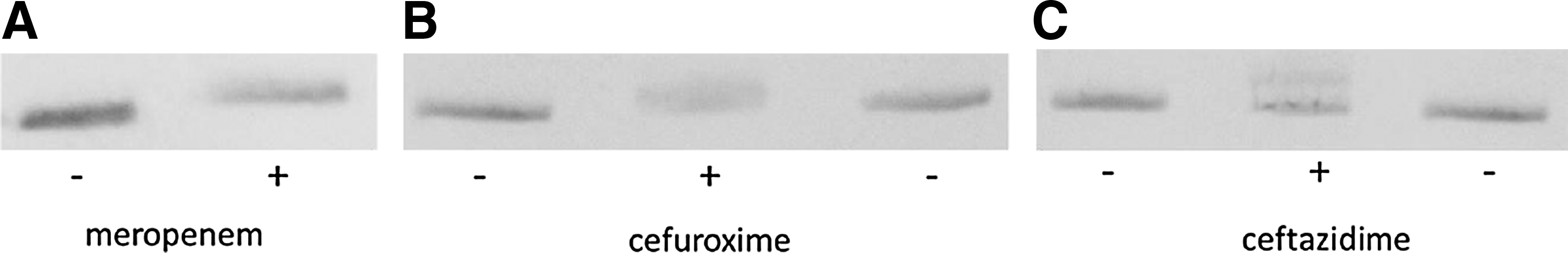
Covalent modification of AmpC with
OMP analysis
It is known that reduced permeability of the outer membrane can also contribute to resistance to cephalosporins. The permeability of the outer membrane to β-lactam antibiotics is largely determined by the presence of a class of abundant channel-forming OMPs, designated porins. E. coli K-12 strains generally produce two porins, OmpF and OmpC, when grown under routine laboratory conditions. To study if the c-AmpC hyperproducing isolates also produced porins, OMPs from various isolates were subjected to SDS-PAGE. The OMP profiles showed abundant bands with similar apparent molecular weights as the porins and OmpA in all isolates examined, except for isolate 3559, which showed only minor bands in this molecular weight range (Fig. 4A, B). Western blot analysis with a porin-specific antiserum confirmed the presence of one to three porins in all isolates and again showed relatively low levels of a porin in isolate 3559 (Fig. 4C).

Analysis of the outer membrane protein (OMP) profiles of E. coli isolates. OMPs were extracted and separated by SDS-PAGE. The proteins were either stained in the gel
Discussion
The purpose of the present study was to investigate the contribution of ampC expression in cefoxitin-resistant ESBL-negative isolates and its impact on third-generation cephalosporin activity, in a collection of clinical E. coli isolates. The phenotypic AmpC tests in nine isolates correlated with the expression level of RNA from the c-ampC gene and with mutations in the promotor/attenuator region of the c-ampC gene found by others to be associated with c-AmpC hyperproduction.2,17,20,29 Since no plasmid-encoded AmpC was found in any of the isolates, all AmpC activity in our isolates was of chromosomal origin. Thus, c-AmpC hyperproduction was found in 9 out of 33 (27.2%) cefoxitin-resistant isolates, that is, in 6 of the 29 (20.6%) ESBL-negative isolates and in 3 of the 4 ESBL-positive isolates.
Since average MICs were higher in c-AmpC hyperproducers than in nonhyperproducers, it is concluded that the c-AmpC hyperproduction affects the susceptibility to third-generation cephalosporins. In the clinical laboratory, this decrease in inhibitory activity may go largely undetected, because the MICs were in the range between WT and the S/I breakpoint of susceptibility, except for one c-AmpC hyperproducing ESBL-negative isolate, which clearly showed loss of susceptibility to the third-generation cephalosporins with MICs of 4 mg/L. In the ESBL-positive isolates, the ESBL enzyme probably overrules the effect of c-AmpC hyperproduction on cephalosporins, since the MICs of all three cephalosporins were significantly higher than in the ESBL-negative isolates and were similar in c-AmpC hyperproducers and nonhyperproducers.
The loss of the expression of porins, which facilitate the transport of β-lactams into the bacterial cell, is also known to contribute to resistance. 3 Nonetheless, the study of porin expression in clinical isolates is highly complex due to the variety of porins that can be expressed in different strains and the number of regulatory genes and external factors involved. All of the c-AmpC hyperproducing and control isolates studied here in detail still produced at least one porin, and five out of the six AmpC-overproducing ESBL-negative isolates (2483, 2529, 2958, 4478, and 3430) produced two or even three porins. Therefore, reduced access to the periplasm probably does not play a role in the decreased susceptibility of these strains to cephalosporins.
Compared to other β-lactamases, c-AmpC β-lactamases have been reported to show poor hydrolysis rates for ceftazidime and compared to other β-lactamases, 25 and this was confirmed in the present study. It has been suggested that cephalosporins may be rendered inactive by mere trapping to periplasmic inducible β-lactamases as speculated in Enterobacter cloacae and Pseudomonas aeruginosa. 27 In this study, we were able to demonstrate that preincubation of periplasmic extracts from c-AmpC hyperproducing isolates with small amounts of cefuroxime or ceftazidime was sufficient to at least partially inhibit the hydrolysis of nitrocefin in suggesting the formation of a stable cephalosporin–enzyme complex. Additional evidence for the formation of a stable acyl–enzyme complex was obtained by SDS-PAGE and Western blotting, which revealed a decreased electrophoretic mobility of the β-lactamase after preincubation with cefuroxime or ceftazidime. Antunes et al. 1 recently also described covalent trapping as the mechanism of resistance to ceftazidime caused by a deacylation-deficient mutant derivative of the class A TEM-1 β-lactamase. Similarly, evidence for the mechanism of antibiotic trapping by plasmid-encoded CMY-2 in carbapenem resistance was found in a porin-deficient carbapenem-resistant E. coli isolated from a liver transplant patient. 8 Based on our results and supported by data described in literature, the reduced susceptibility to cephalosporins can partially be explained by c-AmpC hyperproduction in the E. coli isolates studied. This enzyme, if present in high amounts, is able to trap cephalosporins entering the periplasm into a biologically inactive complex. It is expected that mutant derivatives of such isolates with limited entry of cephalosporins into the periplasm due to the loss of porins will show dramatically decreased susceptibility to cephalosporins. Such mutants might be selected under continuous selection pressure of cephalosporin treatment.
In the present study, c-AmpC hyperproduction was not predominant among cefoxitin-resistant isolates. As porins were found in the four cefoxitin-resistant non-AmpC producers examined, it is suggestive that efflux may be responsible for the resistance phenotype in these isolates. The contribution of efflux in β-lactam resistance has been proven in a set of isogenic E. coli K-12 strains, in which inactivation of the AcrAB efflux pump often resulted in a significant decrease in MICs of cefoxitin and cefuroxime. 16
For clinical microbiologists, proper recognition of AmpC-producing E. coli is important for clinical management. Administration of third-generation cephalosporins has resulted in treatment failure in two patients with c-AmpC hyperproducing isolates. 26 MICs of ceftazidime of 4 mg/L and cefotaxime of 2 mg/L were described in these patients, comparable to the MICs of one c-AmpC hyperproducing isolate (2483) reported in the present study. Recognition of AmpC production is difficult and no accepted guidelines are available. Resistance to cefoxitin can suggest AmpC-mediated resistance, but because we found it in 27.2% of the cefoxitin-resistant isolates only, it is not a specific marker. Although a comparison of phenotypic tests was not the purpose of this study, AmpC production was most reliably detected using the cefoxitin-boronic acid disk test, which appeared completely consistent with the qRT-PCR results. This test is inexpensive and can be used in all clinical laboratories.
It has been suggested to report E. coli with c-AmpC hyperproduction as resistant to third-generation cephalosporins irrespective of in vitro susceptibility. 4 However, although no longer WT strains, the MICs are often still within the susceptibility range and the treatment failures described so far were recognizable by MICs above the S/I breakpoints. In our opinion, the interpretation of third-generation cephalosporins should not be changed to resistant on susceptibility reports to avoid inappropriate use of other classes of antibiotic agents and the emergence of resistance. We do recommend surveillance to monitor any developments in the resistance to third-generation cephalosporins by testing for increased AmpC production in cefoxitin-resistant E. coli isolates and by establishing MICs of third-generation cephalosporins in c-AmpC hyperproducers.
Footnotes
Acknowledgment
This project was, in part, supported financially by a research grant from the Dutch ZonMW organization.
Disclosure Statement
The authors have no competing interests to disclose.