
Abstract
A novel assay for rapid determination of resistance to antibiotic inhibitors of protein synthesis was developed for the gram-positive pathogens, Enterococcus faecalis and Streptococcus pneumoniae. To this purpose, a lytic response was obtained by a brief incubation with lysozyme or a mixture of lysozyme, Triton X-100, and EDTA for E. faecalis (n = 82) and S. pneumoniae (n = 51), respectively. Lysis was quantified by visualizing the released nucleoids. Antibiotic-susceptible bacteria treated with Clinical and Laboratory Standards Institute (CLSI) breakpoint doses of erythromycin, azithromycin, or doxycycline that inhibited protein synthesis demonstrated a large reduction of lysed cells with respect to the control, that is, without antibiotics. However, cell lysis prevention was much lower in nonsusceptible strains, with unsuccessful inhibition of protein synthesis. ROC analysis showed that a reduction value of ≥35.6% and ≥40.4% discriminates susceptible and nonsusceptible strains for erythromycin and for doxycycline, respectively, in E. faecalis, whereas ≥20.0% is adequate for both macrolides and doxycycline in S. pneumoniae. Resistant stains were identified in 90–120 min with sensitivity and specificity between 91.7% and 100%. This is a proof of concept that evaluation of the lytic response may be a rapid and efficient test for determination of resistance to antibiotic inhibitors of protein synthesis.
Introduction
E
Both pathogens show a progressive increase of resistance to many commonly used antibiotics, including most of the bacterial protein synthesis inhibitors, such as macrolides and tetracyclines.4,5 Standard antibiograms usually require over 24 hrs for results. In urgent situations, rapid information regarding antibiotic resistance may lead to more timely use of appropriate antibiotics and improve patient outcomes, decrease healthcare costs, and reduce the spread of antibiotic resistance. 6 S. pneumoniae lyses its cell wall after the addition of detergents such as deoxycholate or Triton X-100, through the activation of the autolysin, LytA.7,8 Moreover, autolysins are involved in the bactericidal action of lysozyme in E. faecalis. This autolytic response is independent of its muramidase activity and relies on its cationic character.9,10 We provide evidence that the autolytic response is influenced by ribosomal protein synthesis, being amenable to be standardized for rapid determination of susceptibility resistance to antibiotics that inhibit protein synthesis.
Materials and Methods
Bacterial strains
Clinical isolates were obtained from the University Hospital A Coruña and from the Carlos III National Heath Institute (Madrid, Spain). The minimum inhibitory concentrations (MICs) were determined by automated microdilution (MicroScan Walkaway; Siemens).
Isolates of E. faecalis (n = 82) were grown on Mueller-Hinton agar at 37°C for 24 hrs. A sample was incubated in 2 ml Mueller-Hinton broth at 37°C for 2 hrs. The bacteria were diluted to an OD600 of 0.15 in Mueller-Hinton broth and incubated at 37°C in 200 μl tubes with 0.5 mg/L erythromycin or 4 mg/L doxycycline for 75 min in a final volume of 30 μl.
Isolates of S. pneumoniae (n = 51) were grown on agar plates with 5% sheep blood at 37°C for 24 hrs. The bacteria were diluted to an OD600 of 0.4 in Mueller-Hinton II broth supplemented with 3% lysed horse blood and incubated for 60 min with 0.5 mg/L azithromycin, 0.25 mg/L erythromycin, or 0.25 mg/L doxycycline. Doses of antibiotics were those indicated by the Clinical and Laboratory Standards Institute (CLSI) as the breakpoint of susceptibility in the standard antibiogram based on microdilution. 11
Lysis assay
For E. faecalis, 1 mg/ml lysozyme was added and shaken for 10 min at 37°C. S. pneumoniae were incubated with 0.05% Triton X-100, 2 mg/ml lysozyme, and 25 mM EDTA (TLE) for 5 min at 37°C. An aliquot of each sample was diluted to a concentration of 5–10 million microorganisms/ml in Mueller-Hinton broth.
The Micromax®-Q variant prototype (Halotech DNA SL) has been employed to evaluate the integrity of the cell wall, as described.12,13 Briefly, 30 μl of the diluted sample was mixed with liquid low-melting point agarose at 37°C. A 10 μl aliquot of the sample–agarose mixture was pipetted onto a precoated slide and covered with an 18 × 18 mm coverslip. The slide was placed on a cold plate to produce a microgel with the trapped intact cells inside. The coverslip was removed and the slide was immediately immersed horizontally in 10 ml of the specified lysing solution at room temperature, incubating 5 min for E. faecalis and 2 min for S. pneumoniae. The slide was washed with distilled water for 3 min, dehydrated in cold (−20°C) ethanol of increasing concentrations (70%, 90%, and 100%) for 3 min each, and air-dried in an oven. The dried slide was stained with 100 μl of the fluorochrome SYBR Gold (Molecular Probes) diluted 1:400 in TBE buffer (0.09 M Tris-borate, 0.002 M EDTA, pH 7.5) for 2 min in the dark, with a glass coverslip. After a brief wash in phosphate buffer pH 6.88 (Merck), a 24 × 60 mm coverslip was applied and the slides were visualized under fluorescence microscopy, and 500 bacteria were scored per experimental condition. This is achieved in 5–10 min, depending on the cell density in the microgel. When the bacteria are lysed, the nucleoid contained inside is released and spread into the microgel. Visualization of the cell nucleoid is a reliable sign of cell lysis.
Results
To establish the procedure for E. faecalis, an erythromycin-susceptible strain (MIC 0.125 mg/L) and a resistant strain (MIC >128 mg/L) were incubated with erythromycin alone, lysozyme alone, and erythromycin, followed by lysozyme (Fig. 1). Incubation with erythromycin alone never resulted in bacteria cell wall modification with respect to untreated cells (Fig. 1a, d). However, after incubation with lysozyme and lysing solution, the erythromycin-susceptible strain showed a high percentage, that is, 92.1% lysed cells (cells releasing nucleoids). Nevertheless, the proportion of lysed cells dropped to 3.9% when the strain was incubated with erythromycin beforehand (Fig. 1b, c). In the erythromycin-resistant strain, 97.4% of cells appeared lysed after incubation with lysozyme. Incubation with erythromycin beforehand produced only a minor change in the percentage, that is, 96.8%, of lysed cells (Fig. 1e, f). Similar results were obtained with S. pneumoniae (Fig. 2). In this case, lysozyme enhanced the lytic response of the bacteria to the detergent.14,15 Without EDTA in the TLE solution, the released nucleoids appeared as a field of dispersed spots of fragmented DNA, making it difficult to count them (Fig. 3a). The presence of EDTA in the solution prevents most of the DNA fragmentation, so the released nucleoids are better preserved and counted (Fig. 3b).

Enterococcus faecalis processed with the Micromax® assay to assess cell lysing through visualization of released nucleoids. Images above
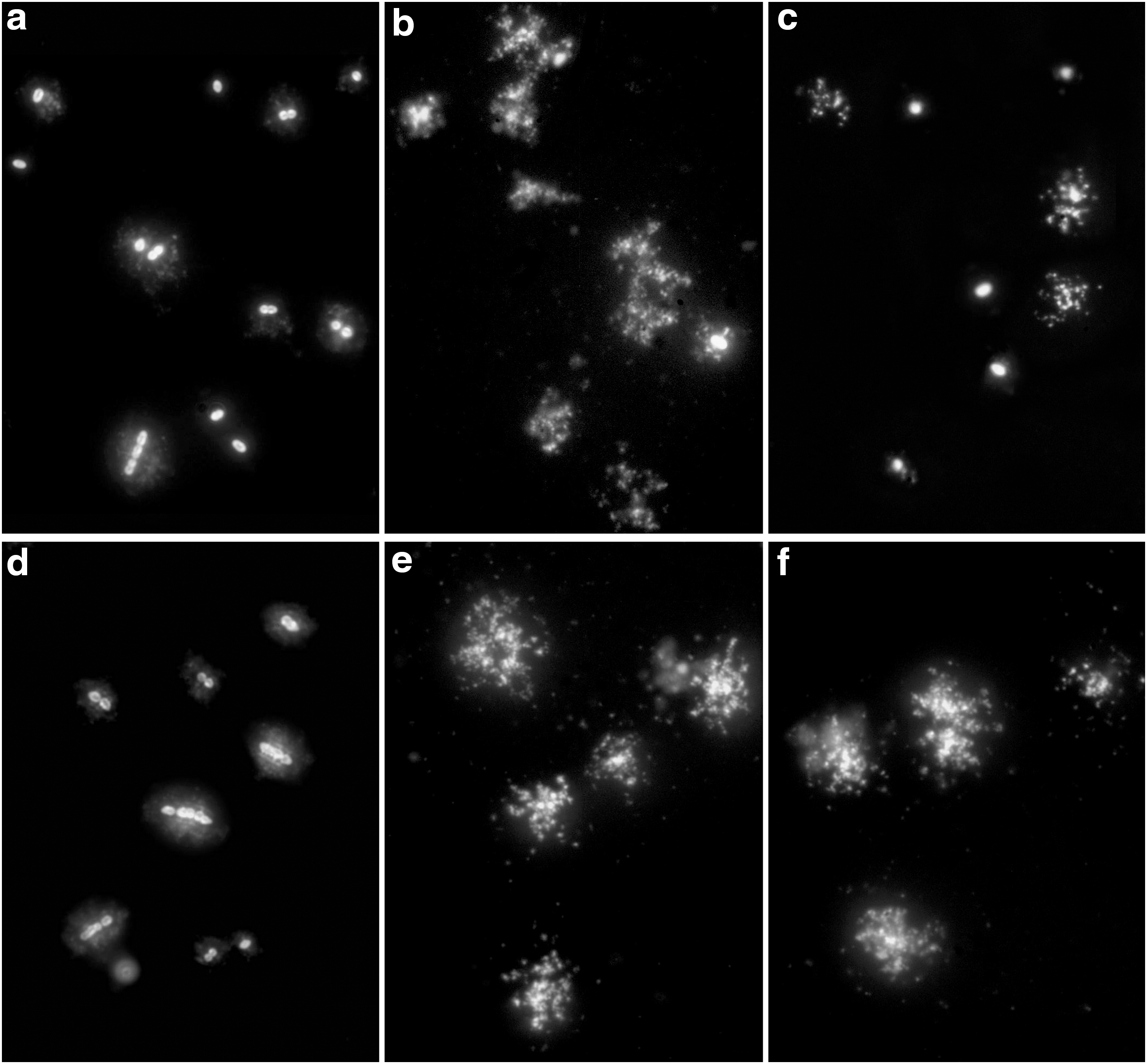
Streptococcus pneumoniae processed to evaluate cell lysis by nucleoid release, using the Micromax assay. Images above
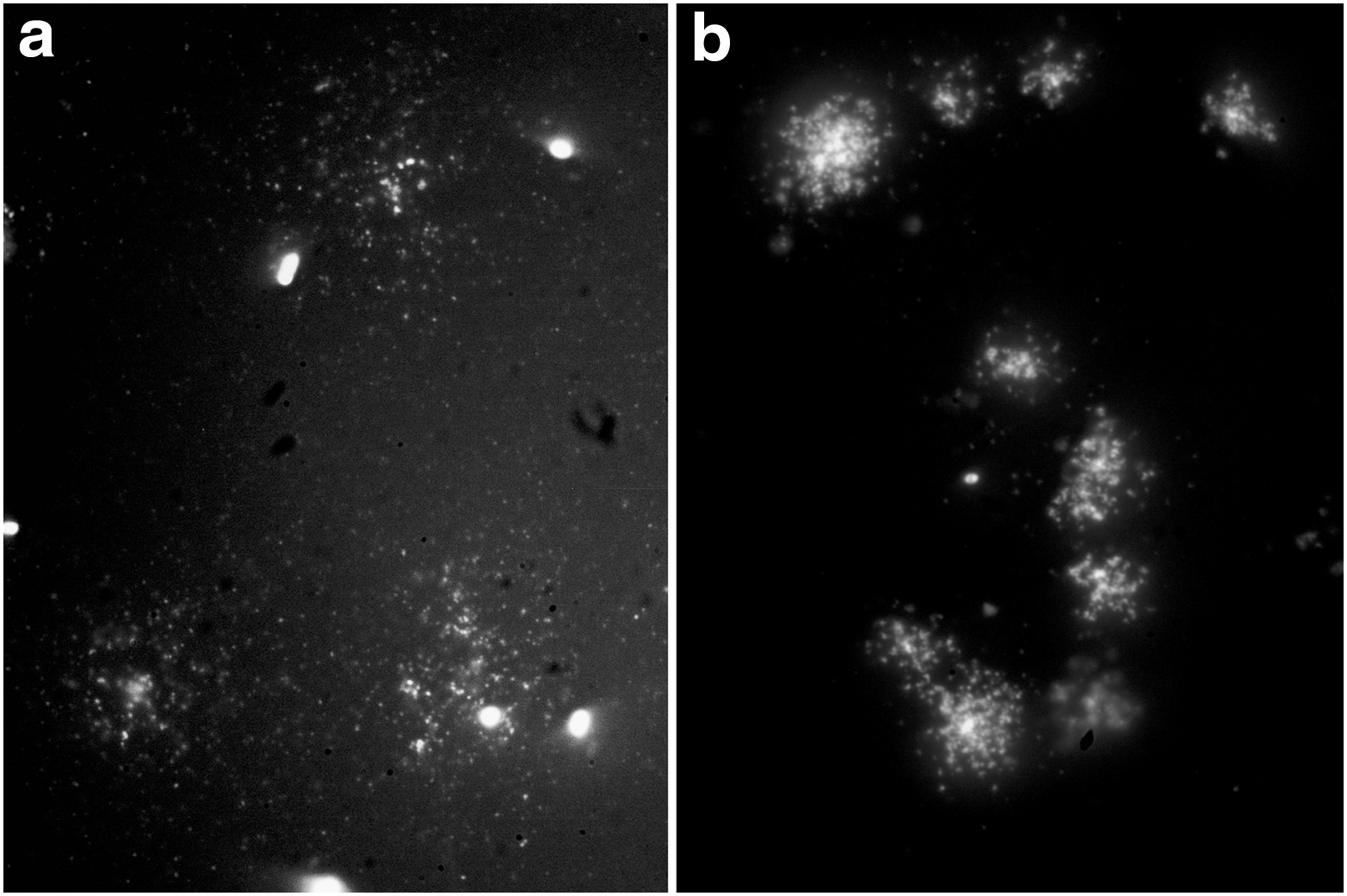
S. pneumoniae incubated with 0.05% Triton X-100 and 2 mg/ml lysozyme, to induce the autolytic response, and processed following the Micromax assay protocol.
Table 1 summarizes results obtained with blindly processed clinical isolates. ROC analysis demonstrates that a reduction of lysed cells ≥35.6% after incubation with 0.5 mg/L erythromycin, and ≥40.4% after incubation with 4 mg/L doxycyclin, accurately discriminates susceptible from nonsusceptible S. pneumoniae strains. False negatives for resistance are most relevant clinically. There was only one S. pneumoniae strain with false resistance to erythromycin, with MIC 1 mg/L, and two for doxycycline, both with MIC 8 mg/L, and they were all close to the breakpoint of susceptibility. For S. pneumoniae, a reduction of lysed cells ≥20% after incubation with the breakpoint of susceptibility of azithromycin, erythromycin, and doxycycline accurately distinguishes susceptible from nonsusceptible strains. Only one false negative of resistance was found for erythromycin. Sensitivity and specificity values ranged between 91.7% and 100%.
Lytic response was induced in susceptible (S) and nonsusceptible (NS) strains, categorized accordingly to the standard antibiogram and CLSI criterion. The percentage of reduction of lysed cells by previous incubation with the antibiotic inhibitor of protein synthesis was very significant in the susceptible strains, unlike in the nonsusceptible strains.
p < 0.0001.
95% CI, 95% confidence interval; AUC, area under the ROC curve; CLSI, Clinical and Laboratory Standards Institute; SD, standard deviation; TLE, Triton X-100, lysozyme, EDTA.
Discussion
Tomasz and Waks previously reported that high-dose chloramphenicol interferes with the autolytic activity of Lyt A in S. pneumoniae, 16 but to our knowledge, there is no report about the decrease of the lytic effect of lysozyme by protein synthesis inhibition in E. faecalis. Our work supports that protein synthesis regulates the autolytic response of these bacteria in the short term. Nonetheless, the potential diagnostic use of this effect had never been explored and standardized to discriminate strains resistant to different types of inhibitors of ribosomal protein synthesis. Erythromycin and azithromycin are macrolides, which bind to the domain V of the 23S rRNA of the 50S ribosome subunit causing premature termination and dissociation of the peptidyl-tRNA. 17 Doxycycline binds to the 30S ribosomal subunit, blocking the entry of aminoacyl-transfer (t)RNAs. 18 Then, different mechanisms of protein synthesis inhibition result in the same effect, that is, a diminished autolytic response to lysozyme or TLE treatment. Importantly, other protein synthesis inhibitors may cause a similar response. In fact, we found that linezolid at 2 mg/L (the CLSI breakpoint of susceptibility) prevented the lytic response in all of the S. pneumoniae strains (data not shown). Antibiotic resistance by the bacteria was not evidenced. Regarding E. faecalis, 14 extra isolates were found (14.6% of the entire 96 strains analyzed) where lysozyme was not effective at inducing lysis. This could be due to modifications of the cell wall, for example, increased levels of O-acetylated peptidoglycan. 19 All of the 14 E. faecalis isolates were nonsusceptible to erythromycin and doxycycline. Although the number of strains is limited, the lack of cell wall lysis suggests nonsusceptibility to the antibiotics studied herein, thus complementing the results of the assay.
Our study is proof of concept that the lytic response may be used for a rapid detection of resistant strains to antibiotics that target the ribosome. If protein synthesis is inhibited by the antibiotic, that is, in the susceptible strains, lytic effect is greatly decreased. However, if protein synthesis is not successfully inhibited, that is, in the antibiotic-resistant strains, cell wall lysis is suppressed much less or not at all. Resistant strains are determined in 1.5–2 hrs. Examination may be automatized using a motorized microscope coupled with image capture and digital image analysis. It must be taken into account that the lysozyme or TLE reagents were optimized for our assay where a lysing solution is required later to release the nucleoids. It is not clear how the antibiotic inhibitors of protein synthesis would work under other lysing conditions. The procedure does not require prior knowledge of the resistance mechanism, unlike the polymerase chain reaction and microarray-based assays. It could be helpful in critical infections where rapid determination of antibiotic resistance may improve patient outcome and also preserve valuable last option antibiotics, while optimizing their medical use and helping to prevent the spread of antibiotic resistance.
Footnotes
Acknowledgments
The authors are grateful to A. Fenoll, ISCIII, for kindly providing S. pneumoniae strains and Prof. M. Kjelland for improving the English style of the article. This work was supported by the Fondo de Investigaciones Sanitarias (PI14/01346) and REIPI, Spanish Network for Research in Infectious Diseases (Instituto de Salud Carlos III, RD12/0015/0014). This work was also funded by IMI 6th call COMBACTE New Drugs 4 Bad Bugs (ND4BB).
Disclosure Statement
J.G. and J.L.F. are advisers of Halotech DNA SL. All other authors declare no conflicts of interest.