
Abstract
An important focus of modern medicine is the search for new substances and strategies to combat infectious diseases, which present an increasing threat due to the growth of bacterial resistance to antibiotics. Another problem concerns free radicals, which in excess can cause several serious diseases. An alternative to chemical synthesis of antimicrobial and antiradical compounds is to find active substances in plant raw materials. We prepared extracts from leaves of five species of the genus Bergenia: B. purpurascens, B. cordifolia, B. ligulata, B. crassifolia, and B. ciliata. Antimicrobial and antiradical features of extracts and raw materials were assessed, and the quantities of phenolic compounds were determined. We also evaluated, using high-performance liquid chromatography, the amounts of arbutin and hydroquinone, compounds related to antimicrobial activity of these raw materials. The strongest antiradical properties were shown by leaves of B. crassifolia and B. cordifolia, the lowest by leaves of B. ciliata. The antiradical activity of extracts showed a strong positive correlation with the amount of phenols. All raw materials have significant antimicrobial properties. Among them, the ethyl acetate extracts were the most active. Antimicrobial activity very weakly correlated with the amount of arbutin, but correlated very strongly with the contents of both hydroquinone and phenolic compounds. Additional experiments using artificially prepared mixtures of phenolic compounds and hydroquinone allowed us to conclude that the most active antimicrobial substance is hydroquinone.
Introduction
A
Today, bacterial resistance is recognized as a grave threat. The problem is especially serious in patients during chemotherapy and after organ transplantation because of immunosuppression.1,7
Bacterial resistance to antibiotics is leading to increased mortality rates due to infections worldwide.8,9 Resistance of microorganisms causes annually about 25, 000 deaths in the European Union and 23, 000 in the USA. 1 The problem is aggravated by the lack of development of new antibiotics. Over the past 25 years, only two new classes of antibiotics have been developed for treatment, namely oxazolidinones and lipopeptides. 1
Due to the increasing resistance of bacteria to antibiotics, it is crucial to develop new strategies to combat microorganisms and new antibacterial and antifungal substances. New compounds with antibacterial activity can be obtained by chemical synthesis or can be isolated from natural raw materials. Natural sources of antibacterial substances include propolis10,11 and plants.12,13 Many fractions derived from plants have strong antimicrobial properties. Plant fractions with antiradical properties include essential oils containing compounds such as phenolics and polyphenols, flavonoids, quinones, tannins, coumarins, alkaloids, terpenoids, lectins, and polypeptides.14–18 Among individual active compounds, we can mention trans-cinnamaldehyde, 19 eugenol, 19 1.8-cineole, 20 and thymol.21,22 Antibacterial substances of natural origin cause membrane disruption, impair the metabolism of bacterial cells, exhibit antibiofilm activity, inhibit formation of the bacterial capsule, reduce resistance of bacteria to antibiotics, reduce production of bacterial toxins, and attenuate bacterial virulence. 14 Very effective antibacterial materials also include raw materials containing hydroquinone and its derivative arbutin. Arbutin is found in pharmacopoeial raw materials, including bearberry leaves 23 and nonpharmacopoeial cowberry leaves. These raw materials are known for their disinfectant activity on the urinary tract. 24 There are more species of plants with a high content of arbutin such as leaves of pear 25 and leaves of Bergenia sp. (badan), 26 which may be a potential source of raw materials used in infectious diseases.
The genus Bergenia is a group of perennial plants growing from Afghanistan to the south of Tibet and the Himalayas. The height is from 30 to 60 cm, the leaves are large, evergreen, and leathery, and the flowers are from pink to dark purple. 27
Species belonging to the genus Bergenia occur in Asia, mainly East Asia, in south eastern areas of central Asia, and in the northern areas of South Asia. They naturally occur at high altitudes in cold regions. 28
Bergenia is rich in chemical compounds of great medicinal importance. These are mainly polyphenolic compounds such as tannins, flavonoids, and quinones. One of the more important substances with medicinal significance is bergenin. Its medicinal use is in alleviating cough, removing mucus from respiratory tracts, and anti-inflammatory activity. 28
Important from the point of view of medicinal uses is also hydroquinone, with strong antibacterial activity, and its derivatives such as arbutin.
Hydroquinone and benzoquinone 29 are formed as a result of the degradation of arbutin in an alkaline environment. 30 Our previous studies confirmed strong antimicrobial activity of extracts containing arbutin and hydroquinone. 31 A limitation is the toxicity of hydroquinone, 32 but it is advantageous that hydroquinone effectively inhibits growth of microorganisms at a concentration that is not harmful for the human body.
In this study, extracts from five species of the genus of Bergenia were obtained. These extracts were further tested for the antimicrobial and antioxidant activity and the content of phenolic compounds, arbutin and hydroquinone, was measured. The antimicrobial and antiradical activity of extracts and raw materials was also studied.
Materials and Methods
Materials
Raw materials
The leaves used in the research were obtained from five species belonging to the genus Bergenia: Bergenia ciliata (Royale) A. Br., Bergenia cordifolia Sternb., Bergenia crassifolia (L.) Fritsch., Bergenia ligulata (Wall.) Engl., and Bergenia purpurascens (Hook. f. & Thomas) Engl.
B. ligulata was obtained from the Botanical Garden of the University of Poznań, and other raw materials were obtained from the Botanical Garden of the University of Wrocław.
Reagents
Methanol gradient grade for chromatography, Merck; ethyl acetate pure for analysis, methanol pure for analysis, Chempur; anhydrous sodium carbonate pure for analysis, Folin–Ciocalteu's reagent Polish Chemical Reagents; potassium persulfate 99%, 2,2′-azino-bis(3-ethylbenzothiazoline-6-sulfonic acid) diammonium salt Sigma-Aldrich.
Instrumentation
High-performance liquid chromatography (HPLC) Thermo Scientific Dionex, model UltiMate 3000, DAD detector, column Cadenza 5 CD-C18 (150 × 4.6 mm, ID 5 μm); spectrophotometer CECIL (CE 3021); spectrophotometer Hitachi U-5100, and glass cuvettes with 1 cm optical path were used.
Methods
Preparation of extracts
The method of extraction was chosen as most effective for extraction of active compounds. Raw materials (50 g) were extracted with 900 ml of 20% methanol in water at 40°C for 48 hr. Extracts were filtered with filter paper (Filtrak 388). One hundred eighty milliliters (20%) of first methanol–water extracts were separated and condensed under reduced pressure to obtain dry extract EA. Methanol was distilled off from the rest of the extract and the water solution was adjusted to 600 ml with water. The solutions were stored for 48 hr at 4°C. The precipitate formed was filtered off with filter paper (Filtrak 388) and dried to obtain extract EB. The water residue after separation of the precipitate was extracted with ethyl acetate (3 × 200 ml, 4 × 100 ml). The obtained ethyl acetate extract and water residue were condensed to dryness to obtain residues EC and ED.
Extracts obtained from leaves of B. ciliata were additionally marked with the letters “ci,” extracts from B. cordifolia with the letters “co,” extracts from B. crassifolia with the letters “cr,” extracts from leaves of B. ligulata with the letter “l,” and extracts from leaves of B. purpurascens with the letter “p.”
As a result of extraction, the following weights of extracts were obtained (mg ± 0.1): from B. ciliata EAci 2463.1, EBci 80.4, ECci 862.2, and EDci 9396.7; from B. cordifolia EAco 3761.0, EBco 1397.6, ECco 3602.7, and EDco 10671.7; from B. crassifolia EAcr 3403.6, EBcr 546, ECcr 3637.3, and EDcr 10321.2; from B. ligulata EAl 2956.3, EBl 260.1, ECl 3203.6, and EDl 11799.4; and from B. purpurascens EAp 3278.0, EBp 1600.7, ECp 2306.1, and EDp 9633.0.
Measurement of phenolic compounds with colorimetric method
Total amount of phenolic compounds was measured with the method of Singleton and Rossi. 33
Seven milliliters of water was added to the test tubes and 0.5 ml of Folin–Ciocalteu's reagent was added. Then 0.5 ml of extract solution at 0.6 mg/ml for extracts EA, EB, and ED, or 0.3 mg/ml for extract EC was added. After 3 min, 2 ml of 20% solution of Na2CO3 in water was added. Then the sample was heated in a boiling water bath for 1 min. After cooling, the absorbance of the sample was measured at 685 nm in a glass cuvette with a 1 cm optical path. All measurements were repeated six times and maximal error was calculated.
Measurement of antiradical activity with ABTS•+
Preparation of reagent ABTS•+
Antiradical activity of extracts from leaves of Bergenia was measured by the method of Re et al. 34 2.2’-azino-bis(3-ethylbenzothiazoline-6-sulfonic acid) diammonium salt (ABTS) was dissolved in water at the concentration of 7 mM. This solution was mixed in the volume ratio of 1: 1 with 2.45 mM aqueous solution of K2S2O8. The solution was left in a dark place for 16 hr in order for the radical cation ABTS+• to form. The final solution was diluted with 50% methanol so that the absorbance was equal to 1.
Measurement of antiradical activity
To measure antiradical activity the EC extracts were dissolved in 50% methanol at the concentration of 0.3 mg/ml, and the rest of extracts were dissolved in 20% methanol in water at 0.6 mg/ml. As a blank, 50% methanol was used. A reagent control was prepared by the addition of 20 μl of 50% methanol to 2 ml of ABTS•+ solution. Absorbance (734 nm) was measured for 6 min with measurements every 10 sec at 25°C using a glass cuvette with an optical path of 1 cm. Each measurement was repeated five times and maximal error was calculated with the total differential method.
Calculation of antiradical activity
Antiradical activity was measured according to Sroka et al. 35 as TAU734 (total antiradical units) units per mg of extracts (TAU734/mg) or g of raw material (TAU734/g).
One unit of antiradical activity is the amount of substance, which in the test conditions (described in Measurement of antiradical activity section) eliminates 1 μmole of ABTS+• radical during the first 1 min of the reaction.
The number of units per mg of extracts was measured using the equation:
where TAU734/mg is the number of antiradical units per mg of extract; A0 is the absorbance of the sample at the beginning of the test; A1 is the absorbance of the sample after 1 min of the reaction; CE is the concentration of the extract in the reaction mixture [mg/ml].
The number of antiradical units was also calculated per g of raw material according to the equation:
where TAU734/g is the number of antiradical units per g of raw material; TAU734/mgEA is the number of antiradical units per mg of extract EA; mEA is the weight of extract EA [mg]; TAU734/mgEB is the number of antiradical units per mg of extract EB; mEB is weight of extract EB [mg]; TAU734/mgEC is the number of antiradical units per mg of extract EC; mEC is the weight of extract EC; TAU734/mgED is the number of units per mg of ED extract; mED is the weight of ED extract; and CR is the amount of raw material taken for extraction [g].
Maximal error was calculated with the total differential method.
The reaction rate constant
Measurement of arbutin and hydroquinone with HPLC
Extracts for analysis were dissolved in methanol (3 mg/ml) and filtered using membrane filters (Millipore, 0.22 μm) and analyzed with HPLC (equipment described above). Analysis was performed at the wavelength of 280 nm, with the injection volume 20 μl, and flow 1 ml/min. Research was performed at the fixed temperature of 25°C. The elution was performed according to the following gradient: (A) methanol and (B) 2% aqueous solution of acetic acid; 0–10 min 3% (A) and 97% (B); 10–12 min 10% (A) and 90% (B); 12–25 min 100% (A); 25–28 min 3% (A) and 97% (B).
The identification and quantitative analyses were done during extract analysis by measuring and comparing the areas and retention times of the identified peaks with the areas and retention times of the peaks of standards (arbutin, hydroquinone). Calibration curves were made for hydroquinone (R2 = 0.99964; relative standard deviation (RSD) = 1.86%) and arbutin (R2 = 0.99987; RSD = 1.15%). The measurement of each extract was repeated five times.
Measurement of antimicrobial activity of extracts
Antimicrobial activity of extracts was determined with the following bacterial and fungal species: S. aureus ATCC 25923, S. aureus MRSA K31 (clinical, antibiotic-resistant strain), Escherichia coli ATCC 25922, E. coli ESBL R194 (clinical, antibiotic-resistant strain), Enterococcus faecalis ATCC 29212, E. faecalis HLAR (clinical, antibiotic-resistant strain), Bacillus subtilis ATCC 6633, and Candida albicans ATCC 90028. All strains were from the Department of Microbiology, Wrocław Medical University, Wrocław, Poland. Antimicrobial activity of tested extracts was evaluated using the disc diffusion method described by Ingolfsdottir et al. 36
The experiments were carried out on Mueller-Hinton agar plates (Oxoid) for bacterial strains and Sabouraud agar plates (Biomed) for fungal strain. Suspensions of bacterial and fungal strains at the turbidity comparable to 0.5 McFarland standard were diluted 1:10 in saline to obtain an inoculum of 107 CFU/ml. The suspensions thus prepared were spread on agar plates with sterile swabs.
Afterward, standard discs with the diameter of 6 mm (Becton) were placed on agar plates aseptically. After 15 min, 10 μl of DMSO solutions of extract were placed on the discs at a concentration of 50 mg/ml. Then plates were incubated at 37°C for 24 hr for bacterial strains or 48 hr for the fungal strain. After incubation, the inhibition zones around the disc were measured [mm].
Erythromycin (15 μg), gentamicin (10 μg), ampicillin (2 μg), penicillin (1 μg), and amphotericin B (10 μg) discs were used as positive controls, whereas DMSO was used as a negative control. All susceptibility experiments were performed in triplicate on separate days. Standard uncertainty was established as ±0.33 [mm].
Measurement of antimicrobial activity of artificial mixtures
The artificial mixtures were prepared so that the hydroquinone and phenolics were applied to the plate in the same amount as in the cases of EC extracts from B. purpurascens, B. cordifolia, and B. crassifolia: mixture I–tannin (94 μg/disc), rutin (94), and caffeic acid (94); mixture II–tannin (94 μg/disc), rutin (94), caffeic acid (94), and hydroquinone (230); hydroquinone (230 μg/disc). The antimicrobial activity was measured in the way described above.
Results
The amount of phenols in extracts and raw materials expressed as a percentage is shown in Table 1. In general, a high amount of phenols was detected for EC extracts, especially ECp obtained from B. purpurascens (67% ± 0.5%) and B. cordifolia (67% ± 1.4%). A low amount was observed for ED extracts; the lowest amount of phenols was observed for extracts obtained from B. ligulata (16% ± 0.5%) and B. ciliata (5% ± 0.2%).
Maximal error was calculated by the total differential method n ≥ 5. Yields of extracts preparation (Y) is given with a parentheses.
When the amount of phenols was calculated per raw material, the highest amount (expressed as a percentage) was found for B. cordifolia (13.5% ± 5.3%) and B. crassifolia (13.4% ± 0.42%). The lowest amount of phenols was noted for B. ciliata (2.7% ± 0.12%).
Antiradical activity (Table 1) of extracts is shown as the amount of antiradical units per mg of extract (TAU734/mg) and g of raw material (TAU734/g). A high number of antiradical units was calculated per mg of EC extracts, with the highest for ECcr extract from B. crassifolia (11.2 ± 0.6). Also strong antiradical activity was observed for ECp extract and ECc extract from B. purpurascens and B. cordifolia, with TAU734/mg values of 10.7 ± 1.03 and 10.4 ± 0.61, respectively. The weak antiradical activity was noted for ED extracts, with the lowest values of TAU734/mg for EDci (1.1 ± 0.11) and EAci (2.2 ± 0.13) from B. ciliata.
The largest number of antiradical units was calculated per gram of dry leaf of B. crassifolia, with the TAU734/g value of 2502 ± 171. The weakest antiradical action was observed for leaf of B. ciliata, with the TAU734/g value of 489 ± 49.
The reaction rate constant k was calculated for the reaction of scavenging of ABTS+• radical, p < 0.0001 (Table 2). All reactions proceeded according to second-order kinetics.
The p-value was always below 0.0001 (p < 0.0001).
The investigated raw materials are rich in hydroquinone and arbutin, which are known for their strong antimicrobial activity. The extracts from leaves of Bergenia were found to be strong antimicrobial substances (Table 3). The activity is presented as the diameter (mm) of the zone of inhibition of bacterial or fungal growth.
Standard uncertainty was equal to ±0.33.
In general, the most sensitive to the action of the extract are the reference strains, especially S. aureus ATCC 25923 (Table 3 and Fig. 1). The methicillin-resistant strain S. aureus MRSA K31 was slightly less sensitive to extracts. For these two strains, the most effective appeared to be EC extracts, with the inhibition zone of 25 mm for ECp and ECco in the case of B. purpurascens and B. cordifolia, respectively. Weak inhibition of growth of S. aureus was exhibited by ED extracts, the weakest by EDci from B. ciliata (inhibition zone 8 mm). Relatively good sensitivity to the action of extracts was shown by the reference strain B. subtilis ATCC 6633 (Table 3). The strongest activity against B. subtilis was exhibited by extract ECp from B. purpurascens, with an inhibition zone of 18 mm. The weakest activity was noted for extracts EBci and EDci for leaves of B. ciliata (inhibition zone 7 mm).
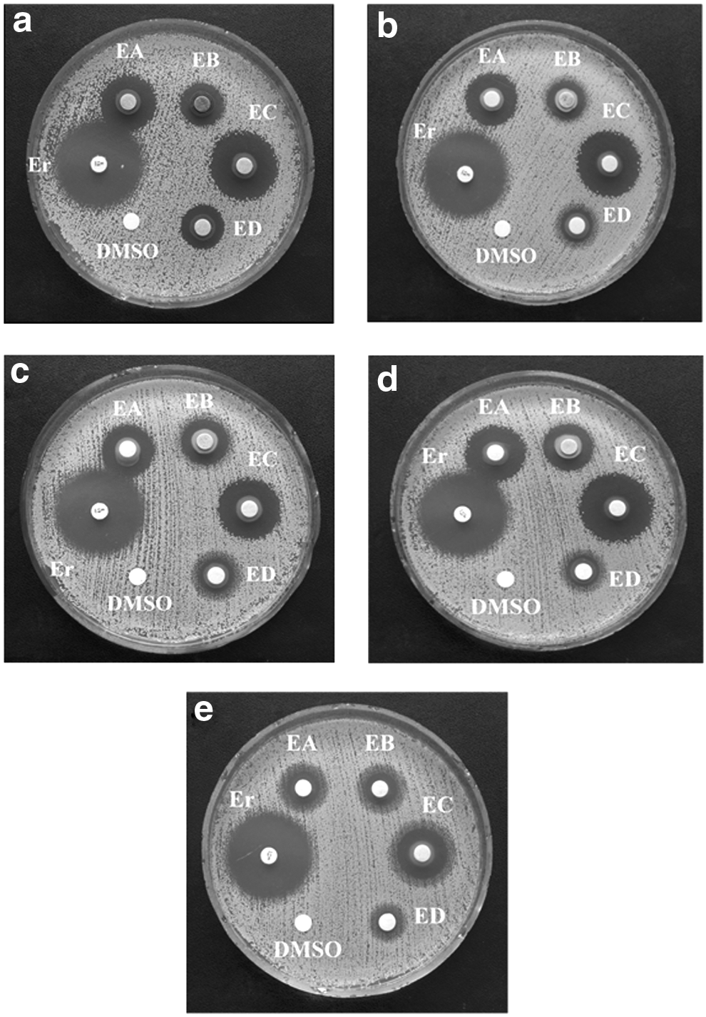
Zones of inhibition of Staphylococcus aureus ATCC 25923 by extracts from leaves:
Gram-positive E. faecalis appeared to be resistant to the extracts. For the reference strain E. faecalis ATCC 29212 the most active were extracts EA and EC, with the highest activity for extract EAcr from B. crassifolia (17 mm). Extracts EB and ED were not active. The strain HLAR E. faecalis appeared to be more resistant to the action of extracts than the reference strain. Only EC extracts exhibited low antibacterial activity (inhibition zones 10–13 mm).
The most resistant to antibacterial activity of extracts appeared to be gram-negative bacteria, facultatively anaerobic E. coli. Both the reference strain E. coli ATCC 25922 and the resistant strain E. coli ESBL R194 were strongly resistant to the extracts. Only EC extracts weakly inhibited the growth of these strains. The inhibition zones for E. coli ATCC 25922 were 11–13 mm, and the inhibition zones for the resistant strain ESBL were 11–12 mm.
The fungus C. albicans ATCC 900028 appeared to be sensitive to the action of extracts. Large zones of inhibition of fungal growth were observed in the presence of EC extracts. The strongest inhibition of growth of fungi was noted in the presence of extract ECcr (31 mm) and the weakest in the presence of extract EDci (9 mm). It is worth noting that the inhibition zone for the antifungal substance amphotericin B was 15 mm.
The amount of arbutin and hydroquinone, which are known for their antiradical activity, was measured with HPLC, and the results are shown in Table 4 and Fig. 2. The amount of compounds is presented as the percentage per weight of extract.
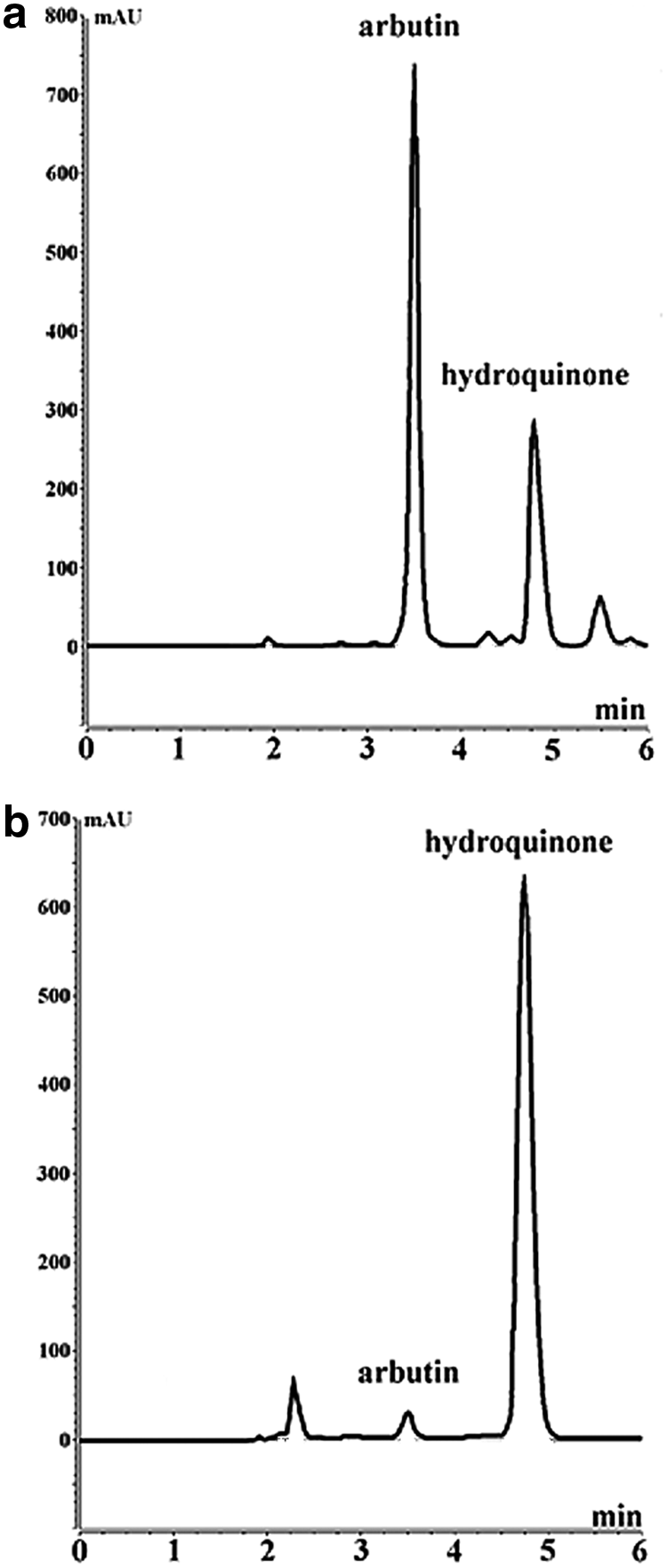
HPLC chromatograms of EA
A high concentration of arbutin was observed in extracts EA and ED and a low concentration in extracts EB and EC. The highest amount of arbutin was observed for extracts EDcr and EDco from B. crassifolia (51 ± 0.002%) and B. cordifolia (45% ± 0.002%), the lowest amount for extract EBci from B. ciliata (0.35% ± 0.00003%).
Hydroquinone was noted mainly in extracts EA and EC. The highest amount was observed for extracts ECcr from B. crassifolia (25% ± 0.002%), ECco from B. cordifolia (24% ± 0.001%), and ECp from B. purpurascens (24% ±0.001%). Extracts EB and ED were very poor in hydroquinone. In these extracts, there were trace amounts or a lack of this compound.
The correlation coefficient was calculated between the amount of arbutin and average antibacterial activity. The correlation was also calculated between the amount of hydroquinone and average antibacterial activity of extracts and between the amount of phenolic compounds and average antibacterial activity of extracts. The highest positive correlation (Fig. 3) was observed between the amount of hydroquinone and antibacterial activity of extracts (r = 0.93). Also, a high positive correlation was observed between the amount of total phenols and average antibacterial activity (r = 0.82). A weak negative correlation was observed between the amount of arbutin and antimicrobial activity (r = −0.31, Fig. 3).
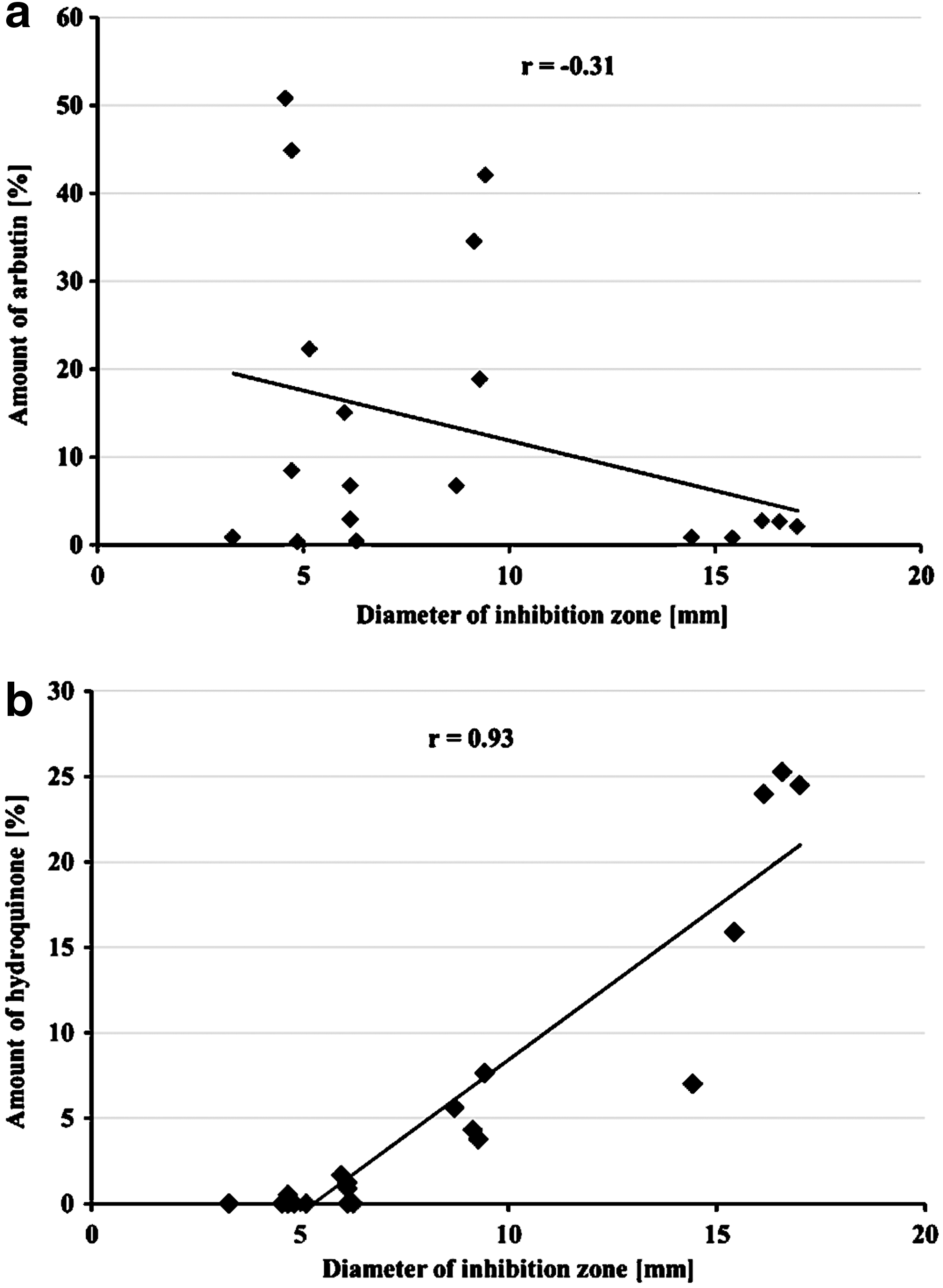
Correlation coefficient between the amount of arbutin
Discussion
The increase in resistance of bacteria to antibiotics is a growing problem worldwide. The problem is serious because many diseases previously treatable by antibiotics are again becoming untreatable. 37
Hence, the search for new methods and substances to combat infectious diseases is one of the more important tasks of modern medicine. In addition to chemical synthesis, an alternative method of obtaining active substances is isolation from plant raw material.
Plant raw materials are rich in phenolic compounds that have strong antiradical activity, but weaker antimicrobial activity. Obtaining extracts from plant raw materials makes it possible to achieve in one formulation both antiradical and antioxidant compounds. This is very important because during infectious diseases, especially chronic oxidative stress takes place. Oxidative stress is advantageous in combating infectious diseases in human and animal organisms, 38 but in chronic cases, when levels of reactive oxygen species persist for a long time, it can cause damage to the body. 39 The effects of chronic oxidation can be relieved with the presence of phenolic compounds. 40
Our research revealed strong antiradical potential of extracts and leaves. We compared the results to our previous studies evaluating a variety of raw materials as well as powerful antioxidants such as green tea leaves. 31 Leaves from Bergenia proved to be the most potent antioxidant of all those we have studied so far. The literature data confirm the strong antioxidant properties of leaves from Bergenia.41,42
The extracts from Bergenia exhibited strong antimicrobial properties. They effectively inhibited the growth of gram-positive bacteria, especially S. aureus, as well as the resistant strain MRSA, which can cause life-threatening diseases. 43 The gram-positive bacterium B. subtilis was also found to be sensitive to the action of the extracts.
Relatively weak activity was shown by extracts against strains of gram-negative E. coli bacteria, both a reference and a resistance strain (ESBL). Weak antibacterial activity of extracts was also exhibited against commensal gram-positive E. faecalis bacteria, especially against the resistant strain HLAR. Both E. coli and E. faecalis can cause life-threatening diseases, especially resistant strains.
A very interesting result is the strong inhibition by the extracts from Bergenia of the growth of the fungus C. albicans. This fungus can cause life-threatening illnesses, particularly in immunocompromised patients. Treatment of fungal infection requires the use of drugs with serious side effects, and a much milder acting extract seems to be an interesting alternative.
The high correlation between the amount of hydroquinone and antibacterial activity suggests that hydroquinone may be a substance responsible for antibacterial activity of extracts (the correlation for arbutin is very low, Fig. 3). There is a problem in deciding whether hydroquinone is the major factor responsible for the antimicrobial activity of extracts because the correlation between the amount of phenolic compounds and antimicrobial activity is also high (r = 0.82), which may indicate an important role of phenols in antimicrobial activity of extracts. To decide whether phenols, hydroquinone, or both are responsible for antimicrobial activity, artificial mixtures were prepared and their influence on the growth of bacteria and fungi was investigated. The composition of mixtures is as follows: mixture I–tannin (94 μg/disc), rutin (94), caffeic acid (94); mixture II–tannin (94 μg/disc), rutin (94), caffeic acid (94), and hydroquinone (230); in the third sample there was only hydroquinone (230 μg/disc). The individual components were used in such an amount that they correspond to the amount of compounds placed on discs with extracts. The results, which are presented in Table 5, clearly show that the antimicrobial activity of phenols is low, but that of hydroquinone is high. The most effective was the mixture of hydroquinone and phenols. The activity of the full mixture II (phenols and hydroquinone) was not the simple sum of activity of ingredients, but slightly higher, which may suggest the existence of positive synergism in the action of these compounds. There are literature reports proving the beneficial synergy of phenolic compounds with antibiotics. 44
The antimicrobial activity was demonstrated with disc diffusion method as diameter (mm) of zones of inhibition of growth of bacteria and fungi. Standard uncertainty was equal to ±0.3 mm.
—, Lack of antimicrobial activity.
There are some literature data related to antimicrobial activity of extracts from Bergenia ciliate Sternb. 45 and B. ligulata (Wall.) Engl. 46
In contrast to our study, the authors noted smaller antimicrobial activity of ethyl acetate extracts. Moreover, the authors connected inhibition of growth of microorganisms with the presence of general phenols, especially flavonoids.
Ethyl acetate extracts in our research exhibited the strongest antimicrobial activity, and the strongest antimicrobial was found to be hydroquinone.
There are many literature data confirming antimicrobial activity of hydroquinone and its derivatives. 47 There are also some reports of antimicrobial properties of plant phenolics. 48 As was mentioned above, our results clearly show that the main substance responsible for antimicrobial properties of extracts from Bergenia is hydroquinone. In previous studies with the leaves of pear we established the crucial role of hydroquinone as an antimicrobial substance. 31
Our research partially confirmed some antimicrobial features of phenols, but because of much stronger activity of hydroquinone we believe that it is the main antibacterial substance in extracts from leaves of Bergenia.
It can be concluded that:
(1) Extracts from leaves of some Bergenia species showed strong antioxidant activity, even greater than green tea leaves, known for their strong antioxidant activity. (2) Ethyl acetate extracts contain the most phenolic compounds and exhibit the strongest antiradical activity. The lowest amount of phenols and the weakest antiradical activity were observed for water residues. (3) The strongest antimicrobial activity was exhibited by ethyl acetate extracts, while the lowest activity was observed for water residues. The level of hydroquinone correlated well with average antibacterial and antifungal activity. (4) Additional investigation using artificial mixtures of phenols and hydroquinone confirmed strong antibacterial activity of hydroquinone, significantly higher than a mixture of phenols. This research showed no antifungal activity of either hydroquinone or phenols. Determining the substance or substances in extracts with antifungal activity requires further research.
Footnotes
Acknowledgment
This research was supported by a grant of Wrocław Medical University number ST-857.
Disclosure Statement
No competing financial interests exist.