
Abstract
In this investigation, data on antimicrobial resistance (AMR) profiles of 213 Gallibacterium anatis isolates were determined from 93 laying hens originating from 39 flocks. Each flock was sampled three times during its life time for the presence of G. anatis. The broth microdilution method was applied comprising 21 antimicrobial substances. Multidrug resistance was found in 96.2% of the G. anatis isolates. Most of the isolates were resistant to tetracycline (89.2%), tylosin (94.8%), enrofloxacin (58.2%), nalidixic acid (77.4%), and sulfamethoxazole (77.0%). Resistance against antimicrobial substances increased significantly with the age of birds. A total of 99 different AMR profiles were detected. On flock level, different AMR profiles were found in 71.8% of the flocks independent of the sampling time point. On bird level, identical AMR profiles were mostly found in isolates originating from the same organ of a single bird, but 22 such paired isolates differed in their AMR profile. Variations of AMR profiles were found within isolates from a single bird, but from different organs. Isolates from systemic organs were significantly more resistant to different antimicrobial substances compared to isolates from the reproductive tract. No influence could be found in regard to an increase of resistance and applied antibiotic treatment.
Introduction
The genus Gallibacterium belongs to the family Pasteurellaceae, 1 and comprises seven species, 2 of which Gallibacterium anatis subsp. haemolytica is most commonly reported in poultry, especially in chickens. These bacteria are considered part of the normal flora from the respiratory as well as from the reproductive tracts of birds. 3
For a long time, there have been doubts with regard to the pathogenicity and importance of G. anatis, although isolation has been reported in different poultry species with septicemia.4–8 Major hindrances to determine the prevalence and pathogenic potential of G. anatis in diseased birds are the facts that these bacteria are often isolated together with Escherichia coli,9,10 and that they are detected at a low rate by the common culture-based methods. 11 An explanation for this might be that G. anatis has a poorer survival rate compared to E. coli in dead birds under certain storage conditions as experimentally demonstrated. 12 However, during the last years, these bacteria were increasingly reported in association with a decrease in the laying performance in layers and breeders because of severe infections of the reproductive tract leading to acute or chronic salpingoperitonitis.9,10,13–16
In addition, experimental infections proved the primary pathogenicity of G. anatis in both male and female birds.17–19 Consequently, G. anatis should be considered a primary pathogen. 20 To minimize clinical signs due to G. anatis infections, increased use of efficacious antibiotics is needed, but treatment failures are frequently reported from field veterinarians and poultry producers. 21 As the success of such a treatment depends on the antimicrobial susceptibility of the pathogen, detailed knowledge is needed in regard to existing variations of antimicrobial resistance (AMR) profiles within a flock, within a bird, and among or even within organs of a bird. For G. anatis, it is assumed that clonal populations of the bacteria are present on flock level.22,23 However, whether these bacterial populations share the same AMR patterns was not elucidated so far.
In general, very limited data in regard to antimicrobial susceptibility of G. anatis are available, and most studies are based on a restricted number of isolates tested.14–16 Therefore, in this investigation, the AMR profiles of 213 G. anatis isolates derived from different organs of layer birds were determined. The intention of the study was manifold: (1) to evaluate the AMR profiles of well-defined isolates, (2) to elucidate a possible association between the age of the birds, (3) to assess the magnitude of antimicrobial use and resistance patterns, and (4) to investigate if antimicrobial profiles of isolates vary within a flock, within a bird, and among or even within organs of a bird.
Materials and Methods
G. anatis isolates
From a recent field study performed from 2013 to 2016 in Austria, a total of 213 G. anatis strains were available for further investigations. 10 The strains originated from 39 layer flocks kept in alternative husbandry systems (comprising barn system, conventional free-range system, and organic system). All flocks were affected at least once by egg peritonitis during their production period. Data on antibiotic treatments were available from 10 flocks, whereas from the remaining 29 flocks, no data were available.
Five birds (birds numbered 1–5) from each flock were sampled during necropsy at three time points for the presence of G. anatis: before beginning of production (pullets), at the peak of egg production, and at the end of egg production. For isolation of haemolytic G. anatis, direct smears from ovary, oviduct, heart, liver, and lung were taken and cultivated on blood agar (Columbia agar with 5% sheep blood; BioMerieux, Vienna, Austria). Incubation was performed aerobically for 24 hours at 37°C. Haemolytic G. anatis isolates were identified by their growth on blood agar within 24 hours, characterized by circular, raised, shiny, and semitransparent colonies with an entire margin and a β-haemolytic zone. The bacterial isolates were confirmed by matrix assisted laser desorption/ionization time of flight-mass spectrometry analysis using a Microflex LT instrument (Bruker Daltonic GmbH, Leipzig, Germany) as previously described. 24
The bacteria were isolated from 93 birds. In case of sufficient bacterial growth on the primary culture, two G. anatis colonies per organ were picked for further investigation. This procedure was successful for 100 organs, resulting in a total of 200 isolates. If the primary culture comprised only one single G. anatis colony, this colony was processed for further investigation. This was the case for 13 organs from which only one isolate was tested (Table 1).
Number of Isolates in Regard to Sampling Time Point and Origin
Birds derived from the same pullet flock.
No isolation of Gallibacterium anatis.
Number of isolates/from which bird (bird number given).
In total, 58 isolates originated from pullets (average age 17 weeks), 73 isolates from birds at peak of lay (average age 38 weeks), and 82 isolates from birds at the end of lay (average age 74 weeks). The majority of strains originated from the reproductive tract (n = 110, 66 strains from the ovary and 44 strains from the oviduct), followed by isolates from lung (n = 60) and heart/liver (n = 43). All G. anatis isolates were stored at −80°C by adding 2 mL of 40% glycerol/10 mL Brain Heart Infusion Broth (Oxoid, ThermoFisher Scientific, Vienna, Austria).
Antibiotic resistance testing, AMR profiles
The broth microdilution method was applied according to CLSI supplement VET06, 25 CSLI supplement M100, 26 CLSI standard VET01, 27 and CASFM recommendations 28 to test the antimicrobial sensitivity of the G. anatis isolates by using an individually designed MICRONAUT-S Veterinary plate (MERLIN Diagnostika GmbH, Bornheim-Hersel, Germany). The antimicrobial substances and their concentrations are given in Table 2.
Antimicrobial Substances and Concentrations Used for Antimicrobial Resistance Testing
Before testing, the isolates were thawed and cultivated on blood agar, at 37°C for 24 hours under aerobic conditions. The preparation of the bacterial test suspensions was done according to the manufacturers' instructions, but as most of the G. anatis strains did not grow in Mueller-Hinton-II broth, LB broth (Lenonox L broth base, Fisher Scientific part of Thermo Fisher Scientific, Vienna, Austria) was used. Briefly, the inoculum for the resistance test was prepared in 5 mL NaCl (McFarland standard 0.5). From this suspension, 50 μL were transferred into 11 mL LB broth and mixed well. Afterward, 100 μL were inoculated into each well of the Micronaut-S Veterinary plate. All inoculated microtiter plates were incubated for 48 hours at 37°C aerobically. Results were evaluated with the MCN6 Software Version 6.00 Release 72 (MERLIN Diagnostika GmbH).
Statistics
All statistical analyses were performed with the SPSS statistical software package (IBM SPSS Statistics; version 24; IBM Corporation, Somer, NY). Descriptive statistical analyses were performed to detect significant differences in AMR profiles of G. anatis isolates within a flock, within a bird, among organs, and within a single organ. Resistance to antimicrobial substances from different sampling points, organs, and flocks was analyzed for significant differences using univariate ANOVA or Mann–Whitney U tests. Similarly, the association between the age of the birds and the AMR profiles was investigated. In all cases, p values <0.05 were regarded as significant.
Results
Antibiotic resistance testing, AMR profiles
Antimicrobial sensitivity data of G. anatis isolates to the different substances are shown in Table 3. Most of the isolates were resistant to tetracycline (89.2%) and tylosin (94.8%). High numbers of resistant isolates were also found with regard to the quinolones enrofloxacin and nalidixic acid with 58.2% and 77.4%, respectively. Similar findings were noticed for sulfamethoxazole (77.0%), trimethoprim (44.6%), and their combination (61.5%). In contrast, the majority of isolates were sensitive to cephalosporins of the second and third generation, colistin, chloramphenicol, the aminoglycosides, as well as imipenem. Multidrug resistance (resistance toward > three drugs) was found in 96.2% (n = 205) of G. anatis isolates.
Antimicrobial Resistance of Gallibacterium anatis Isolates
No specific resistance pattern could be attributed to this finding. The number of G. anatis strains, which were resistant to more antimicrobial substances, increased significantly with the age of birds (p < 0.001; Fig. 1).
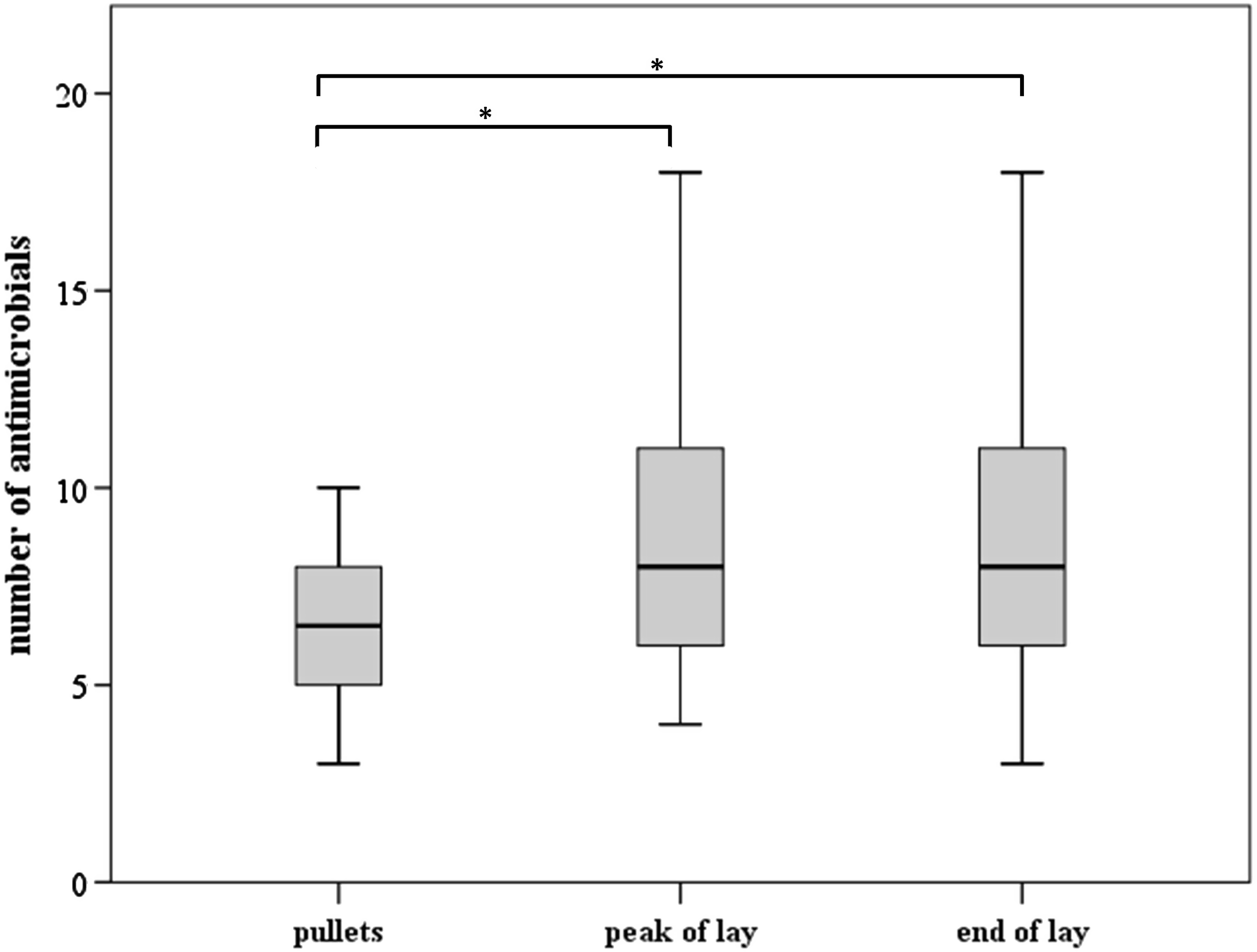
Boxplot diagram showing the number of antimicrobial substances to which Gallibacterium anatis was found resistant, isolated at different sampling points (pullets, peak of lay, and end of lay). Medians are shown with thick lines within the boxplots indicating the interquartile range and whiskers as the maximum and minimum values within 1.5× interquartile range. Asterisks depict significant differences between sampling points (p < 0.001, ANOVA).
In total, the 213 G. anatis isolates showed 99 different AMR profiles. Overall, the majority of isolates (n = 108) could be attributed to two AMR profiles. Three up to ten AMR profiles were detected in 76 isolates, representing the most common resistance phenotypes (Table 4). A unique AMR profile was found in 29 isolates. On flock level, different AMR profiles were found in 28 flocks (71.8%) independent of the sampling time point. The number of different AMR profiles per flock ranged from two to ten with different resistant patterns for up to 15 antimicrobials (Table 5).
Most Common Resistance Phenotypes Comprised by 76 Gallibacterium anatis Strains
AMP, ampicillin; AMX, amoxicillin; CAZ, ceftazidime; CEZ, cefazolin; CMP, chloramphenicol; COL, colistin; COX, cefoxitin; ENR, enrofloxacin; NAL, nalidixic acid; NEO, neomycin; OXA, oxacillin; SMO, sulfamethoxazole; T/S, trimethoprim/sulfamethoxazole; TET, tetracycline; TLS, tylosin; TRP, trimethoprim.
Variation of Antimicrobial Resistance Profiles Within Flocks
Denotes that all Gallibacterium anatis isolates showed identical antimicrobial resistance profiles within a flock.
Number of flocks (number of varying antimicrobial substances between profiles).
n.a., not applicable.
Identical AMR profiles were mostly found in isolates originating from the same organ of a single bird (78.0%), but still 22 such paired isolates differed in their AMR profile. This finding is based on the increase of differences in resistance patterns with the age of the birds: AMR profiles varied in isolates from pullets in regard to four antimicrobials, whereas the number of differing antimicrobials was up to 13 when birds were in production (Table 6).
Differences in Resistant Patterns to Antimicrobials of Paired Isolates from the Same Organ in Consideration of the Age of the Birds
Antimicrobial substance.
Variable AMR profiles were found between isolates from single birds (Table 7). Even isolates from different organs, originating from 31 birds, showed divergent outcomes ranging from one up to thirteen antimicrobial substances (Table 7).
Differences in Resistant Patterns to Antimicrobials of Isolates from the Same Organ and a Single Bird
Antimicrobial substance.
G. anatis strains originating from systemic organs (lungs, heart, or liver) were significantly more resistant than those from reproductive organs (ovary and oviduct) (p = 0.002; Fig. 2).
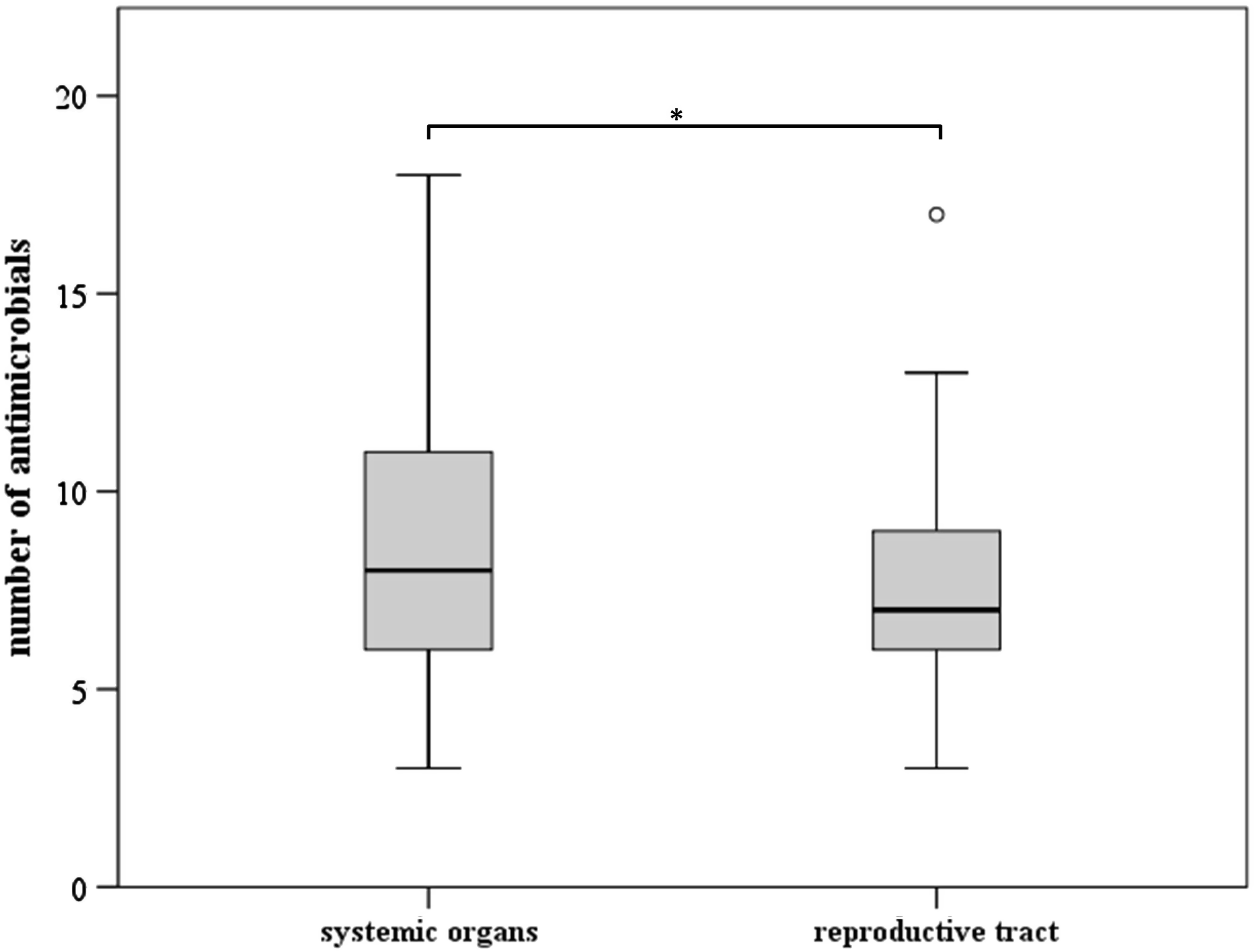
Boxplot diagram showing the number of resistant antimicrobial substances of Gallibacterium anatis isolates from systemic organs (lungs, heart, or liver) and reproductive tract (ovary and oviduct). Medians are shown with thick lines within the boxplots indicating the interquartile range and whiskers as the maximum and minimum values. Outliers are represented by circles (≥1.5× interquartile range). Asterisks depict significant differences between organs (p = 0.002, Mann–Whitney U test).
Development of resistance of investigated G. anatis strains was not significantly influenced by antibiotic treatments given to the animals during the production period (p = 0.238).
Discussion
Members of the highly heterogenous family Pasteurellaceae cause a wide variety of diseases in animals and antimicrobial substances are the most powerful tools to control such infections. For most members of this family, the susceptibility status still looks favourable. 29
In recent years, G. anatis has been increasingly reported as one of the major pathogens associated with reproductive tract lesions in laying hens.9,10,14–16 Over the years, an increase of antibiotic-resistant field isolates of G. anatis has been reported showing remarkable high prevalence of multidrug-resistant strains.14,21 This is in agreement with the current outcome, rating 96.2% of the strains as multidrug resistant. Most prominently, resistance to tylosin and tetracycline was observed. Meanwhile, this alarming development is reported from G. anatis field isolates worldwide.14–16,21 The high number of quinolone-resistant strains is in contrast to previous studies in which most of the G. anatis isolates proved to be susceptible.14–16 However, quinolone resistance in G. anatis with tendency to rise was reported a few years ago. 21
Furthermore, in agreement with previous reports, most of the G. anatis isolates investigated were resistant to sulfamethoxazole.15,16,21 Interestingly, this finding is in clear contrast to the data gained from G. anatis strains isolated in the United States. 14 While high resistance rates were previously reported to penicillins and aminoglycosides,14–16 the majority of the present isolates were susceptible to these antimicrobial compounds, except to oxacillin.
The 213 G. anatis isolates investigated in the actual study diverged into 99 different AMR profiles. This outcome already indicated that the majority of flocks harbor G. anatis strains of different AMR profiles. The finding is also reflected on bird and organ level. Most of the isolates with identical AMR profiles originated from the same organ of a single bird. In general, it is assumed that G. anatis isolates represent a clonal population within a flock and identical AMR profiles would be expected.22,23 In contrast, it was shown for Actinobacillus pleuropneumoniae and E. coli isolates that one animal can harbor clones with different AMR patterns.30–33
The AMR of a bacterial population is often associated with continuous or repeated antimicrobial exposure as this selects for resistant strains. However, it was also reported that resistant strains of bacteria can exist in a population for a long time, even in the absence of antimicrobial use. 34 In this investigation, data of antibiotic medication were available from ten flocks, and no influence on the resistance profiles was found. This is in agreement with previous results, 21 suggesting that the spread of resistance within members of Pasteurellaceae can also be due to horizontal transfer of plasmids beside clonal dissemination, as reported for E. coli.35,36
Interestingly, isolates from systemic organs proved to be more resistant than isolates from the genital tract. This phenomenon was recently reported from Haemophilus influenzae infections in humans.37,38 A possible explanation for this finding might be the presence of niche-adapted genotypes, which appear based upon different ecological opportunities.39–41 Evidence that this might also be the case for G. anatis was previously provided by revealing organ-restricted genetic clonal lineages within the same animals.22,42 Genetic investigations of these isolates were not done so far, but need to be studied in greater detail and aligned with AMR data. Independent of this, the actual finding is of high relevance, considering a targeted use of antimicrobials in the field.
In conclusion, based on a longitudinal sampling scheme, this study illustrates, for the first time, an increase of resistant G. anatis strains with the age of laying birds. Furthermore, the presence of G. anatis isolates with diverse AMR profiles was revealed not only within a flock but also within a single bird and even within the same organ. This finding is of importance in regard to effective treatment regimes revealing the need to test at least multiple isolates from different birds of a flock.
Footnotes
Disclosure Statement
The authors declare no conflicts of interest.
Funding Information
This work was financially supported by the Wirtschaftskammer Wien, Austria: WKO project entitled Antibiotic resistance profiles of selected bacteria from Austrian layers.