
Abstract
Introduction:
Staphylococcus haemolyticus is an acquired opportunistic pathogen causing nosocomial infections. Our previous studies of S. haemolyticus showed a group of isolates that produced a significantly higher disease severity than the others. Further molecular typing showed that the sequence type (ST) 42 was the major clone among the isolates. The main aim of this study was to characterize ST42.
Materials and Methods:
Sixty-one and 36 isolates were collected from burn and nonburn patients, respectively. Molecular typing, antibiotic susceptibility assays, and phenotypic characterizations were performed.
Results:
Thirteen STs, including seven new STs, were established (ST42 to ST48). ST42 was prevalent in burn and nonburn patients, and all the pulsotype C isolates were ST42. Four of the novel STs originated from ST3, suggesting that these clonal lineages evolved locally. ST3 and ST42 showed a significant difference in clindamycin susceptibility; molecular typing showed only one MLST locus variation among seven loci in SH1431, which has been reportedly involved in the regulation of biofilm formation through Zn 2+ binding affinities.
Conclusions:
Seven novel S. haemolyticus STs were identified; phylogenetic analysis suggested the presence of locally evolved clonal lineages. The predominant ST42 showed weak biofilm formation abilities; other factors that cause the clonal lineage change still need further investigation.
Introduction
Staphylococcus haemolyticus is one of the most frequently isolated opportunistic coagulase-negative staphylococci (CoNS)1,2; it is considered one of the predominant staphylococci in nosocomial foreign device-related infections. 3 Over the past decades, studies of this species have shown characteristics of multidrug resistance and biofilm formation abilities, which cause its potential endemic spreading in hospitals.4,5 Furthermore, surveillance reports of S. haemolyticus antimicrobial resistance showed higher levels of drug-resistance among CoNS species; drug-resistance combined with biofilm formation abilities lead to the difficulty of clinical treatments. 6
In 2015, a blast incident in northern Taiwan caused a massive number of burn injury patients, and afterward became a source of a S. haemolyticus outbreak. 7 Our studies found that S. haemolyticus isolated from these patients caused more severe disease compared with other S. haemolyticus isolated from non-burn injury patients. Pulsed-field gel electrophoresis (PFGE) analysis showed that most of these isolates belonged to a dominant group (pulsotype C), and this phenomenon was not observed in non-burned patients. This finding showed the importance of epidemiological surveillance, as the indigenous S. haemolyticus groups were changing. Further molecular epidemiological classification showed that sequence type 42 (ST42) was the predominant clone. Our specific aim was to study these changing S. haemolyticus ST42 isolates and elucidate their pathogenic properties.
Materials and Methods
Bacterial isolates
A total of 97 S. haemolyticus isolates were selected from the previous study. 7 Multiple strains were isolated from the same patient having the same molecular typing results and drug resistance phenotype; however, only one of these isolates was included in the study. Sixty-one isolates (study group) were collected from mass-burn casualty patients hospitalized at the Chang Gung Memorial hospital, and 36 isolates (control group) were collected from the nonmass-burn patients present in the same hospital. General information is provided in Supplementary Table S1. Strains isolated from study group patients were collected mainly from ward 2A (88.5%, 54 of 61) or ward 5A (9.8%, 6 of 61). Strains isolated from control group patients were collected from different wards. Antimicrobial susceptibility testing was performed by the disc diffusion test and followed the CLSI (Clinical and Laboratory Standards Institute) guidelines as in previous studies.7,8 Clindamycin resistance was further confirmed by the D-zone test. 9
Molecular typing and biofilm-related genes
All isolates were grouped by multi-locus sequence typing (MLST), PFGE, and SCCmec typing analysis. The MLST was performed as the previous study. 10 In general, the PCR products of seven house-keeping genes (arc, SH1200, hemH, leuB, SH1431, cfxE, and RiboseABC) were sequenced and compared with reported alleles and STs. The allele data were further analyzed by eBURST to define the evolutionary relationship among each isolate. PFGE analysis was carried out to reanalyze the data collected from the previous study. 7 Gel images used for PFGE analysis and belonging to ST3 and ST42 from a previous study were analyzed with the BioNumerics software (Applied Maths. Kortrijk, Belgium). PFGE analysis parameters were as follows: optimization 0%, tolerance 1%, and Jaccard statistic coefficient method was used for the dendrogram. After recalibration and renormalization, ST3 and ST42 isolates were regrouped in the present study. SCCmec typing was carried out using multiplex PCR as described previously. 11 Biofilm-related gene assays were performed using PCR; the general information is provided in Supplementary Tables S2 and S3.
Biofilm formation assay
Biofilm formation ability studies were performed with slight modifications from previous studies.12,13 Briefly, all isolates were grown at 37°C overnight in tryptic soy broth (TSB) medium with 1% glucose, and further diluted with fresh TSB at 1:100 to adjust the bacterial concentration to 0.5 on the McFarland scale. The biofilm formation assay was performed by adding 200 μL of adjusted bacterial culture per well to a 96-well microplate, and the plate was incubated overnight at 37°C. After incubation, the supernatant was removed and the culture was fixed at 68°C for 90 min. The fixed culture was stained with 0.1% safranin, decolored with 20% acetic acid, and its absorbance was measured at 490 nm (OD490) by SpectraMax M2, Molecular Devices™. Five replicates were analyzed per sample. The cutoff OD value (ODc) was defined as the mean of the negative control OD plus three times of standard deviation (ODc = ODnegative + 3x SD). The biofilm formation strength was defined using the following classification: non biofilm producer (OD ≤ ODc), weak biofilm producer (ODc < OD ≤2ODc, WBF), moderate biofilm producer (2ODc < OD ≤4ODc, MBF), and strong biofilm producer (4ODc < OD, SBF).
Nucleotide sequence accession numbers
Any sequence variations that differed from the reported alleles 10 were applied in the MLST database and assigned a new allele number. Below are the seven new polymorphisms identified in this study, all of them have been submitted to GenBank and received their accession numbers: arcC, allele 8: MT220003 and allele 9: MT220004; SH_1200, allele 10: MT220005; leuB, allele 12: MT220006, and allele 13: MT220007; SH1431 allele 10: MT220008; RiboseABC, allele 10: MT220009.
Results
Molecular typing of S. haemolyticus isolates
The MLST analysis results showed that the 97 isolates belonged to 13 types, except for one isolate that was nontypable (Table 1). Among these 13 sequence types (STs), six STs have been reported in previous studies (ST1, ST3, ST8, ST25, ST29, and ST30), 10 and seven novel STs (ST42–ST48) were identified in this study. The major clone was ST42, with 59.8% (58 in 97) of the total isolates belonging to this type. It was also the most frequent clone in both burn patients (67.2%, 41 in 61) and nonburn patients (47.2%, 17 in 36). The second most frequent clone population was ST3, with nearly 20.6% (20 in 97) of total isolates belonging to this type. ST3 was also the second most frequent clone population in both burn patients (26.2%, 16 in 61) and nonburn patients (11.1%, 4 in 36). The profile of the seven house-keeping genes (arcC-SH1200-hemH-leuB-SH1431-cfxE-Ribose ABC) of ST3 (1-1-1-1-1-1-4) and ST42 (1-1-1-1-5-1-4) showed that only one difference existed in the locus of SH1431. Further analysis of the SH1431 sequence found three nucleotide variations and two of them led to an amino acid change. The nucleotide variations in 212th and 344th bases between ST3 and ST42 caused the 71th and 115th amino acids to change from glycine (G, ggt) and proline (L, cct) into aspartic acid (D, gat) and arginine (R, cgt), respectively (Table 2). In addition to ST42, three of the novel STs identified in this study (ST43, ST44, and ST47) also showed similar patterns like ST3 (Table 2), which suggested that these new STs found in Taiwan may be close to ST3. Further analysis of the phylogenetic relationship by eBURST diagram of 13 STs, combined with STs reported in a previous study, 10 showed nine STs found in this study in one cluster. ST3 was the founder ST for ST42, ST43, ST44, and ST47 (Fig. 1), and the other new STs identified in this study may have evolved independently.

Phylogenetic analysis of the MLST relationship of the 97 isolates in this study and that of Sasmita Panda's data 10 via an eBURST diagram. The circles represent various STs and the circle sizes represent the isolates number. The straight open circle represents the founder ST (ST3), the open circle represents the previous found STs, and the double open circle represents the new STs identified in this study. MLST, multi-locus sequence typing.
Molecular Typing of 97 Staphylococcus haemolyticus Strains, as Determined by Multi-Locus Sequence Typing
Bold type indicated the new STs identified in this study.
MLST, multi-locus sequence typing.
Multi-Locus Sequence Typing Profile of ST3 and Other Novel Sequence Types
The difference of SH1431 between ST3 and ST42 were located on the 71th and 115th amino acid; in ST3 is glycine (71th, G, ggt) and proline (115th, L, cct) and in ST42 is aspartic acid (71th, D, gat) and arginine (115th, R, cgt).
As ST42 and ST3 were the two major clones in this study, PFGE (Fig. 2) and SCCmec typing (Table 3) analysis were performed to distinguish the differences between 58 ST42 and 20 ST3 isolates. There was a total of 28 pulsotypes, including subtypes A through O, and over half of the isolates (80.8%, 63 in 78) belonged to five major pulsotypes [C (32), F (15), A (6), H (5), and K (5)]. The majority of the ST42 isolates belonged to subtype C1 and F2. Among subtype C1, most of them were collected from study group patients (92.6%, 25 of 27). Six of F2 isolates were collected from study group patients and 7 from control group patients. Most of the 58 ST42 isolates were collected from ward 2A (90.2%, 37 of 41 from study group patients; 29.4%, 5 of 17 from control group patients). Unlike ST42, most of the ST3 isolates were grouped to 12 different pulsotypes and were distributed in different wards. The SCCmec typing analysis showed similar distribution between 20 ST3 and 58 ST42 isolates (Table 3). Five different types were found among these isolates, and most isolates belonged to SCCmec III or SCCmec I plus III combination, and SCCmec I was found to be more predominant in ST42 isolates than in ST3 isolates.
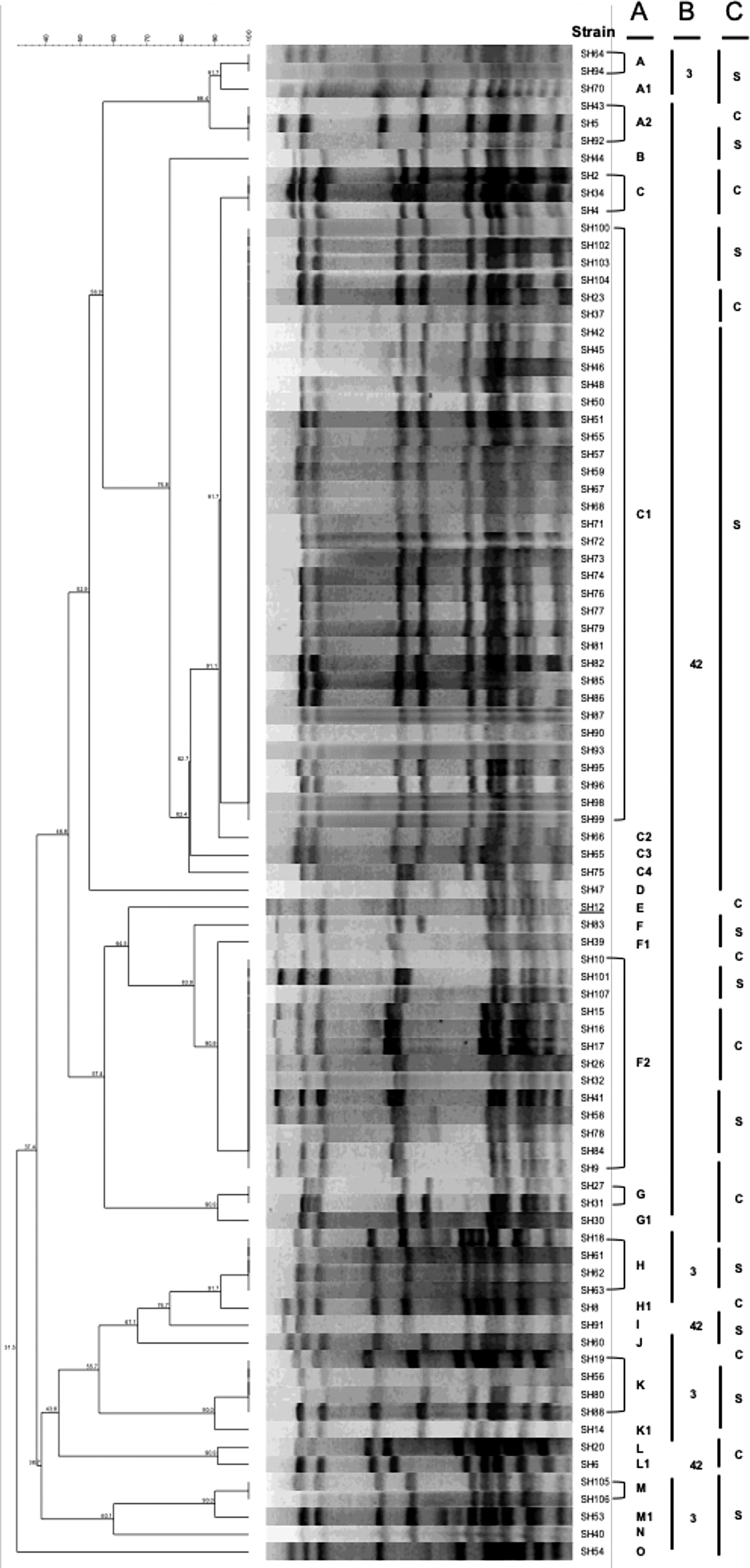
PFGE analysis of the 58 ST42 and 20 ST3 isolates.
SCCmec Types Distribution Among ST3 and ST42 Isolates
Characteristic differences between ST42 and ST3
Previous studies showed a significant difference in drug sensitivity results between the burn and nonburn patients, 7 which indicated that this difference may also exist in ST42 and ST3, and antimicrobial susceptibility analysis was performed to test ST42 and ST3 (Table 4). Our drug resistance analysis results showed that only clindamycin resistance was significantly different between ST42 and ST3. In addition to the drug resistance abilities, the MLST profile showed only one difference existed in locus SH1431, which was assumed to be a putative elastin-binding protein EbpS 14 and reported to have potential involvement in the regulation of bacterial growth and biofilm formation.15–17 The results of biofilm formation abilities assay of these ST42 (58 isolates) and ST3 (20 isolates) isolates were shown in Table 5; 84.5% of ST42 strains belonged to the nonbiofilm producer, and 65% of ST3 strains belonged to the nonbiofilm producer. We also checked the existence of several biofilm-related genes, including atl, aap, cidA, and lyts; all of them were present in both ST3 and ST42 isolates. None of the isolates contain sasX, and only three isolates (SH7, SH9, and SH51) were positive for the icaA gene.
Antibiotic Susceptibility Pattern Comparison of ST42 and ST3 Staphylococcus haemolyticus strains, as Determined by the Disc Diffusion Test
Indicates significant different between ST42 and ST3 (p-value <0.5).
SXT, Trimethoprim/Sulfamethoxazole.
Biofilm Formation Abilities Comparison Between ST42 and ST3
p-value = 0.064.
Discussion
In the previous study, we reported the molecular characteristics of an outbreak of Staphylococcus haemolyticus infection among a mass-burn casualty patient group. 7 In this study, we further performed molecular typing of these isolates and identified new features about these strains. First, we discovered novel multi-locus STs that may have locally evolved in Taiwan; several of these novel STs may have evolved from ST3 which was a predominant strain in other countries. Among these novel STs, we found that one of the novel STs, ST42, may have evolved from ST3 and became a predominant strain in our collection. Furthermore, molecular comparison between ST42 and ST3 showed minor antimicrobial resistance differences and most clones of ST42 and ST3 did not show biofilm forming abilities.
In several studies, the molecular typing of S. haemolyticus has been performed in the past few years; however, due to the genome structure complexity, each group developed their own method to discriminate various isolates. Eventually, loci selected by Panda's group were accepted and became the standard in the MLST database.10,18,19 The studies from Panda's group showed diverse sequence variation distribution of STs among their isolates; 41 STs were found in their analysis, however, only a few isolates belonged to each ST type, which is different from the findings of our present study. Our MLST analysis showed only 13 STs, and most of the isolates belonged to the predominant ST42 and ST3 groups in our collection (Table 1). This suggested that S. haemolyticus may have evolved in the local area over a period of time.
Several studies have shown that coagulase-negative staphylococci (CoNS) exists as a combination of two types,20–23 indicating the complexity and diversity of SCCmec types among staphylococci, and similar mixed types in one strain were also found in our study. Our analysis showed that 44.9% (35/78) of the isolates belong to the mixed types (I + II or I + III) in ST3 and ST42, which suggests that the structure complexity of SCCmec cassette was varied when compared with that of S. aureus, 24 and that the SCCmec typing methods may not be suitable for evaluating the difference between ST3 and ST42.
Previous studies showed that ST3 was one of the major clones in India, 10 which is also consistent with our molecular evolution relation analysis (Fig. 1). The eBURST analysis showed that ST3 was the founder ST for several novel STs identified in this study. Seven of the 13 STs in our collection were novel STs identified in this study, and four types were connected to ST3, which indicated a locally evolved clonal lineage (Fig. 1). Among these novel STs, four STs (ST42, ST43, ST44, and ST47) showed single locus variation in ST allelic profiles between them and ST3 (Table 2), and the only variation found in SH1431 developed to the predominant ST42. Although the allelic profile of ST44 also showed single variation in SH1431, further investigation of the MLST sequence profile composition of ST3, ST42, and ST44 on SH1431 found three differences between ST3 and ST42 and only one difference between ST3 and ST44 (unpublished data). This suggested that ST3 may be similar to ST44 and different from ST42. The PFGE results showed that most of the ST42 isolates collected from ward 2A belonged to pulsotype C1, which suggested the possibility that a predominant clone was already in existence in the hospital.
Previous analysis indicated that SH1431 encoded a putative elastin-binding protein (ebps), several reports indicated it may be involved in the regulation of biofilm formation through Zn2+ binding15–17 ; however, mutation of ebps causes minor effect on biofilm formation, suggesting that further analysis was necessary. The statistical analysis showed no significant difference between these two STs in the context of biofilm formation, even the ST3 has higher biofilm formation with positive rate (Table 5). Furthermore, most isolates of ST3 and ST42 were not biofilm producers (Table 5), which was unlike the previous reports.3,25,26 Several biofilm-related genes (aap, atl, cidA, icaA, lyts, and sasX) were examined for their existence in both STs. Most of them (aap, atl, cidA, lyts, and sasX) showed no difference between ST3 and ST42, except icaA showing tiny difference. Three isolates were icaA positive, and two of them belonged to ST42; however, only one isolate showed weak biofilm formation ability, suggesting that our biofilm formation-positive isolates were mediated by the ica-independent pathway, which agrees with recent findings.27,28 These results suggested that those biofilm-related genes had been examined in the present study may not be directly responsible for biofilm formation phenotype and consequence to the difference between ST3 and ST42. Therefore, the phenotypical differences between ST3 and ST42 may be caused by other factors, due to the clonal distribution change. Previous studies showed that the genome structures of most staphylococci share a similar composition, with most virulence genes and mobile genetic elements being located on core variable or accessory regions.29–31 This feature implies that similar genome structures may exist on S. haemolyticus that could be better understood by whole genome sequence analysis. 1 The analysis showed that 82 insertion sequences were distributed throughout the S. haemolyticus genome, owing to frequent genome structure rearrangements, which consequently led to phenotypic diversification. Therefore, it may be possible that insertion sequence-mediated genome structure rearrangements cause the differences between ST3 and ST42, which enhance ST42 adaptation to the local environment.
In conclusion, this study identified several novel STs and predicted the clonal lineage correlation. Furthermore, the phenotypic diversity between ST3 and ST42 were examined by drug resistance and biofilm formation. Previous studies proposed that genome structure rearrangement mediated by IS elements may cause phenotypic diversification, which requires confirmation by whole-genome sequencing of the various STs of isolates. The molecular level differences between ST3 and ST42 remain to be investigated in the future.
Footnotes
Authors' Contributions
J.J.L. conceived the study, analyzed and interpreted the data, and edited the draft of the article. L.C.L. and T.P.L. analyzed and interpreted the data, and wrote the first draft of the article. T.P.L. and S.C.C. collected and prepared the study material from Chang Gung Memorial Hospital bacterial storage bank. All authors read and approved the final article.
Disclosure Statement
No competing financial interests exist.
Funding Information
This work was supported by grants from Chang Gung Memorial Hospital (CMRPG3G1722 and CMRPG3G1721), the Ministry of Science and Technology, Taiwan (MOST 107-2320-B-182A-021-MY3 and MOST 108-2811-B-182A-513), and Chang Gung Memorial Hospital bacterial storage bank program (CLRPG3E0025).
References
Supplementary Material
Please find the following supplemental material available below.
For Open Access articles published under a Creative Commons License, all supplemental material carries the same license as the article it is associated with.
For non-Open Access articles published, all supplemental material carries a non-exclusive license, and permission requests for re-use of supplemental material or any part of supplemental material shall be sent directly to the copyright owner as specified in the copyright notice associated with the article.