
Abstract
Aims:
Antimicrobial resistance (AMR) spreads not only by pathogenic but also by commensal bacteria, and the latter can become a reservoir for resistance genes. This study was aimed to investigate the AMR patterns along with the presence of mobilized colistin resistance (mcr) genes in commensal Escherichia coli circulating in chickens, farm environments, street foods, and human patients.
Materials and Methods:
By a cross-sectional survey, isolates obtained from 530 samples were tested for their AMR profiles against 9 antimicrobials. Minimum inhibitory concentration (MIC) of the phenotypically colistin-resistant isolates was determined and screened for a set of mcr genes followed by sequencing of mcr-1 gene in the multidrug-resistant (MDR) isolates.
Results:
A total of 313 E. coli strains were isolated and confirmed by polymerase chain reaction. Antimicrobial susceptibility testing revealed that about 98% (confidence interval [95% CI] 95–99) of the isolates were MDR, and 58% (95% CI 52–63) isolates exhibited resistance to colistin. MIC values of colistin against the isolates ranged from 4 to 64 mg/L. Except for human patients, 20.4% colistin-resistant isolates from other sources of isolation had mcr-1 gene.
Conclusions:
There is abundance of commensal MDR E. coli strains with the acquisition of mcr-1 gene circulating in chickens and farm environments in Bangladesh.
Introduction
The emergence and dissemination of antimicrobial resistance (AMR) is an inevitable side effect of the irrational antimicrobial uses. The dissemination of AMR has been attributed not only to pathogenic but also to commensal bacteria, which constitute a reservoir of resistance genes for other nonpathogenic and pathogenic microorganisms.1,2 Escherichia coli is a gastrointestinal commensal for both humans and animals with the ability to cause diseases. A wide range of antimicrobials is used to treat E. coli infections, which include β-lactams, fluoroquinolones, aminoglycosides, tetracyclines, and sulfamethoxazole–trimethoprim. 3 Antimicrobials, such as colistin (a class of polymyxins), tigecycline, temocillin, and fosfomycin, show the best in vitro activity against carbapenemase-producing E. coli. 4 However, the extensive use of antimicrobial agents in animal production would exert a selective pressure to contribute to the emergence and dissemination of resistance genes among pathogenic and commensal microorganisms. 5 Colonization of resistant bacteria in the chicken gut can be a potential source for human infections through environment and food-chain contamination. 6
E. coli strains acquiring resistance to colistin have been described in food-producing and companion animals, 7 wild animals, and in the environment. 8 Emergence of resistance to colistin in naturally susceptible bacteria can be either by the chromosomal mutation or by plasmid encoded resistance genes. Recently, plasmid-mediated colistin resistance gene (mcr-1) has been identified for the first time in China from farm animals, hospital patients, and food sources. 9 Subsequently, mcr-2 to mcr-10 genes have been reported in different parts of the world.7,10 Consequently, it has become a matter of global health concern due to its high transmissibility among the microbial populations.
There is noticeable lack in data on the antimicrobial use and prevalence of resistant-microorganisms in the animals and farm environments in developing countries like Bangladesh. Of the many antimicrobials, colistin appears to be widely used in the commercial poultry production in Bangladesh11,12 despite official ban already imposed on such use. Recent studies from Bangladesh reported mcr-1 carrying E. coli in an urban sludge sample, 13 food, water, hand rinse, and healthy human gut. 14 In another study, E. coli carrying mcr-3 gene has been reported from poultry, house fly, and pond water in Bangladesh. 15 However, information on the prevalence of colistin-resistant genes in commensal microorganisms in chickens, farm environments, and in the human food chain is scarce. Therefore, we aimed to investigate the AMR patterns along with the presence of mobilized colistin resistance (mcr) genes in commensal E. coli circulating in chickens, farm environments, street foods, and human patients in Bangladesh.
Materials and Methods
Study period and population
This cross-sectional survey was conducted during the period of January to October 2018. A total of 30 broiler farms from 6 Upazilas (administrative subdistricts) of Chattogram (previously Chittagong) division of Bangladesh were randomly selected for live chickens and different farm environments to be sampled. Street food samples were collected from different places of Chattogram Metropolitan City area, Bangladesh. Human patients admitted to two medical hospitals in Chattogram district were also randomly sampled.
Sample collection
Cloacal swabs were collected from randomly selected 5 live chickens in each of the 30 broiler farms. In addition, 100 g pooled feed samples, 50 mL pooled water samples, and 200 g pooled soil samples representing different sites of a farm were collected. Around 250 g litter samples were collected from five separate locations of a poultry flock on a farm where the sample consisted of around 25 cross-sectional pinches of litter mixed with feces. Approximately, 250 g pooled litter samples at the dumping site(s) of a farm were also collected. Street food samples, including fast food, handmade juice, salad, and “chotpoti” (a roadside dish consisting of potatoes, chickpeas, onions and is usually topped with additional diced chilies or grated boiled eggs), were collected from small shops/mobile rickshaw vans selling the items mentioned above at different sites of the city. Stool samples were collected from human patients with gastrointestinal disorder admitted to two medical hospitals located in the city. Swabs, as obtained from the sampling matrices mentioned, were kept in Stuart's transport medium (Oxoid), and all solid samples were kept separately in sterile zipper bags. All the samples collected in a day were kept in a cool box before being shipped to the Department of Microbiology and Veterinary Public Health, Chattogram Veterinary and Animal Sciences University, Chattogram, Bangladesh for further analysis.
Isolation and identification of E. coli
For primary enrichment, each cloacal/stool swab, already kept into Stuart's transport medium, was inoculated into 5 mL of buffered peptone water (BPW) (Oxoid) and incubated overnight at 37°C. A solid or a liquid sample other than any swab samples was mixed homogenously, and 10 g of a solid or 10 mL of a liquid sample was inoculated into 100 mL BPW with the same incubation time and temperature. After primary enrichment, the culture was inoculated onto MacConkey agar (Oxoid) and incubated at 37°C for 24 hours. Large pink colonies yielded on the MacConkey agar were inoculated onto Eosin methylene blue (EMB) agar (Oxoid), and after recommended incubation, only distinctive greenish metallic sheen colonies were tested by standard biochemical test, for example, indole test (Kovac's Indole Reagent; HiMedia) 16 followed by detection of a housekeeping gene (adenylate kinase, adk) of E. coli by polymerase chain reaction (PCR) to identify the growth as E. coli. 17
Screening antibiogram of E. coli isolates
The E. coli isolates were screened for antimicrobial susceptibility against a panel of nine antimicrobials comprising seven different groups: β-lactams, tetracyclines, polymyxins, fluoroquinolones, sulfonamides, aminoglycosides, and phenicols. Kirby-Bauer disc diffusion method was used to determine susceptibility patterns. 18 The size of the zone of inhibition (in mm) around a disc was measured using a slide calipers, and the results were interpreted according to Clinical Laboratory Standards Institute (CLSI) guidelines.19,20 However, there are no CLSI guidelines to determine colistin susceptibility, and therefore, we interpreted the results according to the manufacturer's (Oxoid Limited, Basingstoke, United Kingdom) instructions. Multidrug-resistant (MDR) isolates were classified based on their resistance profiles. An isolate was considered as MDR if it showed resistance to ≥3 classes of antimicrobials. 2
Screening of the mcr genes
All the MDR E. coli isolates showing resistance to colistin were investigated for the presence of four plasmid mediated colistin resistance genes, mcr-1, mcr-2, mcr-3, and mcr-4. The primer sequences used for the detection of the selected mcr genes mentioned are shown in Table 1. All the PCRs were conducted with a final volume of 25 μL with 12.5 μL DreamTaq Green PCR Master Mix (2 × ) (Thermo Fisher Scientific Baltics UAB), 1 μL forward primer (20 pmol/μL), 1 μL reverse primer (20 pmol/μL), 8.5 μL nuclease-free water, and 2 μL DNA template. We followed different thermal conditions to detect mcr-1, 9 mcr-2, 21 mcr-3, 22 and mcr-4 23 genes. The PCR products were visualized on a gel documentation system (UVP UVsolo touch; Analytik Jena AG) after electrophoresis with 1.5% agarose gel (SeaKem® LE Agarose from Lonza).
Oligonucleotide Primer Sequences Used to Detect Four Colistin Resistance Genes in the Escherichia coli Isolates
Determination of minimum inhibitory concentration of colistin sulfate to the MDR isolates carrying mcr-1 gene
The minimum inhibitory concentration (MIC) against colistin sulfate was determined by Broth Microdilution Method. 24 Pure colistin sulfate powder (Sigma-Aldrich) was used to prepare the antibiotic solution for broth microdilution. A twofold serial dilution ranging from 0.125 to 64 mg/L was made using cation-adjusted (Calcium and Magnesium ions) Mueller Hinton II Broth (Sigma-Aldrich) to determine MIC, MIC 50, and MIC 90, according to ISO guidelines. 24 E. coli ATCC 25922 and NCTC 13846 were used as a negative and a positive control, respectively.
Gene sequencing
Among the mcr-1 strains three representing three diverse sources of isolation, live broiler chickens, farm environments, and street foods, were randomly chosen for sequencing of the gene. The purified PCR products were Sanger sequenced with BigDye terminator v3.1 Sequencing Kit and a 3730xl automated sequencer (Applied Biosystems, Foster City, CA). Nucleotide sequences were determined on both forward and reverse strands of PCR amplification products at the Macrogen sequencing facility (Macrogen, Inc., Seoul, Korea). The forward and reverse sequences of each of the strains were assembled by CAP3 Sequence Assembly Program. 25 The sequence products were submitted to GenBank of the National Center for Biotechnology Information (NCBI) under accession numbers MK884859, MK884860, and MK884861.
Phylogenetic analysis
BLASTn was performed to compare the mcr-1 sequences from this study and those available in GenBank (Supplementary Data). Based on the results obtained closely related sequences were included to develop phylogenetic trees by the neighbor-joining method. 26 The bootstrap values were calculated using 1,000 replicates. 27 The evolutionary distances were computed using the Maximum Composite Likelihood method. 28 The phylogenetic analyses were conducted using MEGA-X. 29
Statistical analysis
All the available sample data and laboratory results were entered into a spreadsheet program (Microsoft Excel Worksheet 2016) and transferred to R 3.5.1 software 30 and GraphPad prism 7.0 for data summary and analysis.
Results
Prevalence of E. coli
In total, 530 samples were collected from different sources, and E. coli was isolated from 313 of them (59%; confidence interval [95% CI] 55–64). An overview of the samples collected from different sources and status of E. coli in them are shown in Table 2.
An Overview of the Samples Collected from Different Sources, Status of Escherichia coli in Them, and the Distribution of mcr-1 Gene in the Isolates Obtained
AMR profile
Of the isolates obtained, 58% (95% CI 52–63) showed resistance to colistin in disc diffusion method. Irrespective of the source of isolation, all the isolates showed resistance to ≥3 antimicrobials and, therefore, considered as MDR. Their resistance profiles are portrayed in Fig. 1, depicting that, regardless of sources of isolation, most of the isolates were resistant to six to eight antimicrobials and some of them even to nine.

Percentage of isolates showing resistance against the tested antimicrobials
Distribution of the mcr genes
Only mcr-1 gene was detected in the isolates showing resistance to colistin plus different combinations of other antimicrobials. The distribution of mcr-1 gene is shown in Table 2. The susceptibility patterns of the isolates to the nine antimicrobials tested and the distribution patterns of mcr-1 gene in them are illustrated in Fig. 2.
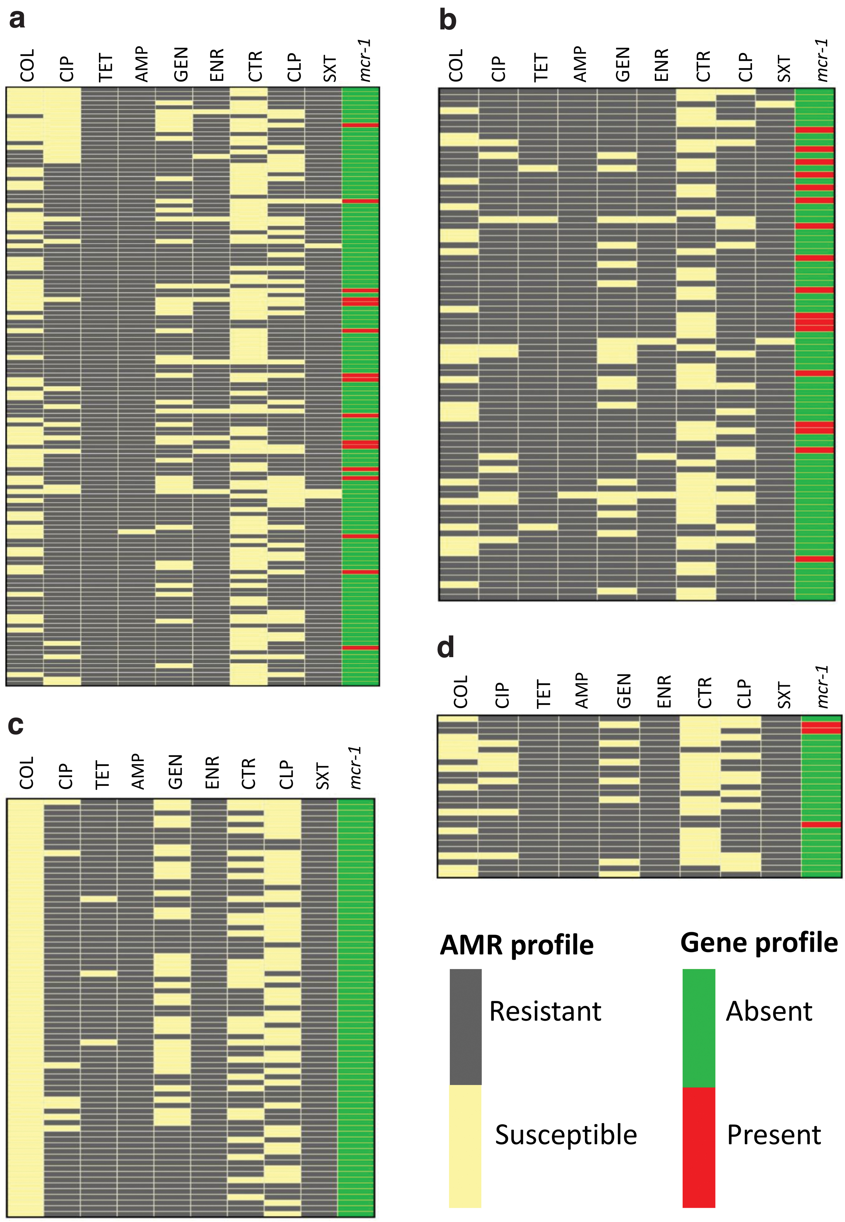
Distribution of antimicrobial resistance phenotype and resistance gene profile of E. coli isolates across the samples
Minimum inhibitory concentrations
The distribution of MICs of colistin sulfate to the mcr-1 bearing colistin-resistant isolates is shown in Fig. 3. The results revealed that 51% of the isolates tested could be inhibited at 16 mg/L colistin sulfate concentration. Overall, the MIC values of colistin sulfate against the isolates ranged from 4 to 64 mg/L where the breakpoint is 2 mg/L. Minimum concentration of colistin sulfate required to inhibit 50% (MIC 50) and 90% (MIC 90) of the isolates, as assessed, was found to be consistent, and it was >16 mg/L.
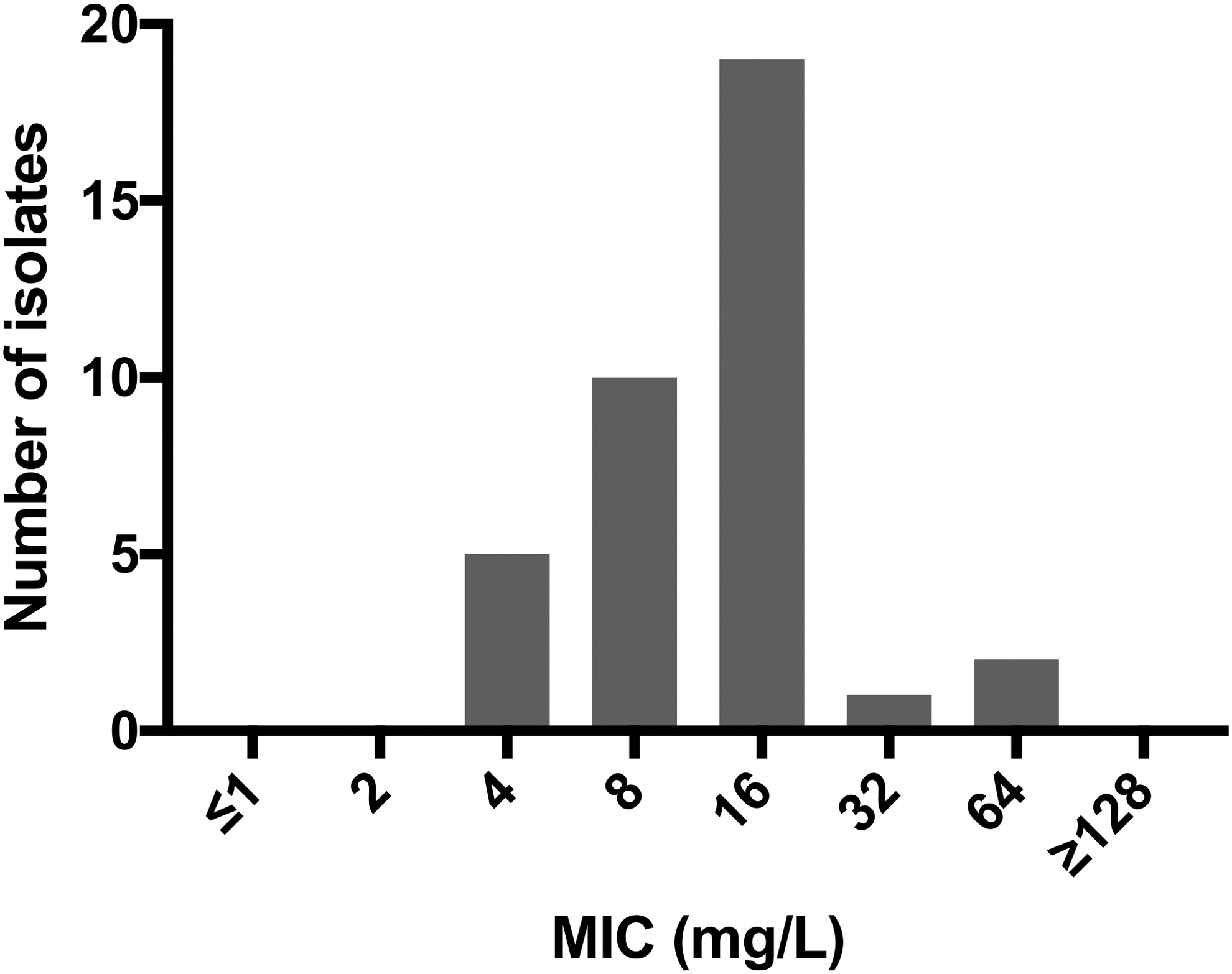
Distribution of MIC values of colistin sulfate to 37 multidrug-resistant E. coli isolates bearing mcr-1 gene where the break point of colistin is 2 mg/L. MIC, minimum inhibitory concentration.
Phylogenetic analysis
The phylogenetic analysis revealed that the sequences obtained from the three mcr-1 possessing MDR strains were closely related to a few strains isolated from Bangladesh in the recent past (Fig. 4). Although the sources of isolation of the three strains were diverse, the identity and similarity percentages of the sequences of mcr-1 gene of the strains showed a close relationship among them.

Phylogenetic comparison of mcr-1 gene sequences. The nucleotide sequences of colistin resistance gene (mcr-1, 309 bp) in broiler chickens, poultry farm environments, and street foods obtained from this study (black filled circles) were compared with those of representative mcr variants obtained from NCBI. NCBI, National Center for Biotechnology Information.
Discussion
The results of the study revealed abundance of commensal MDR E. coli strains circulating in broiler chickens, farm environments, street foods, and human patients in Bangladesh. This finding also supports some other reports indicating the presence and spread of MDR E. coli through broiler chickens31–33 and poultry farm environments.34,35 Their local and global transmissions in human patients and foods were also reported.32–34 The more alarming is that most of the strains isolated in this study had resistance against six to eight antimicrobials which was rarely reported, although the prevalence of MDR as high as 98% was reported in some studies in China, Spain, and Michigan.36–38 Around 50% of the isolates in this study were found to be resistant to colistin by disc diffusion method. The high prevalence of MDR commensal E. coli in live broiler chickens and the farm environments could contribute to the emergence of extensively antimicrobial-resistant strains across the food chain in the future, posing a harder challenge to treat them with the currently available antimicrobials in clinical practices.
Polymyxin E (colistin) is considered as the last resort antimicrobial agent to treat infections caused by MDR pathogens belonging to the family Enterobacteriaceae. 39 What had exactly triggered the emergence of resistance to colistin in commensal E. coli strains isolated in the study was difficult to resolve; however, much anecdotal and increasing factual evidence supports that misuse, and overuse of this antimicrobial in poultry production due to absence of robust surveillance could be the main reason.40,41
Before this study, there were some reports published indicating the possession of plasmid-mediated colistin resistance gene, mcr, in colistin-resistant isolates of a few bacterial species from Bangladesh. Although there are a number of colistin resistance genes reported worldwide (i.e., mcr-1 to mrc-10), only mcr-1 and mcr-3 genes were reported from Bangladesh previously.14,15,42,43 We investigated mcr-1 to mcr-4 genes, and in the present study, only mcr-1 gene was detected from diverse sources, including live broiler chickens (cloacal swab), soil and dumped litter from the farmyards, and the street foods. Such preponderance of mcr-1 gene among the others in different sources was also reported in some other studies in Bangladesh,13–15,42,43 China, and Germany.7,44 Dutta et al. reported a very low prevalence of mcr-1 gene from livestock origin, whereas Ahmed et al. and Islam et al. whose studies were closely related to this study reported a high prevalence, indicating that mcr-1 gene has been disseminated significantly overtime after its initial acquisition in poultry-borne E. coli populations.33,42,43 Irrational use of colistin could be the aggravating factor.11,12 Kawanishi et al. and Shen et al. reported that 5.3% colistin-resistant E. coli isolates harbored mcr-1 gene individually, but overtime the prevalence of mcr-1 and mcr-3 genes increased as high as 30% and 8.3%, respectively, in Japan.45–47 The absence of the other mcr genes namely, mcr-2, mcr-3, and mcr-4, might be due to absence of their acquisition or due to their less abundance in E. coli in the study area.
All the isolates obtained from human patients were found to be free from any of the mcr genes, suggesting that mcr-1 gene circulating in poultry and poultry farm environments might have not been transferred to E. coli in humans yet. However, the presence of mcr-1 gene in street food isolates is worrisome in that the probability of transferring of the gene to the human gut microbes is greater from contaminated foods because they are taken directly without having heated properly unlike eggs and poultry meat.
To our knowledge MIC by broth microdilution method for any colistin-resistant isolates of poultry farm environments and street foods has never been reported from Bangladesh. MICs for 37 colistin resistant, as well as MDR E. coli, strains carrying mcr-1 gene were estimated, and the results revealed that the value varied from 4 to 64 mg/L. Alike sequence information on mcr-1 gene of bacterial isolates from Bangladesh is very limited. However, the phylogenetic information generated from this study by including three sequences of the mcr-1 gene of one strain each from live broiler chickens, farm environment, and street food suggested that the strains were seemingly closely related despite origin divergency.
Determination of MIC value is the recommended method for assessing the resistance profile of any E. coli strains against colistin, according to CLSI and EUCAST guidelines. One of the limitations of the present study was that all of the strains of E. coli obtained from the study were initially screened for their susceptibility to colistin by disc diffusion method due to resource limitation, and the results were interpreted following the manufacturer's guidelines. The break point of MIC to consider an E. coli strain resistant against this antimicrobial is 2 mg/L, according to CLSI, 2018. 18 While acknowledging the limitation mentioned, the results of the 37 MDR strains with resistance against colistin randomly screened eventually by broth microdilution method revealed that they had MIC values ≥4 mg/L, suggesting that they all were indeed resistant to colistin. Due to high MIC value, as observed in the isolates screened, a high prevalence of colistin-resistant E. coli could not be entirely ruled out if all the E. coli strains were screened by the broth microdilution method despite the same result obtained on the 37 isolates tested by disc diffusion and by broth microdilution methods. Another limitation was that we did not use any colistin-added suitable control media to facilitate selective growth of only colistin-resistant E. coli by inhibiting the growth of other competing bacteria. If such control media was used, E. coli strains resistant to colistin could have selectively grown on it from any sample matrices either from animate or inanimate sources containing small quantity of the organism; consequently, its prevalence estimates from the sources of identification could have been higher. Considering these two limitations, the present study could underreport the true prevalence of colistin-resistant E. coli in the sources of investigations. However, the key message from the study on the acquisition of mcr-1 gene in MDR E. coli strains of diverse sources seemed to remain unaffected because its distribution was proportional to the number of resistant strains investigated for the presence of the gene.
In summary, the prevalence of MDR in commensal E. coli circulating in live broiler chickens, inanimate poultry farm environments, street foods, and in human patients is alarmingly high. Except for the strains of the isolation source human patients, 20.4% strains of other sources have already acquired mcr-1 gene with variable prevalence estimates. There seemed to have a close relationship among the MDR strains carrying mcr-1 gene, although their sources of isolation were different. The MICs of colistin sulfate to the MDR strains with mcr-1 gene were 4 to 64 mg/L. The results of the study suggest the need for stopping imprudent use of antimicrobials, particularly colistin in poultry production.
Footnotes
Acknowledgments
The authors gratefully acknowledge all the officials of Veterinary Stations of participating Upazilas for providing cordial support to collect live broiler and poultry farm environmental samples. The authors also thank Dr. Md Saifuddin Siddiki Suja (Department of Urology, Chattogram Maa-O-Shishu Hospital, Bangladesh) and Dr Dipak Sarker (Director, J.K. Memorial Hospital, Bangladesh) for providing the opportunity to collect samples from human patients for the study.
Ethical approval
Ethical Approval for the sampling was taken from Ethics Committee of Chattogram Veterinary and Animal Sciences University, Bangladesh. The Ethical approval No. is CVASU/Dir(R&E)EC/2019/39(2/6/6).
Disclosure Statement
No competing financial interests exist.
Funding Information
This work was supported by Advance Study and Research, Chattogram Veterinary and Animal Sciences University, Bangladesh and National Science and Technology Fellowship under the Ministry of Science and Technology, the People's Republic of Bangladesh.
References
Supplementary Material
Please find the following supplemental material available below.
For Open Access articles published under a Creative Commons License, all supplemental material carries the same license as the article it is associated with.
For non-Open Access articles published, all supplemental material carries a non-exclusive license, and permission requests for re-use of supplemental material or any part of supplemental material shall be sent directly to the copyright owner as specified in the copyright notice associated with the article.