
Abstract
Background:
Polycystic ovary syndrome (PCOS) is a major hyperandrogenic disorder. Many drugs prescribed specifically to treat PCOS have side effects; however, previous studies suggest that natural therapeutics including botanicals may be less invasive and equally effective for the management of PCOS.
Methods:
In the present study, plants were screened for antiandrogenic activity using the RIKILT yeast Androgen bioAssay (RAA). Selected positive plants were subsequently tested for their efficacy against PCOS induced by estradiol valerate (EV) in rat models.
Results:
RAA revealed the antiandrogenic property of Nardostachys jatamansi DC (NJ), Tribulus terrestris L. (TT), and Embelia tsjeriam-cottam DC (EJ), whereas Whithania somnifera Dunal (WS), Symplocos racemosa Roxb. (SR), and Helicteres isora L. (HI) exhibited androgenic properties. EJ also exhibited mild androgenic activity and therefore was excluded from further study. EV administration reduced the weight gain and disrupted cyclicity in all rats. NJ and TT extract treatment normalized estrous cyclicity and steroidal hormonal levels and regularized ovarian follicular growth.
Conclusion:
The in vitro antiandrogenic activity of plant extracts and their positive effects on different parameters of PCOS were proved in vivo.
Introduction
P
Traditional medicines used in the treatment of female reproductive disorders are regarded as safe, have lesser side effects, and possess multiple active compounds. 9,10 In this study, plants traditionally used against reproductive disorders were tested for their antiandrogenic properties by using an in vitro bioassay followed by in vivo evaluation of PCOS in rat models.
Materials and Methods
Chemicals
Ammonium sulfate, dextrose, yeast nitrogen base without amino acids and without ammonium sulfate,
Plant extraction
Plant materials were dried in the shade, powdered, and extracted with methanol (3–4 hr of Soxhlet extraction). The resulting mixture was filtered and evaporated to dryness using rotary evaporator.
In vitro yeast androgen bioassay
The RIKILT yeast Androgen bioAssay (RAA) makes use of yeast cells that are constructed to express the human androgen receptor (hAR) and yeast enhanced green fluorescent protein (yEGFP), which is expressed in response to androgens. 11
RAA bioassay for screening of androgenic and antiandrogenic activities
Media used for the assay were prepared according to Bovee et al.
12
with slight modifications. Either 2 μL of DHT at final concentrations of 3 nM and 100 nM (positive controls) for ensuring the correct run of the assay or 2 μL of undiluted and diluted sample were added to 200 μL of yeast suspension grown in minimal medium with
Aliquots of 200 μL of yeast cell suspension grown in MM/L medium were transferred to each well of a 96-well microplate, and 1 μL of FLU at final concentrations of 3 nM and 100 nM (positive controls) was added. Undiluted and diluted extracts were co-exposed with 1 μL of DHT at 3 nM final concentration for antiandrogenic activity testing. The 96-well microplates were incubated at 30°C and 125 rpm for 24 hr, and fluorescence was measured again at t=24 hr. MM/L medium with DMSO was used as a blank, and yeast cell suspension mixed with DMSO served as a negative control. The concentration of DMSO in the total reaction volume was adjusted to 0.5%, because higher concentrations could inhibit yeast cells. Fluorescence was measured using a Multimode Reader (Biotek Instruments, USA) at t=0 hr with an excitation at 485 nm and emission at 520 nm. The responses obtained with reference compounds were compared with those of extracts for interpretation of results. The plant extracts that displayed antiandrogenic activity were selected for the in vivo studies.
In vivo experiments
Animals
Adult Sprague-Dawley females (150–220 grams body weight) were obtained from the Karpagam Arts and Science College, Coimbatore, India. The studies were conducted in accordance with the guidelines of Committee for the Purpose of Control and Supervision of Experiments on Animals (CPCSEA), India.
Selection of animals and vaginal smear analysis
Estrous cyclicity was monitored by vaginal smears taken between 08:00 and 12:00 hr and assessed by light microscopy for the relative proportion of leukocytes and epithelial and cornified cells found in daily vaginal lavages that change during different stages of the estrous cycle. Animals displaying at least two consecutive normal 4-day estrous cycles were used in this study. Estrous cycles prior to and after treatment were monitored by such daily examinations of vaginal smears. 13 Rats with a normal estrous cycle were selected and divided into six groups of six rats per group: NC, normal control; MC, model control; NJLD, Nardostachys jatamansi DC (NJ) low dose (5 mg); NJHD, NJ high dose (10 mg); TTLD, Tribulus terrestris L. (TT) low dose (5 mg); and TTHD, TT high dose (10 mg). All groups, except the NC group, received 2 mg/0.2 mL of EV for PCOS induction. The NC group received 0.2 mL of sesame oil alone. All EV-treated groups of rats were evaluated for 72 days. From day 60 onward, groups NJLD, NJHD, TTLD, and TTHD received their corresponding dose of the extracts, dissolved in distilled water, by intraperitonial injections until day 72.
Measurement of sex hormone levels
Blood samples were collected from the jugular vein during the subsequent estrous phase or pseudo-estrous phase after the day 72. Serum levels of testosterone, progesterone, and estradiol were determined by competitive immunoassays (ADVIA Centaur CP Immunoassay System, Siemens Healthcare, USA). The intra-assay and interassay coefficients of variation and sensitivity with assay range were: 3.3%, 7.5%, and 10–1500 ng/dL testosterone; 4.8%, 8.0%, and 0.21–60 ng/mL progesterone; and 8.0%, 9.1%, and 0.21–60 ng/mL estradiol.
Ovary histology
Ovaries of the controls and EV-treated animals were removed, cleaned, and fixed in 10% formaldehyde buffer for at least 24 hr. Samples were dehydrated in a graded series of alcohol, embedded in paraffin, and sectioned at 5-μm thicknesses. Serial sections were mounted on glass slides, stained with Hematoxylin & Eosin, and embedded using glass coverslips. All samples were analyzed by light microscopy with an Olympus microscope at 40×magnification. Images were viewed and captured using a MIPS and Magnus Live USB 2.0 viewer. The primary, secondary, atretic, and cystic follicles and fresh corpora lutea were counted on each ovary.
Data analysis
Most of the statistical analysis was done by using SPSS software (v. 17, SPSS Inc.). Comparisons between the controls and extract-treated rats were done by analysis of variance (ANOVA), followed by the Tukey honest significant difference (HSD) post hoc test. All of the values were expressed as mean±standard deviation (SD). A P value less than 0.05 was considered statistically significant.
Results
In vitro androgenic and antiandrogenic properties of extracts
The standard DHT AR-agonist reference compound showed an average fluorescence response of 326±8 at 3 nM. A 3 nM DHT equivalent or near-equivalent response was obtained by extracts of Whithania somnifera Dunal (WS), 330±10, Symplocos racemosa Roxb. (SR) 306±8, and Helicteres isora L.
The standard FLU demonstrated its antagonistic effect at a 3 nM concentration when co-exposed with 3 nM DHT, i.e., FLU reduced the DHT-induced fluorescence response from 326±8 to 237±4.7. Likewise the extracts of NJ, TT, and EJ reduced the DHT-induced fluorescence response from 326±8 to 227±8, 220±7, and 215±8, respectively, at plant extract concentrations of 80 μg/mL, 5.1 ng/mL, and 11.2 μg/mL, respectively. However, although the lowest concentration of the TT extract (5.1 ng/mL) inhibited the DHT-induced response, suggesting it as more potent compared to NJ and EJ, a clear drop in the curve was only visible at 50.9 μg/mL, which is comparable to NJ and EJ (Table I and Fig. 1)
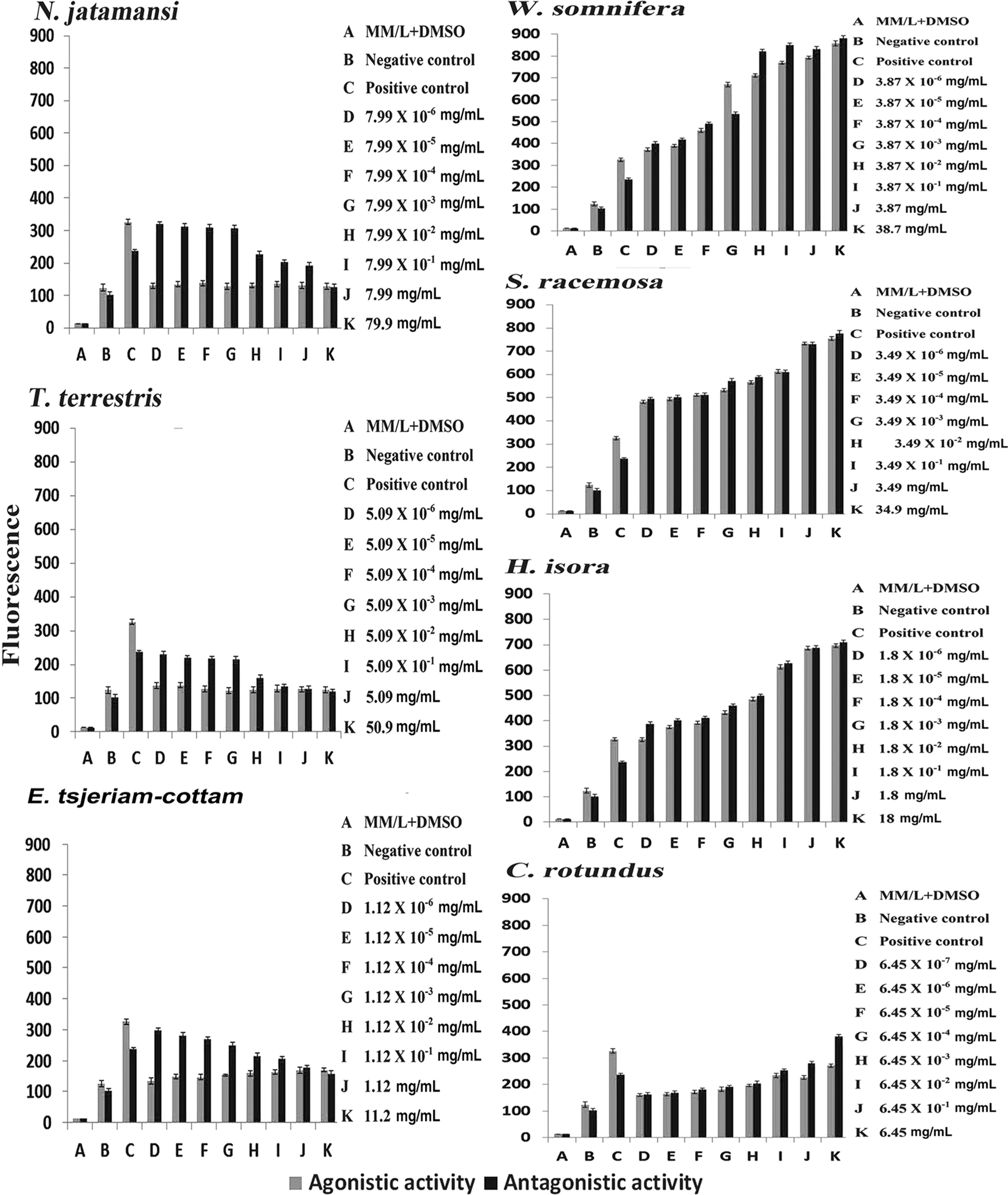
Agonistic and antagonistic activity of plant methanolic extracts in the RIKILT yeast Androgen bioAssay
In vivo experiments
Among NJ, TT, and EJ extracts, EJ was excluded from further studies because of its mixed activities.
Evaluation of vaginal smear analysis
Examination by microscopy showed a persistent vaginal cornification of 100% in the MC group that was treated with EV only, whereas the NC group showed regular estrous cycles with different phases at percentages of 16.6, 41.7, 25, and 16.7 for proestrus (P), estrus (E), metestrus (M), diestrus (D), respectively. The four groups treated with EV and receiving the high and low doses of NJ or TT all showed different percentages in various phases. Even though animals from the NJHD, NJLD, TTHD, and TTLD groups still showed a stressed incidence of estrous phase with percentages of 55.6, 75.0, 63.9, and 70.8, an increase in the metestrous phase was observed with percentages of 19.4, 12.3, 16.7, and 12.3, respectively. Their estrous cyclicity was found to be improved significantly in a dose-dependent manner (Figs. 2 and 3). NJHD or TTHD extracts showed a better reduction in the estrous phase when compared to their lower doses. Of the extracts, TTLD was found to be least effective with phase percentages of 0, 70.8, 12.3, and 16.7 for P, E, M, and D, respectively. The NJHD extract was found to be the most effective, with percentages of 16.7, 55.6, 19.4, and 8.0 for P, E, M and D, respectively, returning it almost to the percentages as obtained with the NC group.
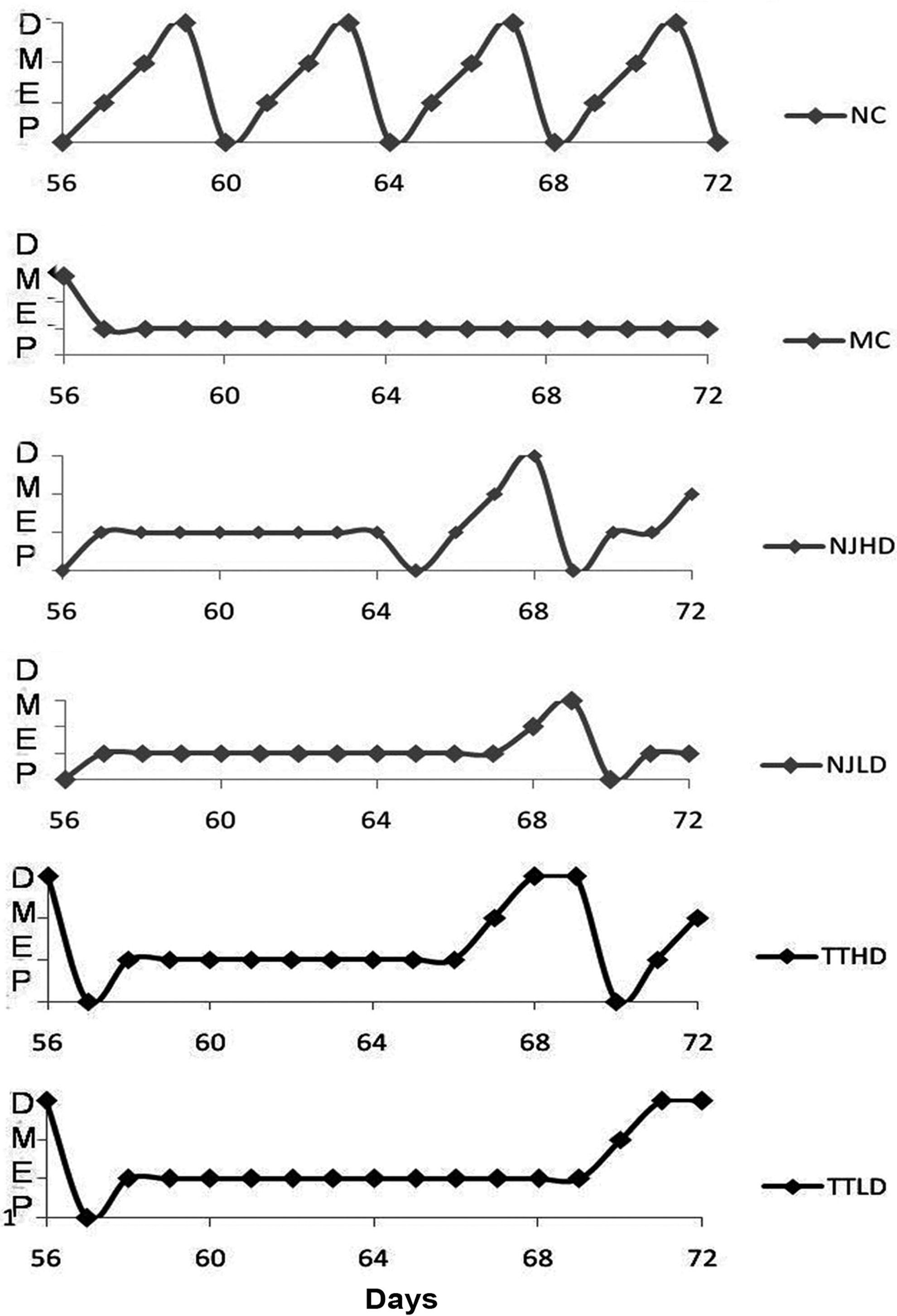
Representative estrous cycle of different experimental groups from day 56 to 72. D, diestrus; E, estrous; P, proestrous; M, metestrous; NC, normal control, MC, model control; NJHD, Nardostachys jatamansi DC high dose; NJLD, N. jatamansi DC low dose; TTHD, Tribulus terrestris L. high dose; TTLD, T. terrestris L. low dose.

Effect of extracts on the maintenance of phase frequency and estrous cyclicity in estradiol valerate (EV)-induced polycystic ovary (PCO) rat models. NC, normal control, MC, model control; NJHD, Nardostachys jatamansi DC high dose; NJLD, N. jatamansi DC low dose; TTHD, Tribulus terrestris L. high dose; TTLD, T. terrestris L. low dose.
Weight gain of PCO-induced rats with extract administration
The weight gained by EV-treated MC rats (3.43±0.15) was found significantly less when compared to NC (6.50±0.55) rats. But the extract-treated rats improved their weight gain significantly with 4.71±0.13, 4.64±0.08, 4.05±0.48, and 4.36±0.08 for NJHD, NJLD, TTHD, and TTLD, respectively. However, the dose-effect relationship was insignificant (Fig. 4).
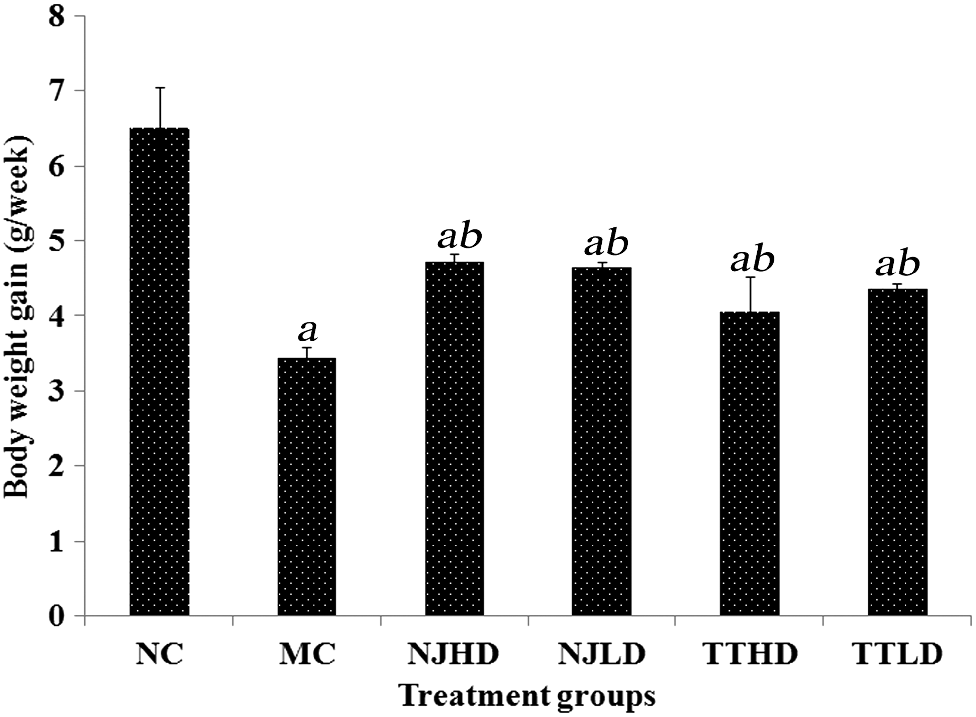
Body weight gain (gram per week) of rats during the treatment period. Label a represents a P value<0.05 between the normal control (NC) group and estradiol valerate (EV) treatment groups. Label b represents a P value<0.05 between the EV control (MC) group and extracts treatment group. NJHD, Nardostachys jatamansi DC high dose; NJLD, N. jatamansi DC low dose; TTHD, Tribulus terrestris L. high dose; TTLD, T. terrestris L. low dose.
Sex hormone analysis
EV treatment markedly increased the progesterone, testosterone, and estradiol levels in the MC group (Fig. 5). Treatment with the extracts resulted in restoration of progesterone levels more or less equal to the NC group. Serum testosterone concentrations for all of the extract-treated groups were lower than MC (49.02±1.37

Circulating levels of sex hormones at the end of the treatment period. Label b represents a P value<0.05 between model control and other treatment groups. NC, normal control, MC, model control; NJHD, Nardostachys jatamansi DC high dose; NJLD, N. jatamansi DC low dose; TTHD, Tribulus terrestris L. high dose; TTLD, T. terrestris L. low dose.
Ovary histology
The MC group exhibited large atreitic follicles, follicular cysts, and a lesser number of corpora lutea compared to the NC group. Ovaries of the NC group had more corpora lutea, several follicles with no follicular cyst, and atreitic follicles. The ovaries of extract-treated groups were markedly different from the MC group. In the NJHD group, the treatment was effective, because corpora lutea and follicles at different developmental stages were observed with the disappearance of follicular cysts and atreitic follicles. The higher number of follicular cysts and corpus luteum status indicate a significant dose-effect relationship and less effectiveness of NJLD extract over NJHD and NC groups. The TT extract also improved the cystic ovarian condition in a dose-dependent manner, i.e., follicular cysts were still observed in all rats of the TTLD group without improvement in corpus luteum status, whereas TTHD group members showed significant improvement (Fig. 6).
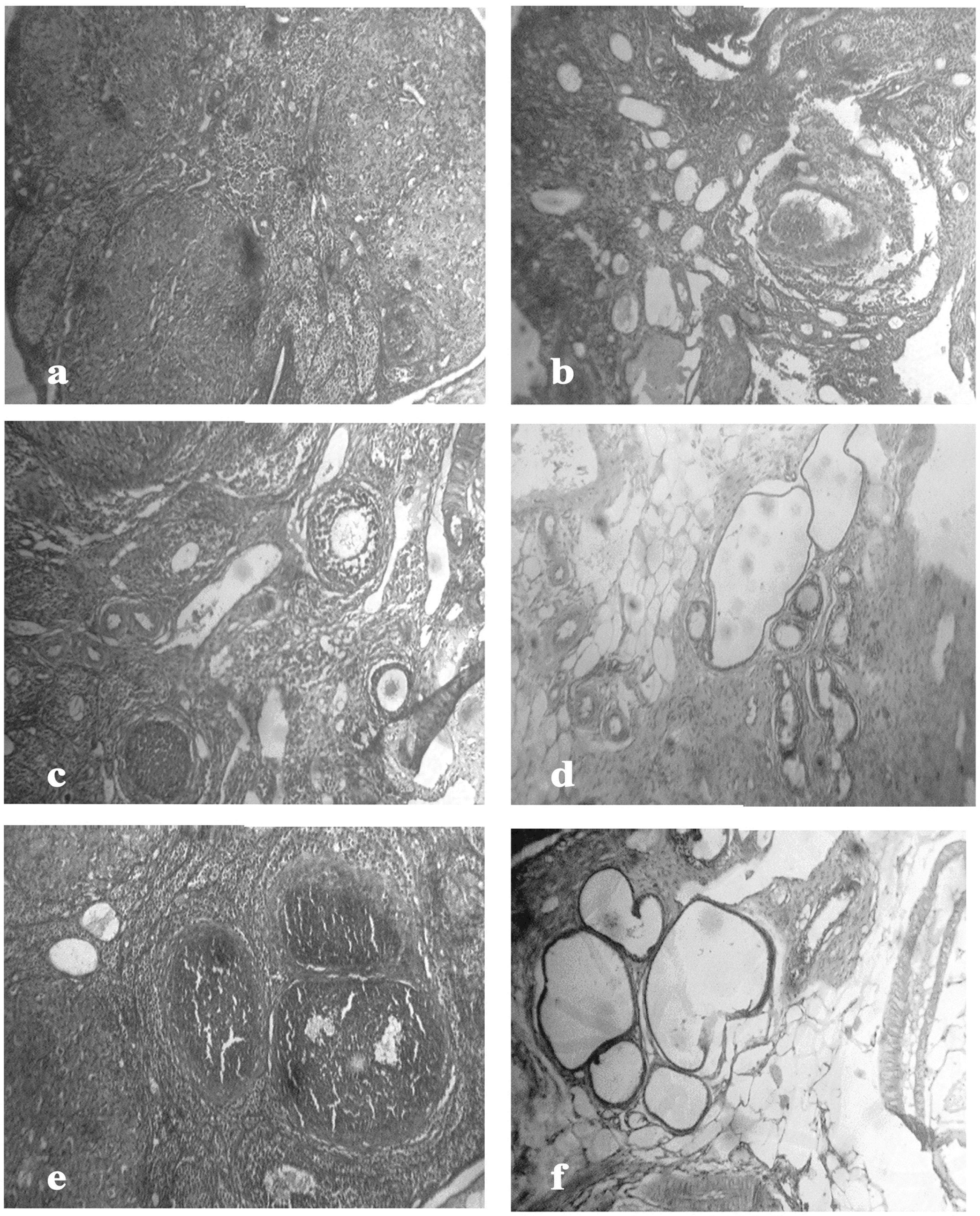
Effect of estradiol valerate (EV) administration and extract treatment on ovarian histology. (
Discussion
Although considered as an imperfect model for the human condition, EV-induced rat models are frequently used for in vivo studies because they exhibit ovarian features of PCOS. The present study aimed to investigate the potential androgenic and antiandrogenic activity of selected traditional medicinal plant extracts and to verify the in vitro–observed antiandrogenic potentials in vivo by the treatment of an EV-induced polycystic ovary–like condition in rats. As previously described, 14 EV injection in rats induced persistent vaginal cornification and anovulatory polycystic ovaries and caused weight loss. NJ and TT showed antiandrogenic activity, whereas EJ showed a narrow androgen agonistic and a higher antagonistic activity. Treatment of rats with the antiandrogenic extracts of NJ and TT influenced the EV-induced PCOS conditions beneficially in a dose-dependent manner. A reduction of the estrous phase was observed with an increase in the diestrous phase along with the appearance of the proestrous phase in the extract-treated groups. Both NJ and TT were able to oppose the EV-induced effects, and rats treated with high doses almost retained normalcy. A similar result was obtained for TT in a previous study. 15
The NJ and TT extracts were able to regulate the elevated serum levels of progesterone and estradiol. This result is supported by previous studies. 16,17 Variation in hormone levels, as suggested elsewhere, may be controlled by neural signals and mediated by the stimulation of β-adrenergic receptors or inhibition of α-receptors. 18 NJ and TT extract treatment reduced progesterone, testosterone, and estradiol levels, suggesting that these extracts are capable of modulating ovarian steroidogenesis at the enzymatic level and thereby improving follicular growth. However, our study showed that phytochemical components of these extracts are capable of inhibiting the transcriptional activity of the androgen receptor, and, as such, might also inhibit the physiological response of androgens in vivo. Histological studies showed that atreitic follicles were prominent in EV-treated rats. Similar results were obtained in the studies conducted by Brawer et al. in 1978. 16 Atreitic follicles were not observed in high- and low-dose NJ and high-dose TT, but a lesser number of atreitic follicles was observed in low-dose TT-treated groups. Thus, it is assumed that NJ and TT can reduce follicular atresia in EV-treated rats.
Additionally, the number of corpora lutea increased and the estrous cycles in the extract-treated rats became normal again. Histological and vaginal smear evaluations showed that treatment with NJ and TT significantly decreased the number of follicular cysts, but some follicular cysts were still found in the low-dose NJ and low- and high-dose TT-treated groups. However, no cysts were observed with high-dose NJ. Together, these data suggest that the antiandrogenic properties of NJ and TT might potentiality be useful for the management of PCOS.
Conclusion
The presence of bioactive constituents in plants might provide an alternative path to modern synthetic pharmaceuticals with fewer adverse effects. In our study, N. jatamansi DC and T. terrestris L. extracts were positive on PCO-induced rat models. These plants normalized estrous cyclicity dose dependently, reduced steroid hormone levels, and improved the ovarian dynamics. Further analysis of the extracts may help in the development of novel therapeutics against PCOS.
Footnotes
Acknowledgments
The authors are indebted to Dr. Rajeevkumar Sukumaran, NIIST, Trivandrum, and Dr. Sangeetha Nayanar, Malabar Cancer Centre, Kerala for providing facilities for instrumentation and analysis respectively.
Author Disclosure Statement
No competing financial interests exist.