
Abstract
Background:
We evaluated structural microvascular alterations in the skeletal muscle and left ventricle, as well as endothelium-dependent microvascular reactivity in the skeletal muscle, of diabetic rats subjected to long-term aerobic exercise training.
Methods:
Diabetes was experimentally induced by a combination of a high-fat diet with a single low dose of streptozotocin (35 mg/kg, i.p.). Animals with diabetes were divided into sedentary (DM+SED) and training groups (DM+TR) and compared with rats without diabetes (CON). We then measured maximal exercise capacity, fasting glucose and insulin, endothelium-dependent microvascular reactivity in skeletal muscle, and structural alterations of microvasculature in the skeletal and cardiac muscles.
Results:
Diabetes induced microvascular rarefaction and reduced endothelium-dependent microvascular reactivity. Physical exercise completely reversed microvascular rarefaction in the skeletal muscle (1.85 ± 0.05 vs. 1.17 ± 0.03 capillary/fiber ratio, P < 0.05) and in the left ventricle (0.48 ± 0.66 vs. 0.25 ± 0.01 Vv[cap]/Vv[fib] ratio, P < 0.05) compared with the DM+SED group and normalized the microcirculatory responses to acetylcholine in skeletal muscle (CON 38.76 ± 5.60 vs. DM+TR 30.47% ± 5.77%). As expected, exercise training increased the maximal velocity and exercise tolerance compared with the DM+SED (P < 0.05) and CON (P < 0.05) groups. Exercise training also reduced fasting glucose (P < 0.05) compared with DM+SED and normalized insulin levels compared with CON.
Conclusions:
Our results suggest that long-term physical exercise reverses skeletal and cardiac muscle microvascular rarefaction, as well as impaired endothelium-dependent microvascular reactivity, induced by diabetes in rats.
Introduction
T
Regular physical exercise has been used as an effective therapeutic strategy for the management of diabetes. 8 It was demonstrated that chronic aerobic exercise is effective in controlling glycemic levels, even when there is no reduction in weight. 9,10 Exercise decreases both insulin resistance and vascular/tissue damage caused by reactive oxygen species; in addition, it also increases the expression and activity of antioxidant enzymes. 11 Several studies have shown the benefits of exercise on microvascular reactivity and in the rarefaction of microcirculation using experimental models of hypertension and metabolic syndrome. 12 –15 However, the effects of long-term aerobic exercise training on structural alterations of microcirculation in the skeletal and cardiac muscles, together with endothelium-dependent microvascular reactivity in the skeletal muscle, are currently unknown in T2D.
In the present study, we used an experimental model of T2D based on the combination of a short period of high-fat diet (HFD) intake and the administration of a low dose of streptozotocin (STZ), as previously described. 16 –18 This model is characterized by hyperglycemia, insulin resistance, reduced bioavailability of nitric oxide (NO), and microvascular endothelial damage 16,18,19 and represents some of the clinical alterations of patients with diabetes. 20
The main objective of this study was to evaluate the effects of moderate aerobic exercise training on structural microcirculation in the skeletal muscle and myocardium of diabetic rats. Additionally, we assessed the influence of exercise training on endothelium-dependent microvascular reactivity in the skeletal muscle.
Methods
Animals
The handling procedures and testing methods used in this study have been previously approved by the ethics committee on animal use (CEUA) by the Oswaldo Cruz Foundation—Fiocruz, number LW-55/14, under the provisions of Brazilian law 11794/08 for the use of laboratory animals in scientific experiments. The experiments were performed on Wistar rats (WKY, Oswaldo Cruz Foundation Animal Facilities, Brazil) that were kept under controlled humidity (60% ± 10%), temperature (21°C ± 2°C), and light (12-hr light/12-hr dark cycle) conditions with free access to food and water.
Study design
At eight weeks of age, 34 animals were randomly divided into two groups and were fed either a standard commercial chow (CON, n = 12) or an HFD (n = 22) for 14 weeks. At the end of the 1st week of the HFD, the animals were injected with STZ. The animals were then subjected to aerobic capacity assessment, and the animals with diabetes were further divided into sedentary (DM+SED, n = 12) and trained (DM+TR, n = 10) groups. At the end of the exercise protocol (12 weeks), all groups were submitted to aerobic capacity, metabolic, and microvascular reactivity assessments. After the animals were sacrificed, gracilis muscle and left ventricular samples were collected for structural capillary microscopy. Figure 1 illustrates the experimental protocol.

Schematic representation of the experimental protocol. CON, control animals with standard commercial diet intake; DM, diabetes mellitus; HFD, high-fat diet; MET, maximal exercise test.
HFD STZ-induced diabetes model
Diabetes was induced by a combination of HFD and a low dose of STZ. 18,21 The CON diet contained 23% protein, 71% carbohydrate, 6% lipid, and 1.3% NaCl (Nuvilab—CR1; Nuvital Nutrients Ltd.), while the HFD contained 14% protein, 56% carbohydrate, 30% lipid, and salt supplementation (standard chow+corn starch+condensed milk+animal fat +0.5% NaCl) as previously described. 22 Saturated fat (lard) was the main fat source in the HFD. After the first week of dietary manipulation, a low dose of STZ (35 mg/kg in 0.5 M citrate–phosphate buffer, pH 4.5) was injected through the intraperitoneal route in the HFD-fed animals. Peripheral blood was drawn from the tail vein 1 week after the STZ treatment, and animals with a fasting glucose level above 250 mg/dL were considered to be diabetic and were included in the study. Animals in the CON group were injected with the same volume of citrate buffer solution.
Assessment of maximal aerobic capacity and training protocol
Rats were habituated to treadmill running on a low-speed, motor-driven rodent treadmill (HT 2.0; Hectron Fitness Equipment). Animals began running at 12 m/min (0% grade) for 15 min/day on three consecutive days. Following this period of habituation, maximal aerobic capacity was measured 1 week after diabetes induction and at the end of exercise training by a maximal exercise test (MET), as previously described. 23 Briefly, MET began at an initial velocity of 10 m/min with 3-m/min increments every 3 min. The test ended when the animals were exhausted and remained at the end of the mat on the shock grid for 5 sec. Time to exhaustion, maximal speed, and maximal distance were measured. The exercise training consisted of 12 weeks, 5 days/week, 1 hr/day at 60% of maximal velocity obtained in MET. 12
Body weight, fasting glucose, and fasting insulin
Body weight and fasting blood glucose levels were determined at the end of the experimental protocol. Animals were weighed using a portable scale. After a fasting period of 8 hr, blood glucose levels were measured using a portable glucose monitor (One Touch Ultra 2®; LifeScan, Inc., Johnson & Johnson), and insulin levels were measured using a commercially available enzyme-linked immunosorbent assay kit (Merck Millipore®).
Evaluation of endothelium-dependent microvascular reactivity
Animals were anesthetized with a combination of ketamine (100 mg/kg, i.p.) and xylazine (10 mg/kg, i.p) and maintained in the supine position with thighs immobilized on the table. Experiments were performed in a temperature-controlled room, and rats were placed on a thermal pad connected to a rectal probe (Harvard Apparatus). Animals' body temperature was maintained at 37.5°C. The gracilis muscle was exposed by an incision in the right thigh, and continuous measurements of skeletal muscle perfusion changes were evaluated using a laser speckle contrast imaging (LSCI) system (PeriCam PSI System; Perimed) at a wavelength of 785 nm and expressed as arbitrary perfusion units. After 5 min of blood flow stabilization, evaluation of endothelial-dependent microvascular reactivity was assessed with topical administration of 150 μL acetylcholine (Ach) 2% solution to the gracilis muscle surface. The image acquisition rate was 12 per sec, and the distance between the laser head and the muscle surface was fixed at 10 cm, as recommended by the manufacturer's manual. Images were analyzed using the manufacturer's software (PIMSoft; Perimed).
Structural analysis of the microvascular density of the skeletal muscle
The gracilis muscle sample was dehydrated in a graded series of ethanol (70%, 95%, and 100%) and stained with fluorescein isothiocyanate (FITC)-conjugated Griffonia simplicifolia I lectin at a 1:150 dilution in a dark humidified chamber at room temperature for 45 min to label capillary endothelial cells. The structural capillary density (number of capillaries per mm2) and the structural fiber density (number of muscle fibers per mm2) were identified and recorded with a fluorescence microscope (Olympus BX51; Olympus) and analyzed using Saisam 5.1.3 software (Microvision). The structural capillary density of the skeletal muscle was evaluated using at least seven photomicrographs of transverse sections of the gracilis muscle, with magnification of ×200 and scale bar of 100 μm. The capillary-to-fiber ratio was calculated by dividing the capillary density by the fiber density and was considered an anatomic index of angiogenesis.
Structural analysis of the microvascular density of the myocardium
The left ventricular structural capillary density was determined using the orientator method as previously described. 24 Briefly, the orientator method describes an approach to generate isotropic, uniform, and random sections of biological specimens, which allows for a quantitative study of three-dimensional anisotropic structures on two-dimensional sections. The myocardium is an anisotropic structure, but isotropic sections are necessary for a stereological study. The technique was performed by cutting the organ using the orthrip method, 24 which turns the samples into uniformly isotropic sections by dividing the fragment thrice consecutively; the first section is random, the second section is orthogonal to the first, and the third section is orthogonal to the second.
The paraffin blocks were cut into 5-μm sections and stained with FITC-conjugated G. simplicifolia I lectin at a 1:150 dilution in a dark humidified chamber at room temperature for 45 min. For each animal, at least seven photomicrographs were randomly examined from the three sections of cardiac tissue, with magnification of ×200 and scale bar of 100 μm. The volume density of the capillaries (Vv[cap]) was calculated as follows: Vv[cap]¼ Pp/PT (%), where Pp is the number of points hitting the capillaries and PT is the total number of test points (PT¼ 64 in the present case). The fiber volume density (Vv[fib]) was similarly calculated. The ratio of the capillary volume density to the fiber volume density (Vv[cap]/Vv[fib]) was calculated to negate any influences of cardiac hypertrophy on myocardial capillary density.
Drugs and reagents
All drugs and reagents were purchased from Sigma Chemical Co., St. Louis, MO.
Statistical analysis
The data are expressed as the mean ± standard error of the mean (SEM). Data normality was assessed by the Shapiro–Wilk test. Body weight, structural capillary density of the gracilis muscle, and capillary-to-fiber ratio of the left ventricle were compared using one-way analysis of variance (ANOVA). Maximal aerobic capacity was compared before and after training using two-way ANOVA. When an overall difference was detected by ANOVA, the Tukey test was used to localize statistically significant differences. Nonparametric Kruskal–Wallis test was used to compare microvascular response to Ach, and Dunn's post hoc test was used to localize statistically significant differences. Differences with P values of less than 0.05 were considered significant. All calculations were performed using a commercially available computer-based statistical package (GraphPad InStat 5.0; GraphPad Software).
Results
Assessment of exercise capacity
No significant differences were observed between groups at baseline. After 12 weeks of training, the DM+TR group demonstrated significant increases in time to exhaustion, maximal velocity, and maximal distance compared with the CON group and with the DM+SED group (Table 1).
P < 0.05 versus CON; b P < 0.05 versus DM+SED.
CON, control group; DM+SED, sedentary animals with diabetes; DM+TR, trained animals with diabetes.
Body weight, fasting glucose, and fasting insulin
Table 2 shows anthropometric and metabolic characteristics of rats after the exercise training protocol. Sedentary rats with diabetes developed marked hyperglycemia, reduction in circulating insulin levels, and severe weight loss compared with the CON group. Aerobic exercise training partially prevented weight loss, significantly reduced blood glucose levels, and normalized insulin levels.
P < 0.05 versus CON; b P < 0.05 versus DM+SED.
Evaluation of endothelium-dependent microvascular reactivity
The endothelium-dependent microvascular reactivity of the skeletal muscle upon topical administration of Ach is presented as blood flow percentage changes between baseline and after Ach. We observed a reduction of 49.22% of microvascular vasodilation induced by Ach in the DM+SED group compared with the CON group. Exercise training restored the Ach-induced microvascular reactivity, normalizing blood flow perfusion, compared with the CON group (Fig. 2).
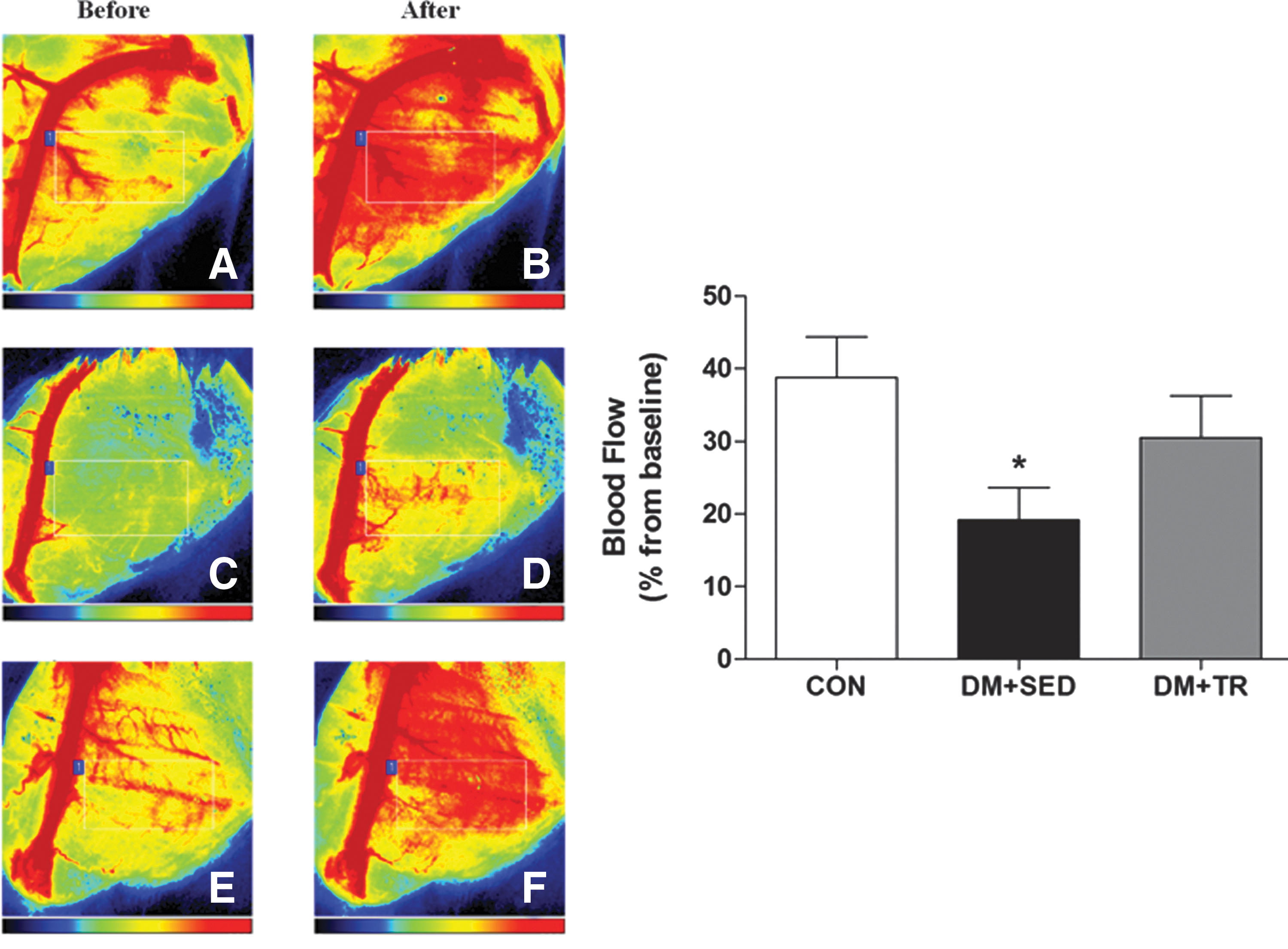
Effects of exercise training on endothelium-dependent microvascular reactivity in the skeletal muscle of rats. Left panel: representative images of microvascular blood flow in the skeletal muscle of the animals before and after topical administration of acetylcholine (2%).
Structural capillary density
As demonstrated in Figs. 3 and 4, respectively, the ratio of the capillary volume density to the fiber volume density in the myocardium (0.25 ± 0.01 Vv[cap]/Vv[fib]; P < 0.05) as well as the capillary density in the skeletal muscle (1.17 ± 0.03 capillary/fiber ratio; P < 0.05) was significantly reduced in animals with diabetes compared with the control group (0.39 ± 0.02 Vv[cap]/Vv[fib] and 1.81 ± 0.03 capillary/fiber ratio, respectively). Figures 3 and 4 also show that exercise training completely reversed diabetes-induced microvascular rarefaction in both the myocardium (0.57 ± 0.04 Vv[cap]/Vv[fib]; P < 0.05) and skeletal muscle (1.92 ± 0.06 capillary/fiber ratio; P < 0.05).
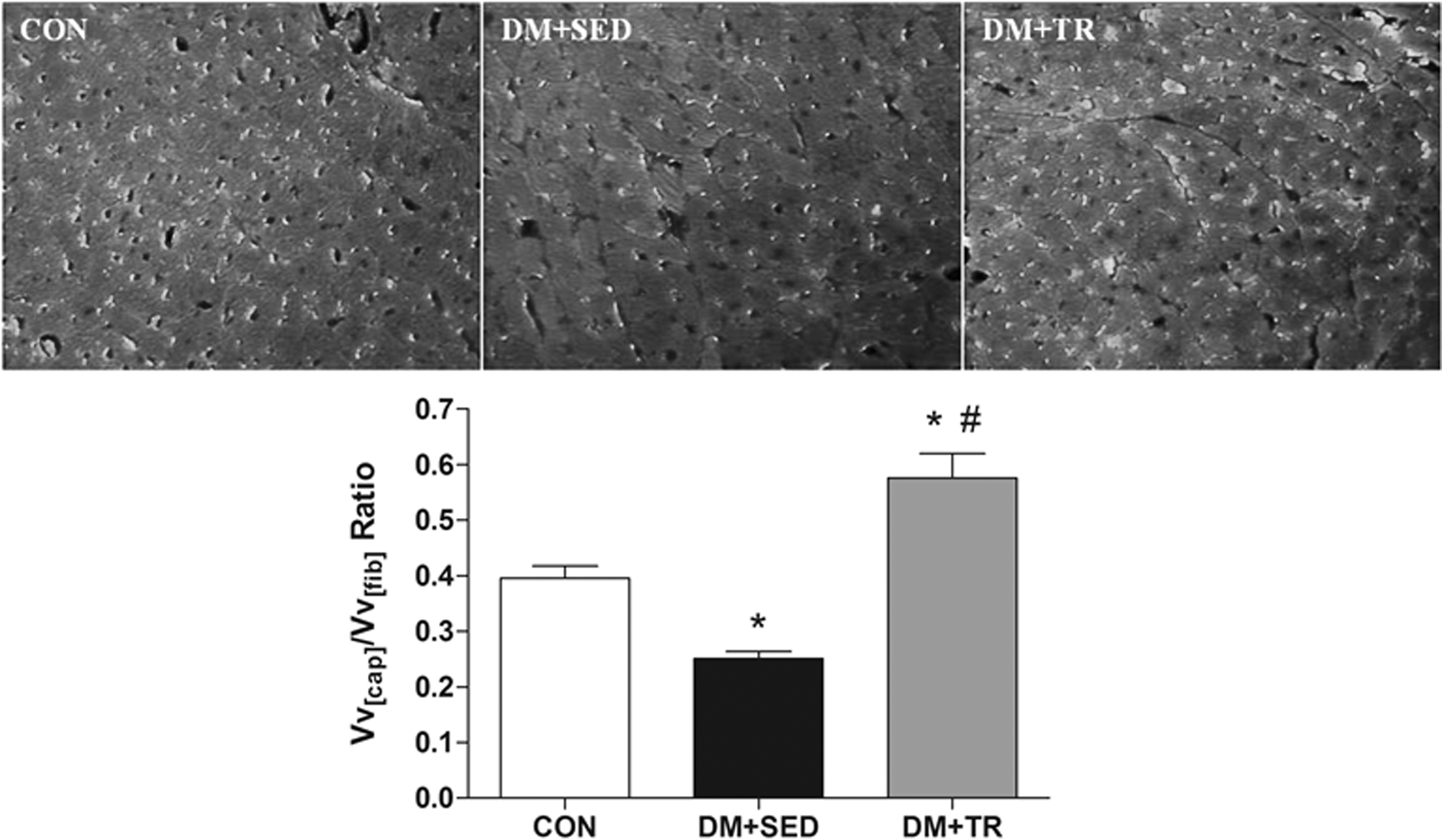
Upper panel: representative photomicrographs of stereological sections of the left ventricle of control animals (CON), sedentary animals with diabetes (DM+SED), and trained animals with diabetes (DM+TR). Images are representative slides used to determine structural capillary density with capillaries stained with FITC-conjugated Griffonia simplicifolia lectin; magnification, ×200. Lower panel: structural microvascular density in the left ventricle after 12 weeks of training in the control group (CON), sedentary animals with diabetes (DM+SED), and trained animals with diabetes (DM+TR). The values represent the mean ± SEM. *P < 0.05 versus CON; # P < 0.05 versus DM+SED. FITC, fluorescein isothiocyanate.

Upper panel: representative photomicrographs of transverse sections of the skeletal muscle of control animals (CON), sedentary animals with diabetes (DM+SED), and trained animals with diabetes (DM+TR). Images are representative slides used to determine structural capillary density with capillaries stained with FITC-conjugated G. simplicifolia lectin; magnification, ×200. Lower panel: structural microvascular density in the skeletal muscle after 12 weeks of training in the control group (CON), sedentary animals with diabetes (DM+SED), and trained animals with diabetes (DM+TR). The values represent the mean ± SEM. *P < 0.05 versus CON; # P < 0.05 versus DM+SED.
Discussion
In the present study, we investigated the effects of exercise training on microvascular function and structure in an experimental model of diabetes. The novel findings in the present study are as follows: (1) moderate intensity exercise training normalizes endothelium-dependent microvascular reactivity in skeletal muscle and (2) structural microvascular rarefaction induced by diabetes in the skeletal muscle and left ventricle was completely reversed by exercise training.
In the present study, diabetes was induced in rats by the combination of the consumption of an HFD with a single low dose of STZ. STZ causes a partial destruction of pancreatic β-cells, which results in insulin deficiency and causes blood glucose to increase. The inability of the tissues to utilize glucose is similar to a state of cellular starvation, activating compensatory responses to increase the release of fuel substrates by lipolysis and proteolysis. 25 In this sense, as expected, we observed a reduction in body weight and plasma insulin levels accompanied by marked hyperglycemia in sedentary rats.
We also observed that exercise training partially restored body weight and reduced plasma glucose levels. The following factors could potentially explain this result: (1) insulin sensitivity; (2) plasma insulin levels; (3) tyrosine kinase activity of the insulin receptor; and (4) translocation of glucose transporters in skeletal muscle (GLUT4). 26 Moreover, exercise training of moderate intensity protected against β-cell damage in STZ-induced diabetes in rats, probably through a reduction of systemic oxidative stress. 27 This is particularly important because STZ is the most commonly used agent in experimental diabetes, causing β-cell destruction and inducing hyperglycemia. 28 Although exercise alone did not fully restore glycemic levels in trained animals with diabetes, it can still be considered an important nonpharmacologic tool to improve glycemic control in patients. 27
Microvascular endothelial reactivity assessment is essential for investigating the pathophysiology of diabetes and cardiovascular diseases. 29,30 In the present study, microvascular endothelial function was evaluated using LSCI, which represents an innovative approach to the noninvasive evaluation of microvascular endothelial function. 31 –33 A major advantage of this technique is that the reproducibility of LSCI is superior to earlier procedures, such as laser Doppler flowmetry and laser Doppler imaging. 34,35 Laser speckle contrast imaging may be coupled with pharmacological tests, such as the topical administration of Ach, to further characterize microvascular reactivity.
It is well established that diabetes impairs vascular reactivity, partly due to an increase in free radicals in the microcirculation that results in decreased bioavailability of NO and impaired microvascular function, which ultimately reduces blood flow to the capillaries. 36 This phenomenon, coupled with the activation of the inflammatory response and increased recruitment of leukocytes in postcapillary venules, may result in capillary obstruction and decreased capillary perfusion. These processes may result in the temporary obstruction of blood flow (functional capillary rarefaction), followed by disappearance of the capillaries (structural capillary rarefaction). 18
Indeed, our results showed markedly impaired endothelial function in skeletal muscle microcirculation and substantially reduced capillary density in skeletal muscle and the left ventricle in sedentary rats with diabetes. On the other hand, chronic exercise normalized endothelial function and completely reversed microvascular rarefaction in skeletal muscle and the left ventricle.
The mechanisms by which exercise improves myogenic tone and structural microcirculation are still controversial. Experimental evidence suggests that the reduction in oxidative stress induced by exercise training as well as changes in blood flow pattern due to increased shear stress on the vascular endothelium may be some of the potential mechanisms involved in this response. 37 In addition, shear stress induces the release of NO and prostacyclin, which are both known to increase endothelium-dependent vasodilatation and inhibit multiple processes involved in atherogenesis. 37,38 NO has a potent vasodilator effect, improving vascular reactivity 39 –41 and stimulating the synthesis of the main regulating factor of angiogenesis—vascular endothelial growth factor (VEGF). 42,43 In the presence of VEGF, angiogenesis occurs. However, in its absence, the capillaries undergo apoptotic regression. 44 Moreover, hemodynamic pattern changes promoted by regular exercise induce structural adaptation responses in the vascular tree, allowing for optimal tissue perfusion. 37,45
In conclusion, our results demonstrate that long-term aerobic physical exercise reverses structural microvascular rarefaction in the heart and skeletal muscle. The impaired endothelium-dependent microvascular reactivity in the skeletal muscle induced by diabetes is also reversed.
Footnotes
Acknowledgments
This investigation was supported by grants from CNPq (Conselho Nacional de Desenvolvimento Científico e Tecnológico), FAPERJ (Fundação de Amparo à Pesquisa do Estado do Rio de Janeiro), and Oswaldo Cruz Foundation (FIOCRUZ), Rio de Janeiro, Brazil.
Author Disclosure Statement
No competing financial interests exist.