




Abstract
The preparation of single-stranded DNA from double-stranded PCR products is an essential step in the identification of aptamers by Systematic Evolution of Ligands by EXponential enrichment (SELEX). The most widely used method for producing single-stranded DNA is alkaline denaturation of biotinylated PCR products attached to streptavidin-coated magnetic beads. Recently, it has been suggested that this method may be unsuitable due to the release of interfering amounts of streptavidin and biotinylated DNA. In this article, the alkaline method is compared with a thermal method that is known to release significant amounts of streptavidin and biotinylated DNA. Results show that trace amounts of streptavidin and biotinylated DNA are released in the alkaline method, but this can be curtailed by preconditioning the beads in aqueous sodium hydroxide. The main product in the alkaline method is single-stranded DNA, which is produced in high yield.
Introduction
Materials and Methods
All solutions were prepared with molecular biology-grade water (Sigma). Approximately 2,125,000 copies of single-stranded library template [5′-AGATGCCTGTCGAGCAT GCT-N60-GTAGCTAAACTGCTTTGTCGACGGG-3′ (where N60 is a 60-mer combinatorial sequence)] in 100 μL of PCR buffer (10 mM Tris-HCl, pH 8.3, at 25°C, 50 mM KCl, 1.5 mM MgCl2, 0.001% gelatin), 0.2 mM nucleotides, and 0.5 μM sense and biotin-5′-antisense primers (5′-AGATGCCTGTCGAGCA TGCT; biotin-5′-CCCGTCGACAAAGCAGTTTAGCTAC), and 5 units of polymerase (MyTaq HS DNA polymerase, Bioline) were amplified (95°C for 2.5 minutes; 25 cycles at 95°C for 30 seconds, 60°C for 30 seconds, 72°C for 30 seconds; and final extension at 72°C for 3 minutes). Next 100 μL of product was added to 1 mg of streptavidin beads (MyOne Streptavidin Dynabeads, Invitrogen) in 1 mL of TE buffer (10 mM Tris-HCl, pH 8.0, 0.15 M NaCl, 1 mM EDTA) and slow-tilt rotated for 1 hour. The beads were then washed with 2×1 mL of TBE buffer and 2×1 mL of water. For the thermal method, beads were resuspended in 100 μL of water and heated to 90°C for 2 minutes. At the end of this time, the beads were precipitated immediately and the supernatant transferred to a fresh tube containing 1 μL of 100×TE buffer (1 M Tris-HCl, pH 8.0, 0.1 M EDTA) and cooled on ice.
For the NaOH methods, the beads were resuspended in 100 μL of NaOH (0.15 M or 20 mM) and slow-tilt rotated at room temperature for 10 minutes; at the end of this time, the beads were precipitated immediately and the supernatant transferred to a new tube containing enough HCl to neutralize the pH (40 mM HCl for the 20 mM NaOH and 0.3 M HCl for the 0.15 M NaOH) and 1 μL of 100×TE buffer. For electrophoresis, supernatants were mixed 4:1 with loading buffer (Type 1 Gel Loading Solution, Sigma). Electrophoresis of 10-μL aliquots was carried out on 15% polyacrylamide gels (Mini-PROTEAN TGX Precast, Bio-Rad) in TBE buffer for 1 hour at 200 V.
After electrophoresis, gels were silver-stained (PlusOne™ DNA Silver Staining Kit, GE Healthcare) according to the supplier's instructions. Ultraviolet/visible (UV/vis) spectra of undiluted supernatants in a 3-mm path-length quartz glass cell (Hellma, Müllheim, Germany) were acquired with a Hewlett Packard 8452A Diode Array Spectrophotometer. For quantitative PCR (qPCR), supernatants were diluted with water to give a final dilution of 1:4×10⁸, and amplified (95°C for 2.5 minutes, 40 cycles; 95°C for 30 seconds, 60°C for 30 seconds) with 0.5 μM primers (the antisense primer was not biotinylated) and KAPA SYBR FAST Master Mix (Kapa Biosystems, Woburn, MA).
Results and Discussion
Lanes 1 and 2 in Fig. 1 show that the single-stranded library template can be distinguished from the double-stranded PCR product on a 15% polyacrylamide gel. Lanes 3, 5, and 6 show results of the three basic strand separation methods. Both NaOH methods (lanes 5 and 6) produced good yields of single-stranded template, but the thermal method did not. All methods produced some bands corresponding to high-molecular-weight structures, especially the supernatant from the thermal method mixed with TBE (buffered saline, lane 3). These bands were much weaker when the supernatant from the thermal method was not mixed with buffered saline (lane 4), and attenuation was accompanied by the appearance of a band corresponding to single-stranded template. Intense bands corresponding to single-stranded template were produced by both NaOH methods, even though the supernatants are buffered. An explanation for these results is shown in Fig. 2. In the thermal method, streptavidin and biotinylated DNA (primers and antisense strands) were released in addition to amplified sense strands. When buffered saline was added, the DNA annealed, leading to a low yield of single-stranded template. In the NaOH methods, only a small amount of streptavidin and biotinylated DNA was released, leading to high yields of single-stranded template.
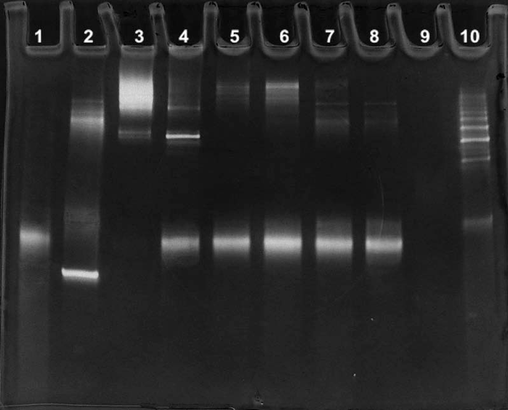
A 15% polyacrylamide gel developed at 200 V for 1 hour and stained with silver. Lane 1, 80 ng of unamplified single-stranded library template; lane 2, unpurified PCR products; lane 3, thermal strand separation method with supernatant transferred to buffered saline; lane 4, thermal strand separation method with supernatant in water; lane 5, 0.15 M NaOH strand separation method; lane 6, 20 mM NaOH strand separation method using unconditioned beads; lane 7, 20 mM NaOH strand separation method using beads preconditioned in 20 mM NaOH for 1 hour; lane 8, 20 mM NaOH strand separation method using beads preconditioned in 20 mM NaOH for 24 hours; lane 9, 2.5 μg of streptavidin treated the same as beads in lane 4; lane 10, 2.5 μg of streptavidin, 80 ng library template, and 40 ng of biotinylated primer treated the same as beads in lane 4.

When PCR products attached to streptavidin beads are heated to 90°C in water, significant amounts of streptavidin and biotinylated antisense strand are released in addition to the sense strand. On cooling, high-molecular-weight aggregates composed of streptavidin and hybridized DNA form in the supernatant. When PCR products attached to streptavidin beads are treated with aqueous NaOH, the main product in the supernatant is single-stranded sense strand. Color images available online at www.liebertonline.com/nat
If the supernatant from the thermal method only contained sense and antisense strands, the addition of buffered saline would result in the appearance of a band corresponding to double-stranded amplicons. The absence of this band in lane 3 is because streptavidin is also released from the beads. Streptavidin is a tetramer in which individual monomers are held together by noncovalent interactions. When streptavidin beads are exposed to high temperature, these interactions are disrupted, leading to the release of streptavidin monomers that reassociate when the temperature is lowered. Similar bands corresponding to high-molecular-weight structures are also produced when a mixture of streptavidin, biotinylated primers, and unamplified single-stranded template (no beads or PCR) is subjected to the thermal protocol, as shown in lane 10 of Fig. 1. No high-molecular-weight structures were visible when the supernatants from streptavidin beads were subjected to the thermal protocol. Preconditioning beads in NaOH released the more labile streptavidin monomers, as can be seen by comparing results of preconditioned beads (lanes 7 and 8) with unconditioned beads (lane 6).
qPCR results showed that the amount of amplifiable template released from the beads was similar in all methods. A 260-nm/280-nm ratio of 1.7–2.0 is indicative of a high-quality DNA sample; lower ratios suggest contamination with protein. UV/vis spectra of the undiluted supernatants gave mean 260-nm/280-nm ratios of 1.57 for thermal methods and 1.95 for NaOH methods. The molarities of DNA in supernatants from NaOH methods (based on an extinction coefficient of ε260=1.1×10⁶ for the single-stranded library template) were: 0.15 μM (0.15 M NaOH method), 0.18 μM (20 mM NaOH method), 0.17 μM (20 mM NaOH method with beads preconditioned for 1 hour), and 0.17 μM (20 mM NaOH method with beads preconditioned for 24 hours).
In summary, methods for the preparation of single-stranded DNA based on streptavidin beads and NaOH produce high yields of single-stranded template. Small amounts of streptavidin and biotinylated DNA are released from the beads, but this does not lead to low yields of single-stranded DNA. Wendel and colleagues have suggested that the streptavidin released from the beads could act as target molecule for the selection of aptamers in SELEX (Paul et al., 2009; Avci-Adali et al., 2010). This would depend on the method used to separate sequences bound to the target molecule from unbound sequences: If a nonspecific method such nitrocellulose filtration was used, sequences bound to streptavidin would be selected, but if a specific method such as target molecules attached to magnetic beads was used, selection of aptamers for streptavidin would not occur.
Selection of aptamers for streptavidin in methods where a nonspecific separation method is used can also be eliminated by employing an appropriate counterselection step. Wendel and colleagues have also reported that streptavidin interferes in other ways with cell-SELEX. The mechanism for this interference is binding of streptavidin to receptors on the surface of certain cells such as platelets. This bound streptavidin promotes cross-linking of the cells and acts as a target for the selection of aptamers. Others have not observed these effects (Kwame et al., 2010), but when target cells display receptors that bind to streptavidin and a counterselection step cannot be used, alternative methods for the generation of single-stranded DNA should be explored. In most cases, however, there is no reason to abandon magnetic bead-based methods for preparing single-stranded DNA in favor of more time-consuming and more expensive methods such as those based on lambda exonuclease (Avci-Adali et al., 2010).
Footnotes
Acknowledgment
Financial support for this work was provided by the United Kingdom's Biotechnology and Biological Sciences Research Council (BBSRC) Grant Number BB/1013245/1.
Author Disclosure Statement
No competing financial interests exist.