




Abstract
A promising strategy for increased intracellular delivery of nucleic acids with the benefit for targeting is photochemical internalization (PCI). PCI relies on the use of a photosensitizing compound that photochemically destroys membranes in the endocytic pathway after illumination, resulting in cytosolic transfer of endosomal content. PCI technology combined with biodegradable polyamino acid carriers and nucleic acids delivers effective targeting and improved biosafety. In an in vitro model system, we have evaluated various poly-
Introduction
Much effort is directed toward the development of delivery systems that offer enhanced biosafety and biocompatibility. Polyamino acids have attracted substantial attention as gene carriers due to their relatively low toxicity, excellent biocompatibility, biodegradability, and pharmacological efficacy. The 3 cationic polyamino acids poly-
Photochemical internalization (PCI) represents a promising strategy for light-directed delivery of macromolecules located in the endocytic pathway (Berg et al., 1999). A prelocalized amphiphilic photosensitizer (PS) resides in the endosomal membrane and is excited by light within its absorbance spectrum. This photoreaction creates reactive oxygen species that interact with nearby molecules in the endosomal membrane. Consequently, the endosomal membrane is damaged and leakage of endosomal content occurs. PCI only has an effect in light-exposed areas, meaning that local illumination of target tissue would only induce biological effect in the desired cells/tissue (Selbo et al., 2010). Interestingly, recent development in photodynamic therapy makes it possible to illuminate tissue more accurately by using ultrasound-guided technology for fiber optics (Jerjes et al., 2009). Previously it has been reported that maximum PCI-mediated transfection is achieved by the use of a light dose that reduces cell viability by approximately 50% (Hogset et al., 2000). However, recent studies have shown that using appropriate conditions may minimize the photocytotoxic effect of PCI (Boe et al., 2007, 2008a). Although the toxicity in most cases is a disadvantage, it may also be beneficial (i.e. in cancer therapy).
To date, several carriers have been investigated for light-directed siRNA delivery, including lipid carriers (Boe et al., 2007; Oliveira et al., 2007; Oliveira et al., 2008), PEI formulations (Boe et al., 2008b), cyclodextrin (Boe et al., 2010b), dextran nanogels (Raemdonck et al., 2010), trimethylated chitosan (Varkouhi et al., 2011), and unmodified chitosan (Jorgensen et al., 2012). In addition, PEI has been used for light-directed mRNA delivery (Boe et al., 2010a). This study describes the first attempt of delivering siRNA molecules and mRNA molecules with unmodified biodegradable polyamino acids with a potential for light-directed targeting. After extensive evaluation of various polyamino acids with different molecular weights, we demonstrate efficient siRNA and mRNA delivery in vitro.
Materials and Methods
Cell lines and culture conditions
The OHS (osteosarcoma) cell line was established at the Norwegian Radium Hospital (Fodstad et al., 1986), while the SK-MEL-28 (melanoma) cell line was acquired from the American Type Culture Collection. Cells were cultured and maintained in Roswell Park Memorial Institute (RPMI) 1640 medium (Bio Whittaker), supplemented with 10% fetal calf serum (PAA Laboratories) and GlutaMAX (Gibco). The cells were maintained at 37°C in a humidified atmosphere containing 5% CO2 and were routinely tested for mycoplasma infections.
siRNA and mRNA
Small interfering RNAs targeting S100 calcium binding protein A4 (S100A4), mitogen-activated protein kinase kinase 1 (MEK1), and mitogen-activated protein kinase kinase 2 (MEK2) were selected from previous studies (Boe et al., 2007; Zhang et al., 2010). siRNA targeting the S100A4 mRNA sequence (Genbank accession No. NM_002961, sequence 5′-UGAGCAAGUUCAAUAAAGA-3′, 3′-ACUCGUUCAAGUUAUUUCU-5′, 481- 499); siRNA targeting MEK1 mRNA sequence (Genbank accession No. NM_002755.3, sequence 5′-AGCAUGAACCAUGAGUUGC-3′, 3′-UCGUACUUGGUACUCAACG-5′, 1534-1552); and siRNA targeting the MEK2 mRNA sequence (Genbank accession No. NM_030662.3, sequence 5′-UGCUGUGAGGCUCUCCUUC-3′, 3′-ACGACACUCCGAGAGGAAG-′5, 1116-1134) were ordered pre-annealed from Eurogentec and employed together with the Silencer® Select Negative Control #2 siRNA from Ambion. Enhanced green fluorescent protein (EGFP) mRNA was synthesized as described previously (Saeboe-Larssen et al., 2002).
Light source and PS
We used the same light source and PS as described previously (Boe and Hovig, 2006). Briefly, we utilized a PS from Porphyrin Products, disulfonated tetraphenylporphine (TPPS2a). When irradiated, cells were exposed to light with a LumiSource prototype (PCI Biotech AS) containing a bank of four fluorescent tubes (Osram 18W/67) with the highest fluorescence around 420 nm. Cells were light protected using aluminum foil to avoid unwanted activation of the PS.
Transfection
Polyamino acid formulations (Sigma-Aldrich) used in this study are described in Table 1. Prior to transfection, cells were seeded and cultivated for 72 hours in 12-well plates. All polyamino acids were diluted to 0.1 μg/μL in sterile water before use. Prior to complex formation, cells were incubated with or without PS (TPPS2a=0.5 (g/mL) in serum containing RPMI 1640 medium (BioWhittaker). All complex formations were carried out in RPMI 1640 medium under serum-free conditions. After 30 minutes of complexing, the complexation solutions were added to the 12-well plates with serum-containing media. Following 18 hours of transfection, the cell culture plates were washed 3 times and re-incubated for 4 hours to remove unwanted PS in the medium and in the cell membrane. Cells were then exposed to blue light (5.1 mW/cm2) and re-incubated before mRNA harvesting (24 hours post PCI treatment). All transfections were conducted at physiological pH under cell confluency ∼50%–70%.
Cell viability
Cells were cultivated for 72 hours in 12-well plates prior to use. Cells were transfected as described under “Transfection” and incubated for 24 hours after light treatment. After incubation, medium was discarded and fresh RPMI 1640 medium containing 3-(4.5-dimethylthiazol-2-yl)-5-(3-carboxymethoxyphenyl)-2-(4-sulfophenyl)-2H-tetrazolium) (MTS) from Promega was added. MTS is a colorimetric method for determining the number of viable cells in proliferation. Absorbance was measured at 490 nm in wells containing RPMI 1640 (with serum and GlutaMAX) and MTS to a final volume of 1200 μL/well (1:6 dilution of MTS/medium).
Real-time reverse transcriptase polymerase chain reaction
Total cellular RNA was isolated with the GenElute Mammalian Total RNA Miniprep Kit (Sigma-Aldrich), and the iScript cDNA synthesis kit (Bio-Rad) was used for reverse transcription. Both kits were used according to the manufacturer's manuals. Real-time detection was obtained by use of SYBR Green 1. For each polymerase chain reaction (PCR), 10 μL cDNA, 30 μL iQ SYBR Green Supermix (Bio-Rad), 300 nM of each primer, and nuclease-free water was added to a final volume of 60 μL. Samples of 25 μL each were then applied to the PCR plate. This method ensures that the parallels are true duplicates. Primer design was performed using the software Primer Express (Applied Biosystems). The primer set used for S100A4 (forward primer 5′-AAGTTCAAGCTCAACAAGTCAGAAC-3′ and reverse primer 5′-CATCTGTCCTTTTCCCCAAGA-3′) amplifies a 79-bp segment covering parts of exons 2 and 3 of the S100A4 sequence. For MEK1, the following primer sequences were used: forward primer 5′- TTCTACAGCGATGGCGAGAT-3′ and reverse primer TCCAGCTTTCTTCAGGACTTG; and for MEK2: forward primer 5′ TTGAACTCCTGGACTATATTGTGAAC and reverse primer 5′ AAGTCGGGGGTGAACACA-3′. Real-time PCR reactions were run on an iCycler machine (Bio-Rad) with the following amplification protocol: 3 minutes initial denaturation at 95°C, 50 cycles of 10 seconds denaturation at 95°C and 35 seconds annealing/extension at 60°C, one hold at 95°C for 20 seconds followed by a hold for 1 minute at 55°C, and finally a melt curve analysis of 80 steps each for 10 seconds, with 0.5°C increases until a final temperature of 95°C. The quality of the RNA samples was verified by amplification of 2 housekeeping genes, the TATA-box-binding protein, with forward primer 5′-GCCCGAAACGCCGAATAT-3′ and reverse primer 5′-CGTGGCTCTCTTATCCTCATGA-3, and the human acidic ribosomal phosphoprotein P0, with forward primer 5′-CGCTGCTGAACATGCTCAAC-3′ and reverse primer 5′-TCGAACACCTGCTGGATGAC-3′. These housekeeping genes were chosen as they were unaffected by the different treatment modalities in this study. The Gene Expression Macro, version 1.1 (Bio-Rad), was used for the quantitative calculations. The program performs calculations based on the delta-delta cycle threshold (ΔΔCT) method (Vandesompele et al., 2002), which allows comparison of cycle threshold values obtained using different sets of primers on the same set of samples. For statistical analyses we compared our 3 controls [negative control−(nc−), A4–/MEK1–/MEK2–, and negative control+(nc+)] with the gene-silencing sample (A4+/MEK1+/MEK2+) in a 2-tailed Student's t-test with equal variance. A significance level of P=.05 was used for the real-time PCR data.
Complex sizes
Complexes of polyamino acids and siRNA or mRNA were prepared in serum-free RPMI medium and measured at 25°C with a scattering angle of 173° on a Zetasizer Nano ZS (Malvern Instruments Ltd.). Complex sizes were determined using the Z-average value from three monomodal samples (particle peak at 100%) with approved instrumental expert advice criteria for measurements.
Fluorescence microscopy
Cells were cultivated for 72 hours in 12-well plates prior to use, transfected with EGFP mRNA as described under “Transfection,” and incubated for 24 hours after light treatment. After incubation, EGFP mRNA transfected cells were visually examined on a Zeiss inverted microscope Axiovert 200 equipped with filter for FITC [450- to 490-nm band pass (BP) excitation filter, a 510-nm farb teiler (FT) beam splitter, and a 515- to 565-nm long pass (LP) emission filter]. Images were composed by the use of Carl Zeiss Axiocam HR, Version 5.05.10, and Axiovision 3.1.2.1 software. Pictures were prepared with Adobe Photoshop 7.0 (Adobe) and Zeiss LSM Image Browser (Version 3).
Flow cytometry
EGFP mRNA transfected cells were quantitatively assessed by flow cytometry. Samples were prepared as described under “transfection” and examined 24 hours after PCI treatment. Prior to analysis, cells were trypsinized and washed once with 500 μL phosphate buffered saline (PBS) at 1000 rpm for 5 minutes and resuspended in 500 μL PBS. Propidium iodine was added to the cell suspensions (to distinguish live cells from dead cells), before they were filtered through a 5-mL round-bottom tube with cell-strainer cap to avoid cell aggregates. Cell fluorescence was examined from 10.000 events and detected on a BD LSR 2 flow cytometer (BD biosciences).
Results
Light-directed gene silencing using siRNA and polyamino acid formulations
We initiated our study by evaluating the gene silencing effect of 7 polyamino acid formulations (listed in Table 1) using 2 concentrations in the OHS cell line (Fig.1A, C). We kept the siRNA concentration (50 nM) and the light dose (153 mJ/cm2) constant, based on previous published and unpublished studies. When using 0.175 μg of PLL no.9404, PLA no.3892, and PLA no.7762, we observed a considerable gene silencing effect of the S100A4 mRNA levels. The gene-silencing effect increased when the amount of polyamino acid was doubled from 0.175 μg to 0.35 μg, and gene silencing was also demonstrated with PLA no.4663. The best achieved gene-silencing effect was observed with PLA no.3892. Using this polyamino acid as siRNA carrier, the S100A4 mRNA level dropped about 90% relative to untreated control, whereas the negative siRNA control demonstrated only a minor gene-silencing effect.

Examination of small interfering (siRNA) transfection ability and cell viability in the osteosarcoma (OHS) cell line using photochemical internalization (PCI) in combination with polyamino acids at 2 concentrations, 0.175 μg
Toxicity after light-directed gene silencing using polyamino acid formulations
To minimize potential toxic effects after PCI treatment and simultaneously obtain optimal gene silencing or gene upregulation, it is important to find the appropriate illumination dose. In general, our data do not show toxic effects from PCI with an illumination dose of 153 mJ/cm2. Instead, the cell viability was in general weakly elevated compared with non-illuminated samples. This was the case for both the 0.175 μg and 0.35 μg polyamino acid samples (Fig. 1B, D). Importantly, our results show the possibility for optimal gene silencing without toxicity when using the appropriate set of parameters.
Light-directed gene silencing of MEK1 and MEK2 in the SK-MEL-28 melanoma cell line
Because we achieved a gene silencing down to 10% of the remaining S100A4 mRNA level with siRNA combined with PLA no.3892 and PCI, we decided to test our delivery system also in the SK-MEL-28 cell line. Here, we used siRNAs targeting MEK1 and MEK2 (Fig. 2A, C). To obtain a significant gene silencing of these genes, we increased the amount of PLA no.3892 from 0.35 μg to 1.4 μg, as 0.35 μg of PLA no.3892 as siRNA carrier resulted in only a modest gene-silencing efficiency (20% with PCI). In addition, we doubled the illumination dose to 306 mJ/cm2 (compared to the OHS cell line), while we kept the siRNA concentration constant (50 nM). Using these transfection parameters, we obtained a gene silencing of both the MEK1 and MEK2 mRNA levels up to 85% of untreated control. In contrast, the negative siRNA control did not seem to affect the MEK1 mRNA level, while the MEK2 mRNA level was slightly upregulated (15%) by the negative control. To examine whether PLA no.3892 and the higher illumination dose was toxic to the SK-MEL-28 cell line, an MTS assay was performed (Fig. 2B, D). The highest cytotoxicity was observed in PCI samples with negative control siRNA, where the cell viability decreased with 4% and 6% compared with untreated control when 0.35 μg and 1.4 μg PLA no.3892 were applied respectively.
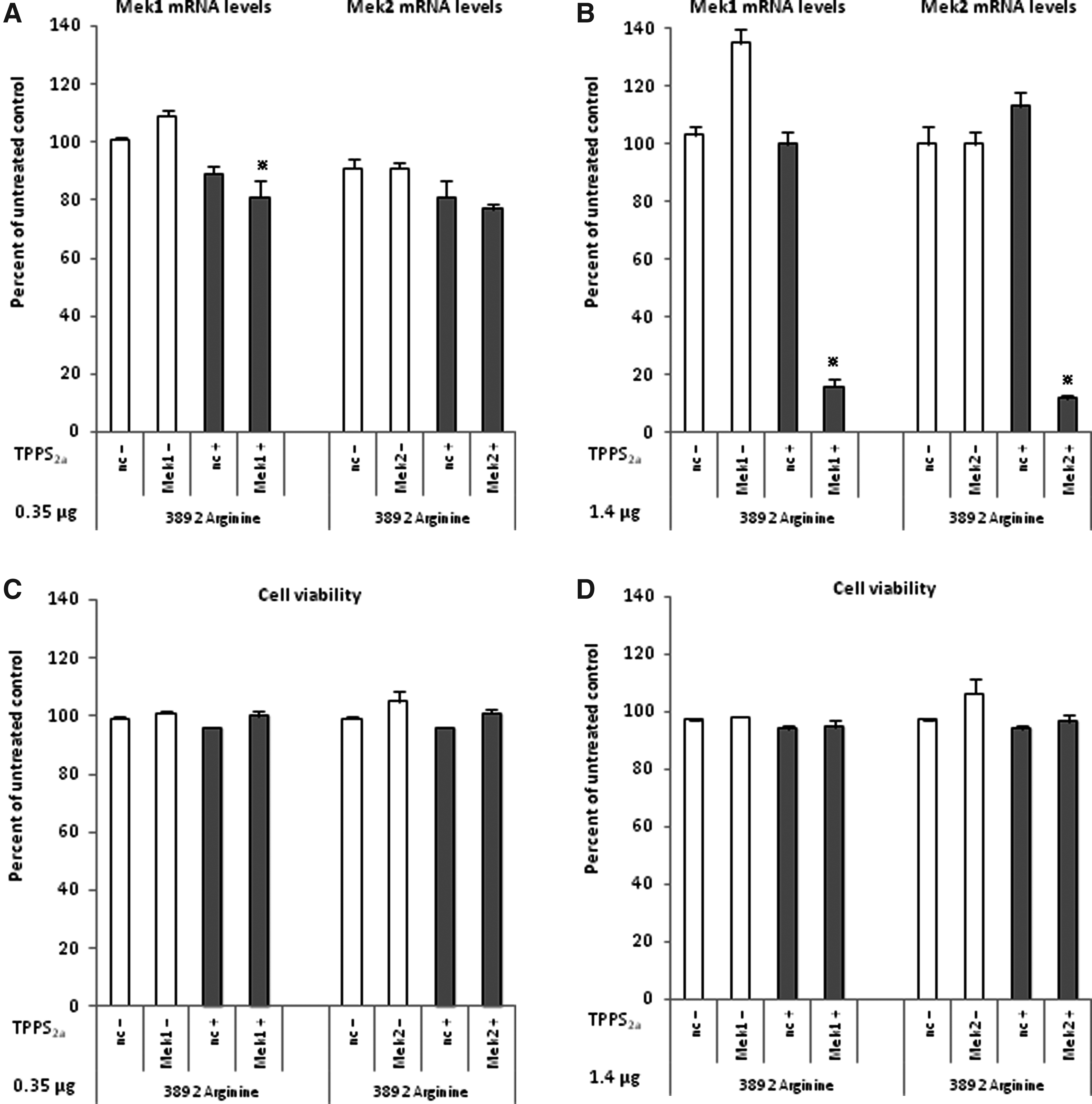
Examination of siRNA transfection ability and cell viability in the SK-MEL-28 cell line using PCI in combination with polyarginine no.3892 at 2 concentrations, 0.35μg
Complex sizes of polyarginines and siRNA
Complex sizes of gene carrier and siRNA were examined for the 3 PLAs used in this study at 3 weight ratios (Fig. 3). PLA no.3892 was measured at 3 weight ratios, 0.175 μg, 0.35 μg, and 1.4 μg, while PLA no.4663 and PLA no.7762 were only measured at the 0.175 μg and the 0.35 μg weight ratios. The Z-average diameter increased with the amount of PLA. Complexes of PLH/siRNA and PLL/siRNA persistently resulted in multiple peaks.
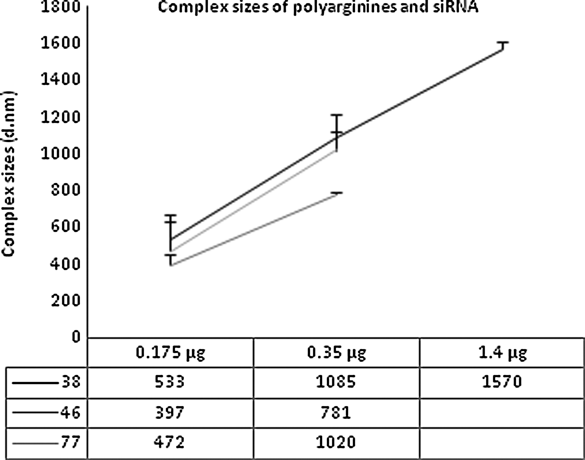
Complex sizes of polyarginines and siRNA. The Z-average diameter (nm) was measured for 2 or 3 concentrations (0.175 μg, 0.35 μg, and 1.4 μg), while the siRNA concentration was kept constant (0.7 μg). Every bar illustrates the mean value of 2 experiments±SEM.
Light-directed EGFP mRNA delivery using polyarginine in OHS cell line
Since polyamino acids no.3892, no.4663, no.7762, and no.9404 demonstrated the highest gene-silencing effects of the tested carriers, we evaluated them for EGFP mRNA delivery (data not shown). PLA no.4663 appeared to be a promising carrier from our initial study, and we continued our EGFP mRNA experiments with this carrier. We used 3 concentrations of PLA no.4663—0.7 μg, 1.4 μg, and 2.8 μg—while the mRNA concentration was kept constant (1.88 μg). PCI-treated and non-treated samples were analyzed by fluorescence microscopy (Fig. 4A) and flow cytometry (Fig. 4B, C). The proportion of EGFP-positive cells raised from 25% to 55% in PCI-treated samples when the concentration of PLA no.4663 increased from 0.7 μg to 1.4 μg. When the concentration of PLA no.4663 was again doubled, from 1.4 μg to 2.8 μg, only a minor difference in the number of EGFP-positive cells was observed. Of the PCI non-treated samples, less than 0.5% of the cell population was EGFP positive at the 3 concentrations. Importantly, the data obtained from the flow cytometry analyses were in agreement with the visible EGFP expression detected by fluorescence microscopy. A cell viability study (Fig.4D) with PLA no.4663 and mRNA was monitored with the same concentrations and light dose as the samples used for mRNA delivery. The highest cell toxicities were detected in PCI samples with 1.4 μg and 2.8 μg PLA no.4663, with a decrease in cell viability of 12% in both samples compared with untreated control.
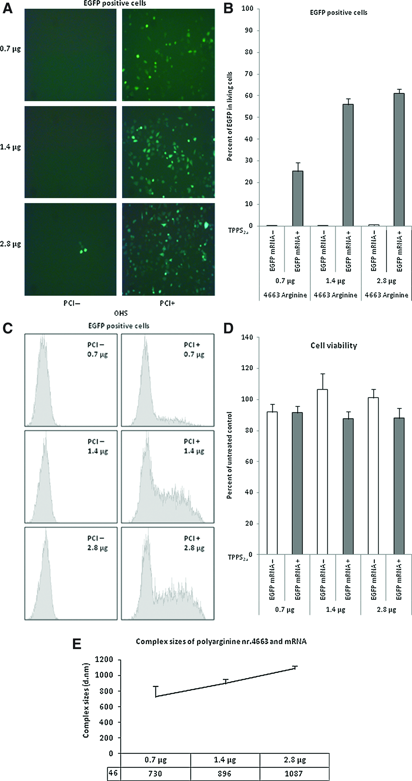
Delivery of enhanced green fluorescent protein (EGFP) mRNA in the OHS cell line using PCI in combination with polyarginine no.4663 at three concentrations: 0.7 μg, 1.4 μg, and 2.8 μg. The transfection efficiencies were examined with both live cell fluorescent images (20×)
Discussion
In this study, we aimed to evaluate nanoparticles consisting of biodegradable polyamino acids and nucleic acids for targeted delivery using PCI technology. We show for the first time that standard unmodified polyamino acid carriers can be utilized for efficient delivery of both siRNA and mRNA molecules when combined with PCI technology. Our results implied that entrapment of nanoparticles in lysosomal vesicles is an important barrier when using polyamino acid carriers. The high gene-silencing effect of 90% that we achieved with polyarginine no.3892 in both the OHS and SK-MEL-28 cell lines was specific for PCI-treated samples. No reduction of S100A4, MEK1, or MEK2 mRNA levels was observed in samples without TPPS2a. This targeting effect was also observed for EGFP mRNA delivery where only 0.5% EGFP expression was detected in non-PCI-treated samples. Arginine has an isoelectric point of 10.76, and is thus positively charged at physiological pH (7.4). PLA carriers are therefore not able to buffer the pH inside the endosomes/lysosomes by uptake of protons in a mechanism described as a “proton sponge” effect. This property has been described for carriers such as PEI (Boussif et al., 1995), PAMAM (Ouyang et al., 2011), and chitosan (Huang et al., 2005). A carrier with intrinsic “proton sponge” properties may enhance the delivery effect of the PCI technology, but may also lead to uncontrolled release of endosomal content to cytosol. The delivery mechanism we observe in our present work is solely determined by illumination. In a study with PLL (isoelectric point 9.74) and a plasmid encoding EGFP, Dietze and colleagues demonstrated that light was the decisive factor in obtaining a significant gene delivery (Dietze et al., 2003).
Despite promising results, earlier siRNA/PCI reports using non-metabolized carriers such as jetSI reagent, PEI, and Lipofectamine 2000, which represent an important tool for effective siRNA delivery, are unfortunately restricted to in vitro applications as they may result in severe toxicity and immune response effects when utilized in vivo. Targeted delivery and biosafety have been main barriers for progress within RNAi therapeutics. Interestingly, systemic administration of cyclodextrin/siRNA nanoparticles targeted toward melanoma has just recently been demonstrated in the first proof-of-concept study that RNAi works in humans. Of note, these cyclodextrin nanoparticles had the benefit for both targeting and reduced long-term accumulation in vivo due to lack of immunogenicity and preferable biocompatibility (Davis et al., 2010). As shown by Davis and colleagues, nanoparticles can be equipped with for example targeting ligands (transferrin) for targeted delivery in vivo.
Indeed, cyclodextrin has been investigated for light-directed siRNA delivery, but very high concentrations of the unmodified cyclodextrin were required to obtain gene-silencing activity of 80%–90% (Boe et al., 2010b). Another barrier for further development of cyclodextrin as a carrier for RNAi therapeutics is the lack of availability for the research community. Noteworthy, this is also the matter for chitosan, another promising biodegradable carrier under evaluation for RNAi-based applications. With cationic PLAs, we show the possibility of light-directed delivery of nucleic acids using a relatively small amount of unmodified polyarginines (0.35 μg and 1.4 μg in OHS and SK-MEL-28, respectively). There was a correlation between gene silencing, complex sizes, and molecular weight. The highest gene silencing was achieved with PLA no.3892, the polyamino acid that measured the largest complex sizes of carrier/siRNA and has the highest molecular weight. We expected PLA no.3892 to be a good carrier for mRNA delivery as well, but another polyarginine formulation, PLA no.4663, was clearly the best mRNA carrier after our initial studies. It was not surprising that peptides of arginine were the most effective gene carriers, as they have been assessed more capable of entering cells than polymers of lysine and histidine (Mitchell et al., 2000). Moreover, in a recent study, complexes of plasmid DNA and arginine were reported to be stable in blood circulation for 6 hours and uptake in organs were detected (Woo et al., 2011).
In conclusion, we have developed an efficient transfection protocol for both siRNA and mRNA molecules with potential for further investigation for use in vivo. Our results with polyarginines in combination with PCI demonstrated a safe and controllable delivery system.
Footnotes
Acknowledgments
The authors are grateful for the PhD grant allocated by the Norwegian Gene Therapy Programme, the Norwegian Radium Hospital. We would also like to thank the flow cytometry core facility at the Norwegian Radium Hospital, Institute for Cancer Research, for assistance with analyses, and Stein Sæbøe-Larssen for providing us the EGFP mRNA molecules.
Authors Disclosure Statement
No conflict of interest exists.