
Abstract
Covalent binding of squalene to siRNA has already been shown to be an interesting way of delivering siRNA in vivo. Whether squalene derivatives could also be used to deliver siRNA in cells without covalent binding similar to usual transfection with cationic lipids is the question addressed in this article. Accordingly, we investigated the activity of two squalene derivatives bearing a quaternary ammonium head group and a guanidinium group, respectively. The second derivative displayed interesting properties for delivering siRNA into cells in vitro.
Introduction
T
Although quite effective, this method required the postsynthetic covalent coupling of the squalene to a 3′-end thiol-modified siRNA. This task turned out to be quite challenging due to the total insolubility of squalene derivatives in water and was achieved only with a moderate chemical yield.
We therefore reasoned that it might be possible to benefit from the self-assembling properties of squalene without making a covalent bond, using the simple electrostatic interaction of the negatively charged siRNA with a cationic squalene derivative (SQ+). We indeed know that cationic lipids have specific topics of application in vivo such as localization in endothelial cells of tumoral angiogenesis [4] or delivery of nucleic acids in lung tumors [5]. Our hope was that cationic squalene derivatives might display the same transfecting properties than other cationic lipids, but with the unique self-assembling properties of the squalene, and might therefore be good candidates for these specific topics of application in vivo. Using either trimethylammonium-substituted (SQ-NMe3+, Cl−) or hydrazinoguanidinium-substituted (SQ-NH(NH2)C═NH2+, AcO−) squalene derivatives, we have studied their nanoassembly formation and stability, their capacity to bind siRNA, and to protect it from nucleases degradation. Cell penetration and in vitro biological activity against EWS/Fli-1 oncogene were also addressed.
Materials and Methods
Cationic squalene synthesis
Material
Squalene was obtained from Alfa Aesar; aminoguanidine bicarbonate and trimethylammonium chloride were obtained from Sigma-Aldrich Chemical Co.
General methods
Infrared (IR) spectra were obtained as neat liquid on the Fourier Transform Bruker Vector 22 spectrometer. Only significant absorptions are listed. The 1H and 13C NMR spectra were recorded on the Bruker Avance 300 (300 and 75 MHz for 1H and 13C, respectively) spectrometer. Recognition of methyl, methylene, methine, and quaternary carbon nuclei in 13C NMR spectra rests on the J-modulated spin-echo sequence. Mass spectra were recorded on the Bruker Esquire-LC. Analytical thin-layer chromatography was performed on Merck silica gel 60F254 glass-precoated plates (0.25 mm layer). Column chromatography was performed on the Merck silica gel 60 (230–400 mesh ASTM). Diethyl ether was distilled from sodium/benzophenone ketyl. All reactions involving air- or water-sensitive compounds were routinely conducted in glassware, which was flame-dried under a positive pressure of nitrogen. Chemicals obtained from commercial suppliers were used without further purification.
Synthesis of the cationic squalene derivatives
{Amino[(E)-2-[(4E,8E,12E,16E)-4,8,13,17,21-pentamethyldocosa-4,8,12,16,20-pentaen-1-ylidene] hydrazin-1-yl]methyldene}azanium acetate (SQ-NH(NH2)C═NH2+, AcO−)
Aminoguanidine bicarbonate (145 mg, 1.06 mmol) was added to a solution of 1,1′,2-trisnor-squalenaldehyde (382 mg, 1.0 mmol) in acetic acid (6 mL). The reaction mixture was heated at 60°C for 1 h. After cooling, the solvent was removed under reduced pressure using dry-ice evaporator. The residue was taken up into diethyl ether (50 mL) and was washed with water (3×2 mL). The organic layer was dried over magnesium sulfate and concentrated under reduced pressure to leave the title compound as colorless viscous oil (475 mg, 95%). IR (neat, cm−1) ν=3500-2700, 2964, 2928, 2913, 2854, 1682, 1626, 1520, 1449, 1405, 1388, 1376, 1108, 1012, 907; 1H NMR (300 MHz CDCl3) δ=11.00-7.50 (broad s, 4H, N═C(NH2)NH2), 7.44 (t, J=4.5 Hz, 0.87 H, HC═N anti ), 6.63 (t, J=4.5 Hz, 0.13 H, HC═N syn ), 6.50-5.75 (broad s, 1H, ═NNH), 5.20-5.06 (m, 5H, CH═C(CH3)), 2.36-2.28 (m, 2H, N═CHCH2CH2), 2.20-1.90 (m, 21H, N═CHCH2CH2, ═C(CH3)CH2CH2, CH3CO2), 1.67 (s, 3H, ═CH(CH3)2), 1.59 (s, 15H, ═CH(CH3)CH2); 13C NMR (300 MHz CDCl3) δ=178.3 (C, CH3CO2), 156.0 (C, NC═N), 151.2 (CH, HC═N), 135.1 (C, CH═C(CH3)CH2), 134.9 (2C, CH═C(CH3)CH2), 133.3 (C, CH═C(CH3)CH2), 131.1 (C, CH═C(CH3)2), 125.4 (CH, CH═C(CH3)CH2), 124.4 (CH, CH═C(CH3)), 124.3 (CH, CH═C(CH3)), 124.2 (2CH, CH═C(CH3)), 39.7 (2CH2, C═C(CH3)CH2), 39.6 (CH2, C═C(CH3)CH2), 35.9 (CH2, ═C(CH3)CH2CH2), 31.3 (CH2, ═C(CH3)CH2CH2), 28.2 (2CH2, ═C(CH3)CH2CH2), 26.7 (2CH2, ═C(CH3)CH2CH2), 26.6 (CH2, ═C(CH3)CH2CH2), 25.6 (CH3, CH═C(CH3)2), 23.6 (CH3, CH3CO2), 17.6 (CH3, ═C(CH3)CH2), 16.0 (2CH3, ═C(CH3)CH2), 15.9 (2CH3, ═C(CH3)CH2); MS (ESI+): m/z (%)=441.5 (100) [M]+.
22-Chloro-2,6,10,15,19-pentamethyl-docosa-2,6,10,14,18-pentaene
Methanesulfonyl chloride (205 mg, 1.8 mmol) was added dropwise at 0°C to a mixture of trisnorsqualence alcohol 5 (582 mg, 1.5 mmol), triethylamine (224 mg, 2.2 mmol) and DMAP (20 mg) in anhydrous CH2Cl2 (7 mL). The mixture was then slowly raised to room temperature and stirred for 2 h. The reaction was then quenched with brine and the mixture was extracted with CH2Cl2 (4×50 mL). The combined organic extracts were washed with brine, dried over MgSO4, and concentrated in vacuo. The crude 1,1′,2-trisnorsqualenyl methanesulfonate (695 mg, 85%) was used directly without further purification. LiCl (456 mg, 10.8 mmol) was added to a solution of the above mesylate (500 mg, 1.08 mmol) in anhydrous DMF (5 mL). The reaction mixture was heated at 80°C for 2h. After cooling to room temperature, the mixture was concentrated under reduced pressure. The residue was taken in water and then extracted with diethyl ether (5×12 mL). The combined organic phases were dried over MgSO4, filtered, and concentrated in vacuo. The crude product was purified by chromatography on silica gel (cyclohexane/EtOAc, 98/2, v/v) to give the chlorotrisnorsqualene as colorless oil (377 mg, 86%); IR (neat, cm−1) ν=2855–2970, 1441, 980, 832; 1H NMR (300 MHz, CDCl3) δ=5.02–5.25 (m, 5H, CH═C(CH3)), 3.56 (t, 2H, J=6.7 Hz, CH2Cl), 1.91–2.20 (m, 18H, ═C(CH3)CH2CH2), 1.85 (m, 2H, ═C(CH3)CH2CH2), 1.52–1.73 (m, 18H, CH3); 13C NMR (300 MHz, CDCl3) δ=135.1 (C, CH═C(CH3)CH2), 134.9 (C, CH═C(CH3)CH2), 134.8 (C, CH═C(CH3)CH2), 133.0 (C, CH═C(CH3)CH2), 131.2 (C, CH═C(CH3)CH2), 125.6 (C, CH═C(CH3)CH2), 124.5 (CH, C, CH═C(CH3)CH2), 124.4 (CH, CH═C(CH3)CH2), 124.3 (2 CH, CH═C(CH3)CH2), 44.5 (CH2, CH2Cl), 39.8 (2 CH2, ═C(CH3)CH2CH2), 39.7 (CH2, ═C(CH3)CH2CH2), 39.6 (CH2, ═C(CH3)CH2CH2), 36.6 (CH2, ═C(CH3)CH2CH2), 30.7 (CH2, ═C(CH3)CH2CH2), 28.3 (2 CH2, ═C(CH3)CH2CH2), 26.8 (CH2, ═C(CH3)CH2CH2), 26.7 (CH2, ═C(CH3)CH2CH2), 26.6 (CH2, ═C(CH3)CH2CH2), 25.7 (CH3, CH═C(CH3)2), 17.7 (CH3, ═C(CH3)CH2), 16.0 (CH3, ═C(CH3)CH2), 15.9 (2CH3, ═C(CH3)CH2), 15.8 (CH3, ═C(CH3)CH2); MS (ESI): m/z (%)=456.4 (100) [M-H]+; Anal. calcd for C27H45Cl (%): C 70.22, H 11.40, N 2.60. Found: C 70.71, H 11.21, N 3.13.
Trimethyl[(4E,8E,12E,16E)-4,8,13,17,21-pentamethyldocosa-4,8,12,16,20-pentaen-1-yl]azanium chloride (SQ-NMe3+, Cl−)
A 50% aqueous sodium hydroxide solution (20 mL) was added dropwise by mean of a dropping funnel to trimethylamine hydrochloride (3.0 g, 31.4 mmol) placed in a distilled flask. The evolved trimethylamine gas was passed through a washed bottle containing sodium hydroxide pellets and allowed to bubble through 4.0 g of anhydrous ethanol placed in another wash bottle and exactly weighted. The obtained Me3N (2.2 g, 37.2 mmol) ethanol solution was added to 1-chlorotrisnorsqualene (300 mg, 0.71 mmol) in a screw cap-sealed tube equipped with a stirred bar. The reaction mixture was stirred at 100°C for 72 h. After cooling to room temperature, the mixture was concentrated under reduced pressure to provide the tertiary ammonium salt as pale yellow oil (294 mg, 98%). The crude product was used without further purification. 1H NMR (300 MHz, CD3OD) δ=5.26 (t, J=6.0 Hz, 1H, CH═C(CH3)), 5.25-5.15 (m, 4H, CH═C(CH3)), 3.40-3.30 (m, 2H, Me3NCH2), 3.21 (s, 9H, (CH3)3N+), 2.20-1.80 (m, 20H, ═C(CH3)CH2CH2), 1.69 (s, 6H, ═CH(CH3)2, ═CH(CH3)CH2), 1.62 (s, 12H, ═CH(CH3)CH2);13C NMR (300 MHz, CD3OD) δ=136.8 (2C, HC═C(CH3)CH2), 1.36.7 (C, HC═C(CH3)CH2), 134.7 (C, HC═C(CH3)CH2), 132.8 (C, HC═C(CH3)CH2), 128.1 (CH), 126.45 (CH), 126.4 (CH), 126.3 (2CH), 68.5 (CH2, CH2NMe3), 54.5-54.4 (3CH3, N(CH3)3), 41.7 (2CH2), 41.6 (CH2), 37.9 (CH2), 30.1 (2CH2), 28.7 (CH2), 28.4 (CH2), 26.8 (CH3, HC═C(CH3)2), 23.1 (CH2), 18.7 (CH3), 17.1 (3CH3), 16.7 (CH3); MS (ESI): m/z (%)=428 (100); Anal. calcd for C30H54ClN (%): C 77.62, H 11.73, N 3.02. Found: C 77.21, H 11.71, N 2.90.
Nanoparticles formation and characterization
Nanoparticles formation was obtained by the nanoprecipitation–solvent evaporation method [2]. Practically, cationic squalene derivatives (SQ+) in acetone (0.5 mL, 4 mg/mL) were slowly added dropwise into 1 mL of milliQ water under stirring at room temperature. Formation of nanoparticles occurred immediately without the use of any surfactant. The stirring was pursued for further 15 min, and the solvent was then evaporated at 37°C under vacuum using the Rotavapor®. Then, NPs were characterized by dynamic light scattering (DLS) size measurement and zeta potential determination (zetasizer nano ZS; Malvern), as indicated in Table 1. The DLS measurements for the size distribution were performed in water by the intensity method. The zeta potentials were determined in 1 mM NaCl solution by Laser Doppler Velocimetry. The organic solvent-free colloidal dispersions were stored at 4°C.
NPs, nanoparticles; Pdi, polydispersity index.
siRNA synthesis
The siRNAs were produced by Eurogentec and supplied as a double-stranded sequence. Silencing siRNAs are targeted toward the junction sequence between EWS and Fli-1 part of the mRNA. Anti EWS-Fli-1 siRNA was as follows: antisense strand 5′-GGG UUC UGC UGC CCG UAG C d(GT)-3′ and sense strand 5′-GCU ACG GGC AGC AGA ACC C d(TT)-3′. Control siRNA was as follows: antisense strand 5′-GAU AGC AAU GAC GAA UGC GUA d(TT)-3′ and sense strand 5′-UAC GCA UUC GUC AUU GCU AUC d(TT)-3′. A 20 μM solution was used to prepare any samples. We have used the same EWS-Fli-1 siRNA labeled at the two 3′-extremities by FITC (provided by Eurogentec).
siRNA loading onto squalene nanoparticles
A simple method to measure the nucleic acids/squalene interaction by ethidium bromide fluorescence measurement of the solution after mixing both components has been developed. Fixation of siRNA on SQ+ NPs was determined by fluorescence quenching of siRNA-bound ethidium bromide. Briefly, fixed quantity of siRNA (2 μg/mL) was incubated with different amount of SQ+ NPs expressed in charge ratio from 0 to 6 in 10 mM HEPES pH 7.2, 100 mM NaCl buffer in 10 μL final volume in 96-well plate for fluorescence measurement. Then, 10 μL of 1 μg/mL ethidium bromide was added and fluorescence measurement was performed on Glomax Multi+fluorescence plate reader (Promega) with the green filter (ex: 525 nm, em: 580–650 nm). Experiments were performed in triplicate.
Cell culture
Human Ewing sarcoma cells, A673, were grown in the Dulbecco's modified Eagle's medium (DMEM; Gibco) containing 10% bovine calf serum (Gibco) and 1% solution of penicillin/streptomycin (Gibco). The cells were incubated at 37°C in a moist atmosphere containing 5% CO2.
Cytotoxicity of cationic squalene nanoparticles
To determine the cytotoxicity of cationic squalene derivatives, 2×10³ A673 cells were seeded in 96-well plate 1 day before treatment. Then, the medium was discarded and 100 μL of fresh medium containing increasing quantity of SQ+ NPs or of siRNA/SQ+ NPs was added. The cells were incubated for 48 h, and cell surviving was determined by the MTT test. Then, 10 μL of 5 mg/mL MTT (Sigma-Aldrich Chemical Co.) in phosphate-buffered saline (PBS) buffer was added to the cells and incubated for 2 h at 37°C, 5% CO2 in moist atmosphere. Cells lyses and formazan solubilization were obtained by adding 100 μL of 10 mM HCl, 10% sodium dodecyl sulfate (SDS) solution followed by overnight incubation at 37°C. Produced formazan was quantified by measurement of absorbance at 570 nm in plate reader (EL808; BioTek). Experiments were performed on six independent wells and expressed as the percent of untreated cells.
siRNA delivery to cells
To evaluate the capacity of SQ+ NPs to deliver efficiently siRNA to cells, a 3′-labeled FITC siRNA was used. One day before treatment, A673 cells were seeded on 12 wells plate containing a cover slide rinsed once in ethanol bath and once in sterile water. Fifty nanomolars of siRNA (final concentration) was complexed with different amounts of SQ+ NPs to obtain a charge ratio N/P [ratio between the amount of ammonium groups of the squalene derivative (N) and the phosphates of the siRNA (P)] of 0, 2, 4, or 6. Then, the cell medium was eliminated, and 450 μL of Opti-MEM (Gibco), a serum-free medium, or 450 μL of DMEM containing 10% fetal bovine serum was added. Afterward, 50 μL of the different siRNA/SQ+ NPs was added and incubated for 4 h at 37°C, 5% CO2 in moist atmosphere. The cells were then washed with PBS, and 1 mL of 4% formal solution in PBS was added for 20 min at room temperature. The cells were washed three times with PBS and mounted on slide with DAPI fluoromount G (SouthernBiotech) before being observed with an epifluorescence microscope (Observer; Zeiss) or a confocal microscope (TCS SPE; Leica) to confirm the presence of intracytoplasmic siRNA. Untreated cells present only a blue fluorescence due to nucleus coloration.
Nucleases protection of siRNA by cationic squalene nanoparticles
To study the capacity of cationic squalene to protect siRNA, we first determined the condition for the siRNA/SQ+ NPs to dissociate. It was observed by agarose gel electrophoresis that siRNA/SQ+ was not detectable by ethidium bromide fluorescence. Nevertheless, because siRNA are very sensitive to nuclease degradation, the solution used for complex dissociation needs to inhibit nuclease activities. We have then selected a stop solution containing a high concentration of SDS to destroy the lipid nanoparticles and heparin for nucleases inhibition.
To determine the siRNA protection by SQ+ NPs, free siRNA or siRNA/SQ+ NPs at N/P ratio of 6 have been incubated in DMEM containing 10% bovine calf serum. At various times, 10 μL of aliquots was stopped by 5 μL of 1% SDS, 4 mg/mL heparin, and 0.1 M EDTA solution. The siRNAs were analyzed on 2% agarose gel electrophoresis in 0.5×TAE buffer, and their detection was performed by ethidium bromide coloration. The intensity of bands was monitored with a gel analyzer (Ingenius; Syngene).
Inhibition of EWS-Fli-1 by siRNA/SQ+ NPs
The efficiency of siRNA/SQ+ NPs has been determined by the inhibition of targeted EWS-Fli-1 mRNA expression by reverse transcriptase-quantitative polymerase chain reaction (RT-qPCR).
Cell treatment
One day before treatment, A673 cells were seed in 12-well plate at 4×10⁴ cells per wells in the DMEM containing serum. Then, the medium was discarded, and 450 μL of OptiMEM serum-free medium (Gibco) was added to the cells. Then, 50 μL of siRNA/SQ+ NPs prepared in 10 mM HEPES buffer, pH 7.2, 100 mM NaCl at charge ratio N/P of 0, 2, 4, and 6, was added to the cells and incubated for 4 h at 37°C, 5% CO2 in moist atmosphere. The medium was then discarded and 500 μL of DMEM containing 10% calf bovine serum was added for a further 20 h of culture.
RNA extraction
Total RNAs were extracted by Trisol technique (Invitrogen). Briefly, the cells were washed with PBS, and then, 400 μL of Trisol was added before cells scraping and transferred in a centrifuge tube. After homogenization, 60 μL of chloroform:isoamyl alcohol (49:1, v:v) was added and the tubes were centrifuged for 15 min at 12,000 g. One hundred fifty microliters of the superior aqueous phase was withdrawn and 150 μL of isopropanol was added before RNA precipitation for 10 min at room temperature. After centrifugation for 15 min at 12,000 g, the pellet was washed two times with 75% ethanol and dried before to be dissolved in 10 μL of water containing 0.5 U of RNasin (Promega). Total RNA was quantified by spectrophotometry (Nanodrop 2000; Thermo).
Reverse transcription
cDNA was synthesized by heating 1.5 μg of total RNA with 2 μL of random primer (Promega) in 12.5 μL of total volume for 3 min at 70°C. Then, 4 μL of M-MLV RT 5×buffer (Promega), 0.5 μL of 20 mM dNTP, 0.5 μL RNasin (Promega), 0.5 μL of 200 U/μL M-MLV RT (Promega) were added and the tube was incubated for 10 min at room temperature followed by 1 h at 42°C.
PCR quantification of EWS-Fli-1 mRNA expression
Quantitative PCR was processed on 5 μL of cDNA diluted 20 times, 10 μL SYBR green mix 2×(Syber fast; Kapa BioSystem), 0.4 μL of 10 μM forward primer, and 0.4 μL of 10 μM reverse primer in 20 μL final volume. The EWS-Fli-1 gene was amplified with the EWS-forward primer: 5′-AGC AGT TAC TCT CAG CAG AAC ACC-3′ and Fli-1-reverse primer: 5′-CCA GGA TCT GAT ACG GAT CTG GCT G-3′. As a control, we used the human 18S rRNA gene with the 18S forward primer: 5′-CGT TCA GCC ACC CGA GAT-3′ and 18S reverse primer: 5′-TAA TGA TCC TTC CGC AGG TT-3′ (Eurogentec) with a real-time PCR system (StepOne Plus; Applied Biosystems). The comparative Ct method was used to normalize the target Ct by the 18S control gene Ct.
Results
Synthesis of the cationic squalene derivatives
Two cationic squalene derivatives (SQ+) were straightforwardly prepared from squalene either with a quaternary ammonium head group (SQ-NMe3+) or with a guanidinium head group (SQ-NH(NH2)C═NH2+). The squalenaldehyde hydrazinoguanidinium acetate salt (Fig. 1, SQ-NH(NH2)C═NH2+, AcO−) has been obtained as a 87:13 anti/syn mixture by condensation of 1,1′,2-trisnor-squalenaldehyde with aminoguanidine bicarbonate in acetic acid [6]. The aldehyde was in turn prepared in three steps from commercially available squalene according to the van Tamelen procedure [7,8]. On the other hand, squalenyl-trimethylammonium chloride salt (Fig. 1, SQ-NMe3+, Cl−) was obtained by alkylation of trimethylamine with 1-chlorotrisnorsqualene in a seal tube in ethanol at 100°C. 1-Chlorotrisnorsqualene was prepared in 73% yield from trisnorsqualenol by mesylation, followed by nucleophilic displacement of the mesylate group by lithium chloride, as described previously [9].

Nanoparticles formation and characterization
Nanoparticles were obtained by the nanoprecipitation procedure previously described for squalene derivatives [2]. As indicated in Table 1, the nanoparticles obtained from the quaternary ammonium squalene (SQ-NMe3+) were found to have a size of 90 nm, but with a polydispersity index of 0.45 indicating a heterogeneous suspension with probably a large size distribution. These nanoparticles were cationic with a zeta potential of+20 mV compatible for nucleic acid complexation. On the other hand, siRNA/(SQ-NH(NH2)C═NH2+) NPs were found to have an average size of 170 nm with a polydispersity index around 0.1 indicating a narrow size distribution. They were found to be round nanoparticles as shown by electron microscopy (see Supplementary Data; Supplementary Data are available online at www.liebertpub.com/nat). The global surface charge was cationic (+70 mV), indicating a nanoparticle organization presenting the guanidinium head group at the surface. These suspensions were found extremely stable since 1-year-old preparation stored at 4°C displayed unchanged size and zeta potential.
When a nucleic acid was bound to these SQ+ NPs, we observed a slight increase of the nanoparticles size, whereas the zeta potential became fairly negative (−50 mV for siRNA/SQ-NH(NH2)C═NH2+), corresponding to the presence of an excess of negatively charged nucleic acids at the nanoparticles surface (Table 1).
siRNA loading onto squalene nanoparticles
The binding of the siRNA on cationic squalene derivatives has been evaluated by the addition of a given concentration of siRNAs to increasing quantities of cationic squalene derivatives and fluorescence measurement after ethidium bromide labeling. In these conditions, only the unbound siRNA produced a fluorescence signal, which is proportional to the concentration of the free siRNA fraction. Results are expressed in Fig. 2 according to increasing values of the charge ratio N/P. The concentration of free siRNA decreased when the amount of cationic squalene derivative increased and reached a minimum value when N/P was 3.
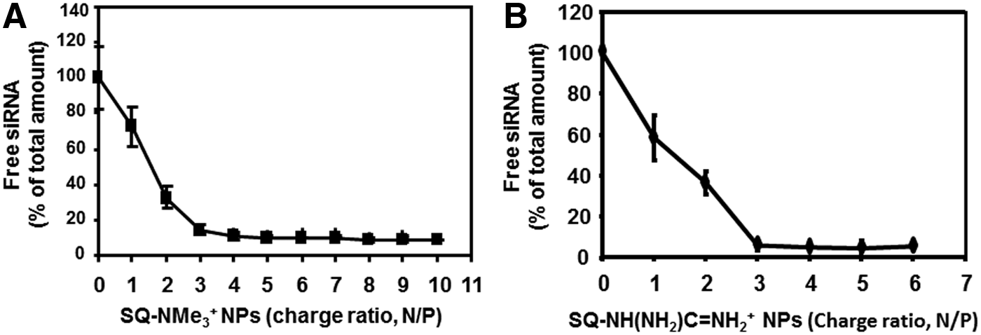
Binding of siRNA on SQ-NMe3+ NPs
Protection of siRNA against nucleases by squalene nanoparticles
Free siRNA or siRNA/SQ-NH(NH2)C═NH2+ were incubated in 10% fetal bovine serum and analyzed by agarose gel electrophoresis at different incubation times. As depicted in Fig. 3, free siRNA was completely degraded by the nucleases present in the serum after 3–4 h. On the other hand, the siRNA associated to SQ-NH(NH2)C═NH2+ NP was found to be mostly unchanged after 6 h in the same conditions with less than 6% of degradation. In the same conditions at 6 h, nearly 70% of siRNA associated to SQ-NMe3+ NP is degraded (see Supplementary Data).
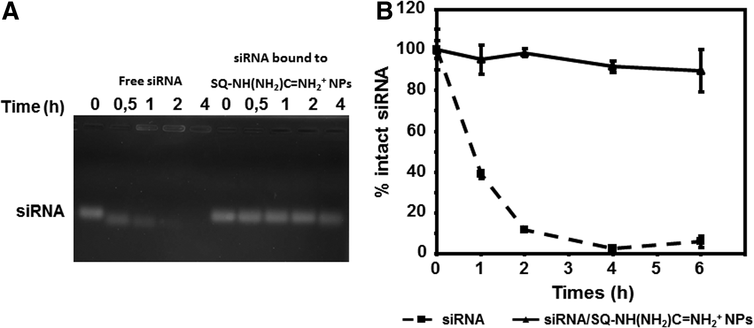
Protection from nuclease degradation of siRNA bound to SQ-NH(NH2)C═NH2+ NPs. Free siRNAs or siRNA/SQ-NH(NH2)C═NH2+ were incubated in 10% fetal bovine serum and then analyzed by gel electrophoresis
Cytotoxicity of squalene nanoparticles
To evaluate the cytotoxicity of these cationic squalene nanoparticles, A673 cells were incubated with an increasing amount of SQ+ NPs, either free or bound to 50 nM EWS/Fli-1 siRNA. Both squalene derivatives NP display a rather similar inhibition of cell growth after 3 h time whatever the culture medium used (Fig. 4). However, some morphological modifications can be seen on A673 cells treated by SQ-NMe3+ (Fig. 6).
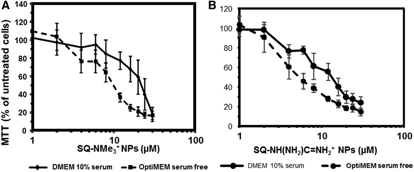
A673 cell survival after 3 h incubation with SQ+ NPs measured by the MTT test. Nanoparticles were tested either free or associated to 50 nM siRNA. SQ-NMe3+
Cell distribution of siRNA/SQ+ NPs
The capacity of SQ+ NPs to deliver the EWS/Fli-1 siRNA into A673 human Ewing sarcoma cells was studied using 3′-FITC-labeled nucleic acids. Untreated cells present only a blue fluorescence due to nucleus coloration (Fig. 5). This indicated that no background fluorescence was detectable in our imaging conditions. When the cells were treated 4 h with free 50 nM fluorescent FITC-labeled siRNA, no green fluorescence was detected, confirming that free siRNA does not penetrate into cells. When fluorescent siRNAs were complexed with various amounts of SQ-NH(NH2)C═NH2+, at charge ratio N/P=2, 4, or 6, an increasing number of green dots into the cell cytoplasm corresponding to internalized siRNA were visible (Fig. 5). Transfection therefore already takes place for 50 nM siRNA at N/P=2, which corresponds to 4 μM in squalene. According to Fig. 4, this is associated with 25% cell inhibition of MTT production. Similar experiments were performed with the association of siRNA-SQ-NMe3+, and we found that no significant increase of green dots is then observed in cells (Fig. 6).
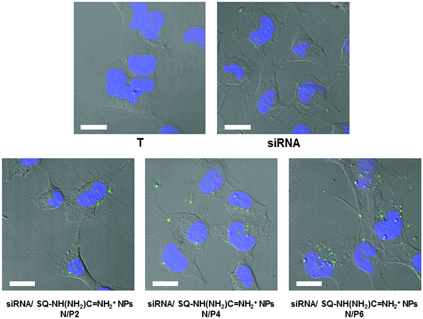
Transfection of fluorescent siRNA by SQ-NH(NH2)C═NH2+ NPs in A673 human Ewing sarcoma cells. A673 cells were treated for 4 h in serum-free OptiMEM medium by labeled siRNA-FITC (green) either free or bound to SQ-NH(NH2)C═NH2+ NPs at a charge ratio (N/P) of 2, 4, or 6 (corresponding to 4, 8, or 12 μM). The cell nucleus was labeled by DAPI (blue). The slides were observed by confocal microscopy. Scale bare is 20 μm.
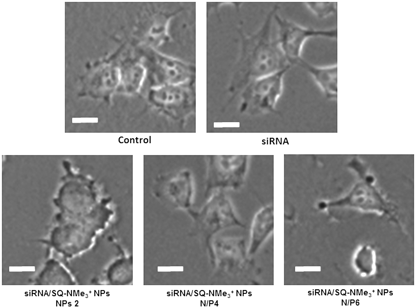
Transfection of fluorescent siRNA by SQ-NMe3+ NPs in A673 human Ewing sarcoma cells. A673 cells were treated for 4 h in serum-free OptiMEM medium by labeled siRNA-FITC either free or bound to SQ-NMe3+ NPs at a charge ratio (N/P) of 2, 4, or 6 (corresponding to 4, 8, or 12 μM). The cell nucleus was labeled by DAPI. The slides were observed by epifluorescence microscopy. Scale bar is 20 μm.
Inhibition of EWS-Fli-1 expression by siRNA/SQ+ NPs
Transfection efficiency was evaluated by the capacity of cationic SQ-NH(NH2)C═NH2+/siRNA to inhibit specifically the targeted EWS-Fli-1 oncogene expression. The A673 human Ewing sarcoma cells were treated with an already validated siRNA [10], either free or bound to SQ-NH(NH2)C═NH2+ nanoparticles for 24 h before total RNA extraction. Then, RT-qPCR was performed to evaluate the inhibition of expression of the targeted EWS-Fli-1 mRNA (Fig. 7). An irrelevant siRNA sequence was used as a control. We observed that both free control and specific siRNA have no effect on EWS-Fli-1 gene expression in the absence of nanovectors. When the same EWS/Fli-1 targeting siRNA was used bound to SQ-NH(NH2)C═NH2+, EWS/Fli-1 mRNA expression inhibition was observed. This effect was maximum for N/P=4 and 6, with around 50% inhibition. In the same conditions, the control siRNAs was found to be devoid of effect. Inhibition of EWS/Fli-1 is not related to the modification of cell growth shown by MTT since the control siRNA has no effect in the same conditions.
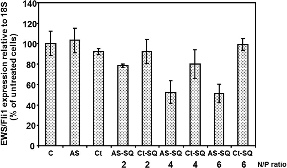
Inhibition of EWS-Fli-1 expression by siRNA/SQ-NH(NH2)C═NH2+ NPs in A673 human Ewing sarcoma cells. siRNAs either free or bound to SQ-NH(NH2)C═NH2+ NPs (at various charge ratio N/P from 2 to 6) were incubated with A673 cells for 24 h. EWS-Fli-1 expression was then evaluated by RT-qPCR as described in the Materials and Methods section. Results are expressed as the percent of untreated cells expression and normalized by 18S RNA. Experiments were performed in triplicates. C, control; AS, EWS/Fli-1 siRNA; Ct, control siRNA; AS-SQ, EWS/Fli-1 siRNA associated to SQ-NH(NH2)C═NH2+ NPs; Ct-SQ, control siRNA associated to SQ-NH(NH2)C═NH2+ NPs.
Discussion
In this study, we have evaluated the capacity of cationic squalene derivatives to form nanoparticles that are able to deliver efficiently siRNAs into cells. For this purpose, two types of ammonium head groups were introduced on the squalene chain, and we have evaluated their ability to form nanoparticle in aqueous solution and to bind short nucleic acids. The first one containing a quaternary ammonium group with a permanent positive charge (SQ-NMe3+, Cl−) was prepared with a good yield, as described previously [9]. The second type of SQ+ molecule was obtained by grafting a guanidyl-hydrazone (SQ-NH(NH2)C═NH2+, AcO−) on the polyisoprenyl chain. This type of cationic squalene has a protonable nitrogen group triggering a pH-dependent cationic charge. Both SQ+ derivatives were able to self-assemble as submicronic nanoparticles when an acetone solution was dispersed in aqueous solution followed by solvent evaporation. For instance, the SQ-NH(NH2)C═NH2+ NPs have a size of 170 nm. Their zeta potential was found cationic with+80 mV. When siRNA was added, the size of SQ-NH(NH2)C═NH2+ nanoparticles increased to 190 nm (Table 1) and their zeta potential became negative indicating that siRNA bound to the nanoparticles were mainly located at their surface, masking the overall cationic charge. It has to be noted that 1-year-old SQ-NH(NH2)C═NH2+ nanoparticles conserved at 4°C have unchanged size and zeta potential (Table 1). This indicates that such easily formulated squalene nanoparticles obtained by simple nanoprecipitation procedure form very stable and reproducible nanoparticles. This monocomponent transfection agent might therefore be of simpler use than bicomponent agents such as classically formulated cationic vectors comprising the cationic lipids and DOPE or cholesterol in different proportions to stabilize the particles and to control their size [11,12].
We have then determined the siRNA binding capacities of these nanoparticles. Free siRNAs were directly quantified in the reaction sample, taking advantage of the ethidium bromide exclusion for bound siRNA, as already described for other vectors [13]. It was confirmed by gel analysis that free and bound siRNA to SQ+ derivatives show no fluorescence. By this method, for a charge ratio of 3, we have observed that all the siRNAs bound to both SQ-NMe3+ and SQ-NH(NH2)C═NH2+ (Fig. 2). These results indicate a strong binding between siRNA and cationic squalene derivatives. This is favorable to the protection of bound siRNA toward the nucleases present in the biological medium, such as culture medium containing 10% serum (Fig. 5). In these conditions, the half-life of a free siRNA was around 1 h, whereas only some percent of the SQ-NH(NH2)C═NH2+-bound siRNA were degraded after 6 h. Protection of siRNA by the other squalene derivative was much lower. We did not measure the size of SQ-NMe3+ NP with siRNA. However, the size of SQ-NMe3+ NP goes from 90 to 130 nm with a double-strand DNA oligonucleotide (see Supplementary Data). It is therefore likely that the lack of transfecting activity of these NPs is not related to their size but more likely to their low siRNA protection.
We have studied the cell growth inhibition at 3 h by both SQ-NMe3+ and SQ-NH(NH2)C═NH2+ NPs on Ewing sarcoma cells (A673). In the concentration range used for siRNA transfection, both squalene made NP display a rather similar concentration-dependent cell growth inhibition. However, only SQ-NH(NH2)C═NH2+ allow the uptake of siRNA in A673 cells in conditions where their cell growth inhibition is low. It is to be noted that the antisense siRNA might have an inhibitory effect on cell growth when the EWS-Fli-1 gene is inhibited, but this effect is observable only after a long treatment such as two transfections of 96 h each [14].
The capacity of NPs to deliver siRNA to cells was demonstrated using fluorescein-labeled siRNA by epifluorescence and confocal microscopy for various N/P ratios. We observed using epifluorescence microscopy that in the same conditions, SQ-NH(NH2)C═NH2+ delivered siRNA into cells, whereas SQ-NMe3+ NP associated to siRNA did not. The penetration efficiency was increasing with the N/P ratio and was less effective when cells were treated in the serum-containing medium. In epifluorescence microscopy, where global distribution in cells was observed, siRNA were detected in cells incubated with siRNA/SQ-NH(NH2)C═NH2+ NPs for N/P>2. This result was further confirmed by confocal microscopy where green cytoplasm dots were observed on optical cell slides (Fig. 6). In the same conditions, free siRNA did not interact with cells. The lower ability to protect siRNA against degradation might play a role in the fact that we did not obtain transfection using SQ-NMe3+ NP.
To evaluate the efficiency of transfection by the SQ+ NPs, the inhibition of the targeted mRNA by RT-qPCR has been measured using an EWS/FLI-1 siRNA, previously described as inhibitor of EWS/Fli-1 [14]. The free siRNA did not promote any gene inhibition. This result confirmed that vectorization is necessary to get cellular effect triggered by siRNA. The treatment of cell with control siRNA/SQ-NH(NH2)C═NH2+ nanoparticles was found to have no effect, whereas EWS/Fli-1 siRNA/SQ-NH(NH2)C═NH2+ NPs led to the inhibition of the EWS/Fli-1 mRNA expression (Fig. 7). This effect was low for the N/P=2 ratio for which a poor transfection efficiency was observed by microscopy. These conditions corresponded to an excess of siRNA compared to SQ-NH(NH2)C═NH2+. For higher SQ-NH(NH2)C═NH2+ concentration (N/P=4 or 6), 50% inhibition of EWS/Fli-1 was obtained. This suggests that the efficient siRNA is the fraction bound to nanoparticles since the same concentration of siRNA was used in all these experiments. The same siRNA, vectorized by Lipofectamine 2000 agent, was found to have no effect on EWS-Fli-1 expression in A673 cells.
In this study, we have synthesized a new type of nanovector, made only of one squalenic component able to self-organize in nanoparticles. Noncovalently bound squalene has already been described, but as a helper lipid, instead of cholesterol, for cationic niosomes for gene delivery [15]. This new type of nanovector, made of one component, formed complexes with siRNAs and protected siRNAs from nucleases. These complexes allowed an efficient delivery of siRNAs in cells where their interfering activity indeed occurred. This constitutes a new strategy for siRNA vectorization with promising applications for animal experiments or therapeutic developments.
Footnotes
Acknowledgments
The research leading to these results has received funding from the Agence Nationale de Recherche ANR, Programme P2N, NanoSqualOnc, grant no. NANO 00301 and from the European Research Council, Programme FP7/2009–2014.
Author Disclosure Statement
No competing financial interests exist.
References
Supplementary Material
Please find the following supplemental material available below.
For Open Access articles published under a Creative Commons License, all supplemental material carries the same license as the article it is associated with.
For non-Open Access articles published, all supplemental material carries a non-exclusive license, and permission requests for re-use of supplemental material or any part of supplemental material shall be sent directly to the copyright owner as specified in the copyright notice associated with the article.