




Abstract
Abstract
Sepsis is a leading cause of morbidity and mortality in critically ill patients. OMICS and systems pharmacology approaches offer the promise of new therapeutic candidates for the treatment of patients with sepsis. Qin-Re-Jie-Du (QRJD) and Liang-Xue-Huo-Xue (LXHX) are two traditional Chinese herbal medicine (CHM) formulas with putative effects in sepsis treatment. The present study aimed to assess their efficacy in an experimental model of sepsis in rats (cecal ligation and punctures) and investigate their mechanism of action using a 1H-NMR metabolomics approach. Rats were randomly divided into four groups (i.e., model group, sham control group, and two CHM treatment groups). Water extracts of QRJD and LXHX were orally administered to the two CHM treatment groups at a dose of 24 g/kg of body weight, once daily for 3 consecutive days. The same volume of 0.9% saline solution was orally administered to the model and sham surgery groups. Plasma samples were collected and measured using 600 MHz 1H-NMR spectroscopy. As a result, 18 potential metabolite biomarkers involved in multiple metabolic pathways, including increased energy metabolism, fat mobilization, and disrupted amino acid metabolism, were identified in septic rats. The principal component analysis (PCA) and partial least squares discriminant (PLS-DA) plots of the metabolic state correlated well with the mortality and clinical biochemistry results. An analysis of potential biomarkers verified the holistic effects of the two CHM formulas. The Cori cycle was positively regulated in the QRJD-treated formulas treatment group but also inhibited in the LXHX-treated group, which demonstrates the different efficacies of these solutions in septic rats.
Introduction
Chinese herbal medicine (CHM) has been used as a regular treatment for many diseases in many Asian countries, including China, Korea, and Japan, for thousands of years (Normile, 2003). CHM is superior in the treatment of complex multifactorial diseases because of the global therapeutic effects of combination drug formulas (Wang et al., 2008; Zimmermann et al., 2007).
Systems biology, especially metabolomics, is an in vivo profiling technique that simultaneously monitors changes in the levels of multiple endogenous metabolites in response to a metabolic stressor, such as drug administration or disease onset. Furthermore, systems biology provides a global overview of the integrated response of an organism to a stimulus (Nicholson et al., 2003). Thus, metabolomics is considered to be aligned with the integrity and systemic features of CHM. Metabolomics might provide a promising tool with which to elucidate the therapeutic effects and molecular mechanisms of CHM.
High-resolution 1H-NMR spectroscopy has been widely used in metabolomics studies because it provides a rapid, nondestructive, and high-throughput method for bio-fluid investigations (Lenz et al., 2007; Ogegbo et al., 2012), and it has become an important tool for assessing the effects of herbs and CHM formulas (Tang et al., 2012; Zhou et al., 2011).
Two CHM formulas, Qin-Re-Jie-Du (QRJD) and Liang-Xue-Huo-Xue (LXHX), which were designed by Professor Xianzhong Wu (Tianjin Institute of Acute Abdominal Diseases, Tianjin, China), have been effectively used in clinics to treat sepsis for many years. To date, investigations of these two CHM formulas have focused on their effects on cells and organs (Yang et al., 2010; Yu et al., 2010) and little work has been conducted on its metabolomics profile.
In the present work, metabolomics study based on 1H-NMR was applied for the first time to investigate changes in the metabolic regulatory networks in an experimental sepsis model in rats and to investigate the effects of sepsis treatment with two CHM formulas (QRJD and LXHX). The present study will help researchers better understand the underlying effects and mechanisms of CHM in the treatment of sepsis.
Methods
Preparation of the decoction of two CHM formulas
The QRJD formula was prepared from a combination of Radix et Rhizoma Rhei, Radix Scutellariae, Anemone chinensis, and China Ixeris (at a ratio of 2:2:3:3). The LXHX formula was a combination of Caulis Sargentodoxae, Radix Paeoniae Rubra, Moutan Cortex, and Rhizoma Corydalis (at a ratio of 4:3:3:2). These eight herbs were provided by Sanjiu Medical and Pharmaceutical Co. Ltd., and were identified by Dr. Wen-Jin Zhang. Voucher specimens were deposited at the Tianjin Institute of Acute Abdominal Diseases, Tianjin, China.
The preparation of the decoction of the two CHM formulas was as follows: the herbs (one kilogram) were boiled twice in distilled water (in 8000 mL for 2 h and in 6000 mL for 1 h), and the supernatants were filtered, combined, and concentrated to a volume equivalent to 3 g of raw material per milliliter. The concentrated decoctions were stored at 4°C until use.
Animal handling and CLP model
All relevant national legislation and local guidelines were followed for all animal experiments, and the experiments were performed at the Centre of Laboratory Animals, Tianjin Institute of Acute Abdominal Diseases, Tianjin, P. R. China. Male Wistar rats weighing 210–230 g were purchased from the animal center of the National Institute for the Control of Pharmaceutical and Biological Products (Beijing, China). The animals were individually housed in metabolic cages and certified standard rat chow and tap water were provided ad libitum. Room temperature and humidity were regulated at 24±2°C and 50±15%, respectively, with a 12 h light/12 h dark cycle, and the lights were turned on at 08:00
The cecal ligation and puncture (CLP) model was used as a model of systemic sepsis syndrome, as described in the literature (Wichterman et al. 1980). Briefly, under ether anesthesia, a 2-cm midline incision was made and the cecum was divided carefully while avoiding all blood vessels. The distal two-thirds of the cecum (approximately 2.0 cm) was punctured through-and-through twice with an 18-gauge needle and ligated tightly with a 1# silk suture. Then, the cecum was placed back in the peritoneal cavity, and the abdominal cavity was closed. In sham surgical controls, the cecum was exposed but not punctured and ligated before being returned to the abdominal cavity. All animals were administered 4 mL of sterile saline s.c. for fluid restoration and given free access to normal diet and drinking water after surgery. The postoperative survival rate was recorded over the following 3 days.
Sample collection and clinical chemistry
Seventy rats were randomly divided into a sham-surgery group (n=10), model group (n=20), and two CHM formula intervention groups (n=20 each). In the two CHM formula intervention groups, the test decoctions of the QRJD and LXHX formulas were orally administered at a dose of 24 g/kg of body weight once a day. The timing of the first CHM administration was 12 h after CLP surgery, and the same volume of 0.9% saline solution was administered to the model group and sham-surgery control group by oral gavage once a day. Drug or vehicle was always administrated between 9:00 and 10:00
Clinical chemistry was performed on an Olympus 2700 analyzer. The following plasma parameters were measured: alanine aminotransferase (ALT), aspartate aminotransferase (AST), creatinine (Cr), C-reactive protein (CRP), total biliary acid (TBA), total bilirubin (T-BIL), urea, lipopolysaccharide (LPS), lipopolysaccharide-binding protein (LBP), and monocyte chemoattractant protein-1 (MCP-1). The means±SDs were calculated with Microsoft Excel 2003 (Microsoft Co., Redmond, WA).
1H-NMR measurement of plasma samples
Plasma samples were thawed and 400 μL plasma aliquots were mixed with 200 μL of D2O and centrifuged at 15,000 rpm for 10 min. The supernatants were transferred into φ5 mm NMR tubes. NMR measurements were performed on a Bruker AV II-600 NMR spectrometer operating at a 600.13 MHz 1H-frequency at 298 K. A 1D NOESY (RD-90°-t1-90°-tm-90°-acquire) pulse sequence was used to suppress the water signal. In addition, a T2 relaxation-edited Carr-Purcell-Meiboom-Gill (CPMG) spin-echo experiment ([90 - (τ -180 - τ) n - acquisition]) was used to attenuate broad signals from macromolecules, such as proteins and lipoproteins, which produced spectra with only the signals from small metabolites because of their long transverse relaxation time. For each sample, 128 transients were collected into 64 K data points with a relaxation delay of 2 s. A spectral width of 8000 Hz and an acquisition time per scan of 3.32 s were used. Prior to Fourier transformation, the free induction decay (FID) was multiplied by an exponential weighting function corresponding to 0.3 Hz line broadening. The chemical shift of the anomeric proton signal of α-glucose (α 5.233) was used as a reference.
1H-NMR spectral data reduction and multivariate data analysis
All 1H-NMR spectra were phased and baseline corrected, and the spectral region from δ 0.0 to 10.0 ppm was integrated into regions with equal width (0.04 ppm) using MestRe-C 2.3 (www.mestrec.com). The regions δ 4.12–5.21, which were distorted by imperfect water saturation, were discarded along with the regions containing urea signals. The integrated data were normalized to the total integrals of each spectrum and exported to text files for statistical analysis using SIMCA-P plus software (version 11.0, Umetrics, Umeå, Sweden).
Results and Discussion
Cecal ligation and puncture resulted in a marked septic response in model rats, including lethargy, decreased movement, piloerection, huddling, and tachypnea. The mortality rates of the model group and sham-operated group were 65% and 0%, respectively. Clinical chemistry (Tables 1 and 2) test results showed that the levels of all indicators of the model group were significantly increased compared to those of the sham-operated control group. ALTS, AST, T-BIL, TBA, urea, and Cr are clinical indicators of the dysfunction of important organs (e.g., liver, lung, and kidneys), and increased levels of LPS, LBP, CRP, and MCP-1 are correlated with sepsis (Bozza et al., 2007; Herzum and Renz, 2008; Opal et al., 1999). After CHM intervention, all of the increased indicators were significantly downregulated, and the mortality rate decreased sharply (the mortality rates of the QRJD- and LXHX-treated groups were 10% and 20%, respectively).
ALT, alanine aminotransferase; AST, aspartate aminotransferase; Cr, creatinine; TBA, total biliary acid; T-BIL, total bilirubin; *p<0.01 vs. model group; ▴p<0.001 vs. sham surgery group.
CRP, C-reactive protein; LBP, lipopolysaccharide-binding protein; LPS, lipopolysaccharide; MCP-1, monocyte chemoattractant protein-1. *p<0.01 vs. model group; ▴p<0.001 vs. sham surgery group.
Four representative 600-MHz 1H-NMR spectra of the plasma samples that were obtained from the control, CLP model. and CHM-treated groups are shown in Figure 1. The peaks were assigned to specific metabolites based on data from the literature (Martin et al., 2006; Tang et al., 2004; Teague et al., 2007; Yap et al., 2006). The identified major metabolites include glucose, amino acids (alanine, glutamine, leucine, isoleucine, and valine), lipids, organic acids (lactate and pyruvate), and N–acetyl cysteine (NAC) groups from glycoproteins. Visual inspection of the 1H-NMR spectra of the plasma suggested that the plasma glucose/lactate ratio of septic model rats was lower than that of the sham controls, however, increased after treatment. To extract more information about CLP-induced metabolic changes, multivariate statistical analysis of the NMR data were performed.

Representative 600 MHz high resolution 1H NMR spectra of rats blood plasma.
CLP-induced changes in metabolomics
Principal component analysis (PCA) and partial least squares discriminant analysis (PLS-DA) are two multivariate statistical analysis tools that are routinely used to find differences in the biochemical compositions of endogenous metabolites from body fluids to discriminate different groups of living objects (Price et al., 2008). PCA, an unsupervised method, is applied as the first step in the separation procedure to filter out the noise and reduce the number of dimensions of the dataset. PLS-DA, a supervised method that is based on principles similar to those of PCA, is used to enhance the classification performance (Aa, 2010). Scores plots indicate the similarity of metabolic profiles from various samples. Each data point on a scores plot represents one NMR spectrum (sample). The clustering of data points indicates that the samples have similar metabolite compositions and vice versa. In the present study, PCA was first used to detect any inherent trends within the data. An obvious separation can be observed between the control group and model group, which indicates that the inherent metabolites have been significantly perturbed in the model group. (Supplementary Fig. 1; Supplementary Data are available at www.liebertonline.com/omi). To maximize this distinction, the data were analyzed with PLS-DA to further study of the differences between the controls and models as well as to identify potential biomarkers. R2Y (cum) and Q2 (cum) parameters were used to evaluate the fitness and prediction ability of the statistical models (Qiu et al., 2009; Yin et al., 2009). The classification results showed distinct clustering between the controls and models (Fig. 2). The R2Y (cum) and Q2 (cum) parameters were indicative of robust statistical models and good predictive ability. To guard against model over-fitting, permutation tests with 200 iterations were performed. These permutation tests compared the goodness-of-fit of the original model with the goodness-of-fit of randomly permuted models (Wiklund et al., 2007). The criteria for validity are that all the permuted R2Y (cum) and Q2 (cum) values to the left are lower than the original point to the right and that the blue regression line of the Q2 (cum) points has a negative intercept (Mahadevan et al., 2008; Pasikanti et al., 2010). The validation plot strongly supported the validity of the model (Supplementary Fig. 2).

PLS-DA score plots of plasma of control (▲) and model group (■) (R2X=0.908, R2Y=0.985, Q2=0.90).
To compare the regions of the spectra that contributed to the classification, variable importance parameters (VIP>1) were used to select biomarkers according to the orders of their contributions to the separation of clustering. A loading plot (Fig. 3) was used to generate the list of endogenous metabolites that were most responsible for the distance between different groups. Based on the VIP and corresponding loading plot, 18 endogenous metabolites were selected as biomarkers (Table 3). These endogenous metabolites showed that increased energy metabolism (lactate and alanine), fat mobilization (acetoacetate, 3-D-hydroxybutyrate, and lipids) and a disruption of amino acid metabolism occurred in septic rats. The increased level of lactate indicated enhanced anaerobic metabolism, which suggests that gluconeogenesis was inhibited and the TCA cycle was disrupted in the model groups. The levels of alanine and glucose are associated with the glucose–alanine cycle. The mitochondrion is the site at which the glucose–alanine cycle and Cori cycle occur. Hence, changes in the levels of alanine and glucose indicate that mitochondrial function was impaired in the septic rats (Karbowski et al., 1999). Mitochondrial impairment is related to microcirculation dysfunction (Ince et al., 2005), which leads to tissue ischemia and hypoxia, as well as the inhibition of a number of enzymatic activities. Thereafter, an increased level of lactate was produced during the transition from aerobic oxidation to anaerobic glycolysis in the mitochondria. However, the lactate could not be utilized by cells because of the decreased activity of pyruvate dehydrogenase (PDH) in sepsis (Vary et al., 1996). Therefore, intracellular acidosis induced by the accumulation of lactate will cause large amounts of Ca2+ to enter the cell and lead to Ca2+ overload in the cells (Tani and Neely, 1990). The increased Ca2+ can accelerate the depletion of cellular ATP and directly contribute to lethal ischemic cell injury (Steenbergen et al., 1990).
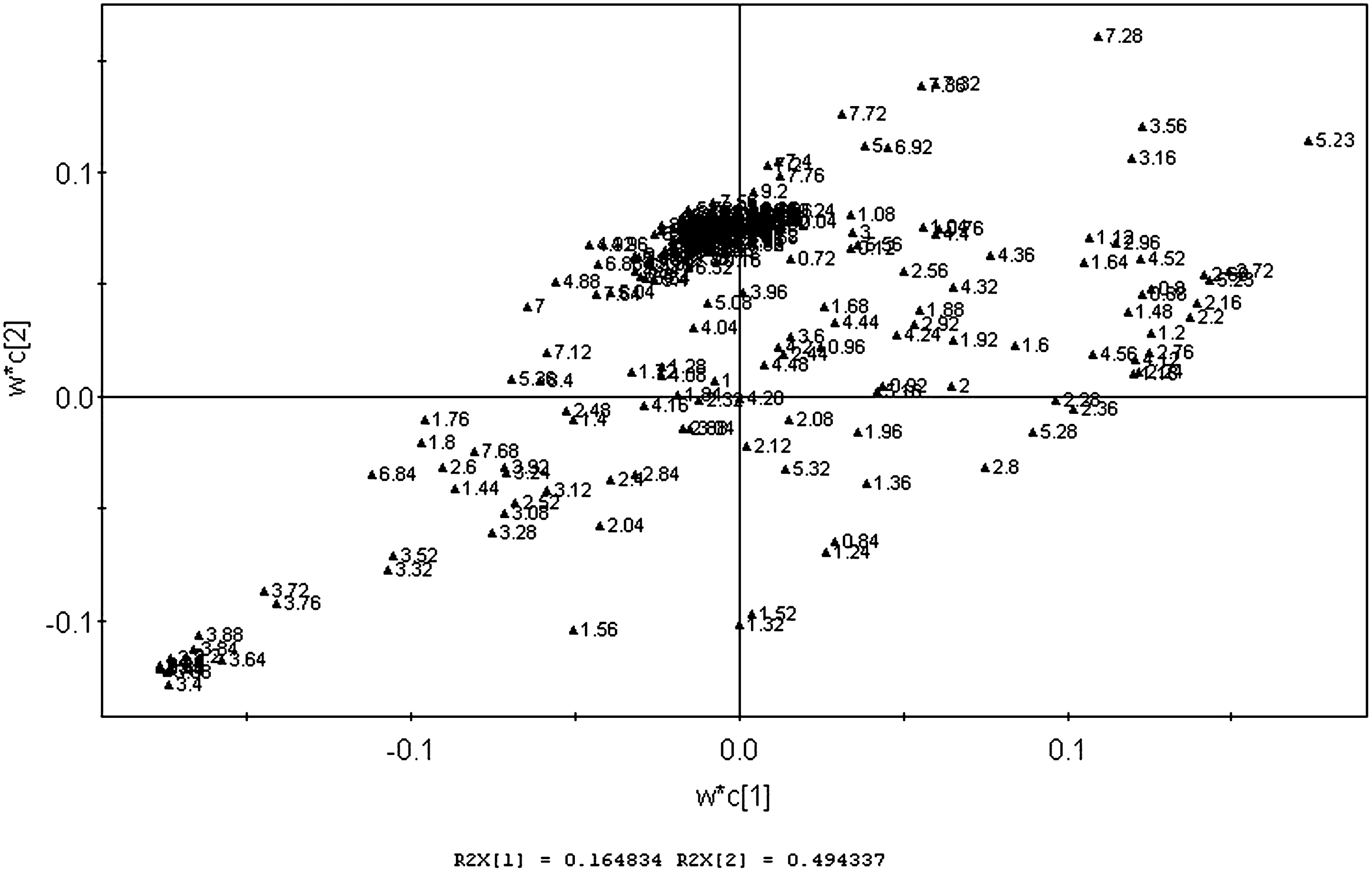
PLS-DA loading plots of plasma of control and model group.
A, QRJD group; B, LXHX group; C, control (sham surgery group); M, model.
↑=higher level of metabolites, ↓=lower level of metabolites.
The metabolic effects of CHM formula intervention
The PLS-DA score plot was used to investigate the metabolic effects of intervention with the two CHM formulas. As shown in Figure 4, the clusters representing the two treatment groups were located between the clusters representing the model and control, which demonstrated that the disturbed metabolic states of the CLP-induced model rats were in the process of returning to the normal state after CHM intervention. The score plot also showed that the QRJD-treated group cluster was closer to that of the control groups than the LXHX-treated group cluster, which demonstrates that QRJD has better therapeutic effects than LXHX on septic rats and is consistent with the clinical chemistry and mortality data.
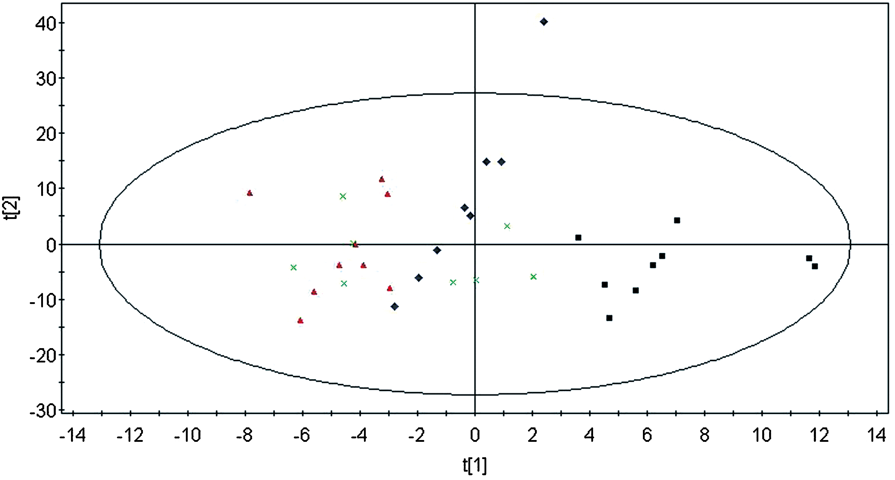
PLS-DA score plots obtained from plasma analysis of control (▲), model (■) and two treatment groups (QRJDF,♦; LXHXF,×) (R2X=0.915, R2Y=0.888, Q2=0.623).
PLS-DA loading plots were used to analyze which metabolites were regulated after the intervention. Treatment with either of the two CHM formulas attenuated the CLP-induced decreases in praline, taurine, lysine, arginine, isoleucine, creatine, choline, and trimethylamine-N-oxide (TMAO), and increases in alanine, aspartic acid, 3-hydroxylbutyrate, and acetoacetate, which suggests that these two CHM formulas regulate various metabolic networks. The plasma concentrations of lactate and threonine in septic rats were reduced by QRJD but not by LXHX, whereas the plasma concentration of valine in septic rats was increased only by LXHX. Lactate is one of the most important endogenous metabolites in several biochemical processes; it is produced by the anaerobic glycolysis of glucose because of the weak gluconeogenesis in muscle. Under normal circumstances, lactate is transported to the liver to regenerate glucose. The dysregulation of lactate demonstrated that the Cori cycle was also inhibited in the LXHX-treated group.
Conclusions
This study used 1H-NMR metabolomics technology to describe changes in the metabolic regulatory network in response to experimental sepsis in rats and CHM intervention. The potential metabolite biomarkers identified suggest that multiple metabolic pathways were disturbed in septic rats, as increased energy metabolism and fat mobilization, as well as disrupted amino acid metabolism, were observed. Metabolic profiling and biomarker analyses verified the therapeutic efficacy of two CHM formulas. These findings further our understanding of sepsis and the mechanisms of the QRJD and LXHX formulas. This work also confirms the feasibility of using the 1H-NMR metabolomics platforms to evaluate the therapeutic effects and mechanisms of Chinese herbal medicines.
Footnotes
Author Disclosure Statement
No competing financial interests exist.
References
Supplementary Material
Please find the following supplemental material available below.
For Open Access articles published under a Creative Commons License, all supplemental material carries the same license as the article it is associated with.
For non-Open Access articles published, all supplemental material carries a non-exclusive license, and permission requests for re-use of supplemental material or any part of supplemental material shall be sent directly to the copyright owner as specified in the copyright notice associated with the article.
