




Abstract

The COVID-19
Although a limited number of studies have reported direct identification of virus from human tissues based on viral RNA (Majumdar et al., 2022), viral proteins by immunohistochemistry (IHC) (Roden et al., 2021), or electron microscopy (Bullock et al., 2022), direct evidence of viral proteins from host tissues using mass spectrometry has not been reported thus far. In this study, using mass spectrometry, we sought to detect SARS-CoV-2 virus-encoded proteins from lung tissues obtained at autopsy from patients confirmed to have COVID-19. All samples were collected after prior approval from Mayo Clinic's Institutional Review Board and a written informed consent from the decedent or a family member.
All patients were tested for SARS-CoV-2 virus using the routine RT-PCR testing on nasopharyngeal or oropharyngeal swab samples. Formalin-fixed paraffin-embedded (FFPE) lung tissue sections from six patients were subjected to bottom-up proteomic analysis which included protein extraction followed by overnight trypsin digestion (Fig. 1A). Digested peptides were fractionated using basic pH reversed-phase liquid chromatography followed by LC-MS/MS analysis on a high-resolution mass spectrometer coupled to a nanoLC platform. Database searching was performed against a combined database of human and SARS-CoV-2 viral protein sequences using Sequest search engine. Detailed methods employed for mass spectrometry analysis are provided in Supplementary File S1.
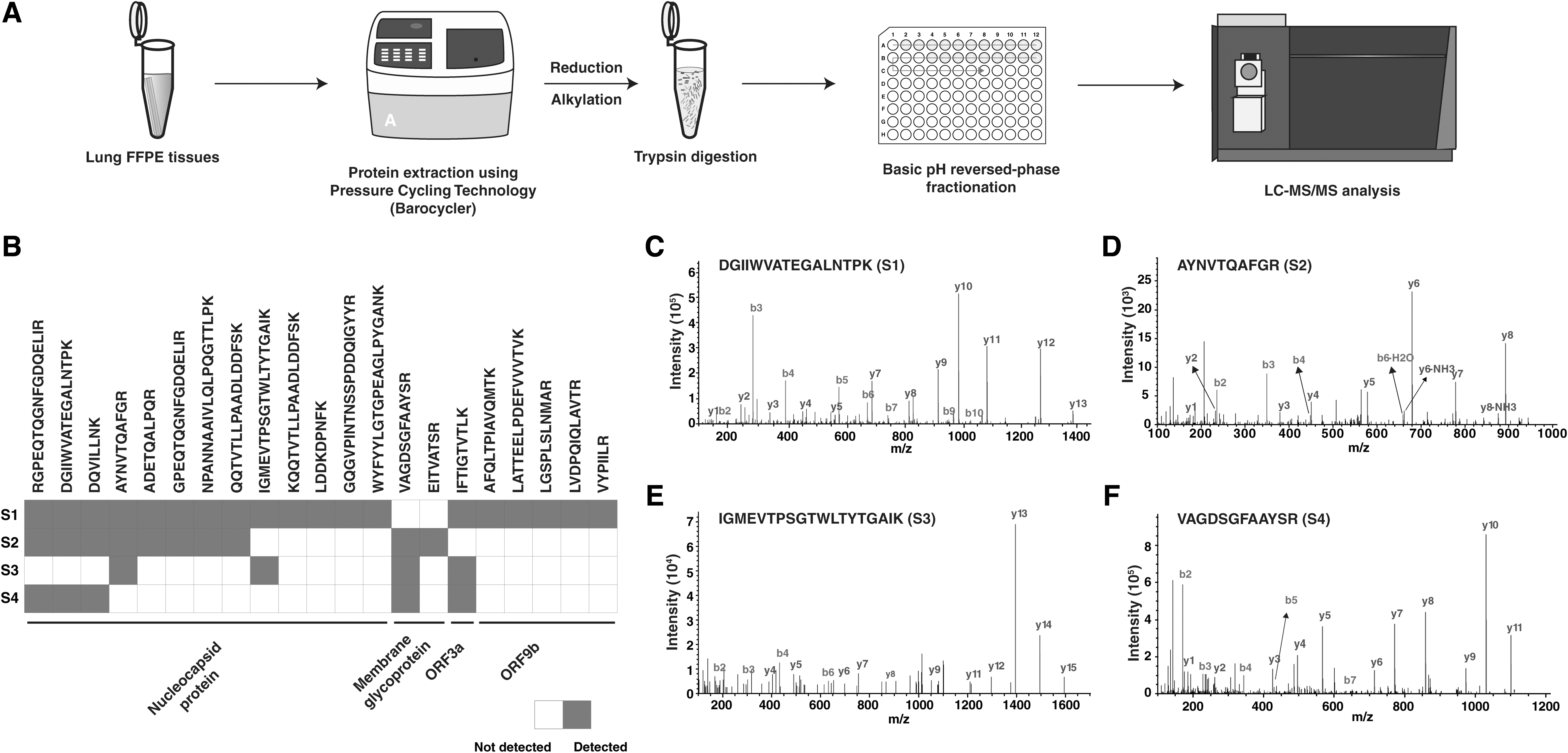
SARS-CoV-2 detection in human lung FFPE tissues using mass spectrometry.
From the six FFPE samples tested, we identified a median of nearly 7000 proteins, and nearly 54,000 peptides (Supplementary Table S1). SARS-CoV-2 peptides derived from four different viral proteins (nucleocapsid, membrane glycoprotein, ORF3a, and ORF9b) were identified from four out of six autopsy tissue samples (Fig. 1B). Nucleocapsid protein was identified from all four positive samples owing to its high expression levels in SARS-CoV-2. Representative tandem mass (MS/MS) spectra of viral peptides identified from each sample were shown in Figure 1C–F that includes DGIIWVATEGALNTPK, AYNVTQAFGR, IGMEVTPSGTWLTYTGAIK peptides from nucleocapsid protein and VAGDSGFAAYSR from membrane glycoprotein. The FFPE samples were also tested for presence of viral RNA using RNAScope in situ hybridization technology and ddPCR (Supplementary Table S2).
Of the four samples that were positive for SARS-CoV-2-encoded peptides by mass spectrometry, three samples were positive by both RNAScope and ddPCR. Notably, although several peptides from SARS-CoV-2, that is, three peptides from nucleocapsid protein and one peptide each from membrane glycoprotein and ORF3a, were identified from the fourth sample (S4), it was negative by RNAscope and contained barely detectable viral RNA copies by ddPCR. We did not identify SARS-CoV-2 derived peptides from two of the samples (S5 and S6)—these two samples also tested negative by RNAscope and barely detectable viral RNA as determined by ddPCR.
To our knowledge, this is the first demonstration of peptides detected from lung tissues from patients with COVID-19, although reports employing mass spectrometry-based platforms for detecting viral proteins from nasopharyngeal swabs (Mangalaparthi et al., 2021) and urine samples (Chavan et al., 2021) have been published. Importantly, viral peptides identified using limited material from FFPE tissues as shown in this study might enable opportunities for advancing our understanding of viral infections beyond SARS-CoV-2. Overall, this study and findings highlight the potential of mass spectrometry as a promising method for identification of viral proteins including SARS-CoV-2 or other pathogens from clinical tissue samples.
Footnotes
Authors' Contributions
A.P. and A.C.R. designed the study. K.K.M and S.S. carried out the sample preparation. K.K.M. carried out the LC-MS/MS analysis and data analysis. K.G. and K.K.M. prepared the figures. B.R.K. supervised ddPCR experiments. K.K.M., A.C.R., J.J.G., B.R.K., and A.P. performed data analysis and interpretation. A.P. edited, critically read, and revised the manuscript. All authors made a critical intellectual contribution and have read and approved the final manuscript.
Author Disclosure Statement
The authors declare they have no conflicting financial interests.
Funding Information
This study was supported by a grant from NCI (P30CA015083) and the philanthropy and generosity of Eric and Wendy Schmidt.

Abbreviations Used
References
Supplementary Material
Please find the following supplemental material available below.
For Open Access articles published under a Creative Commons License, all supplemental material carries the same license as the article it is associated with.
For non-Open Access articles published, all supplemental material carries a non-exclusive license, and permission requests for re-use of supplemental material or any part of supplemental material shall be sent directly to the copyright owner as specified in the copyright notice associated with the article.
