




Abstract
Introduction
Recently, we established a colony of descendants of Mexican hairless dogs, which show age-related changes in skin color and histologic features. 18 Hairless dogs more than 1.5 y old develop a spotty pigmentation similar to that of senile lentigines in humans. As reported in another study, 19 we found that successive UVB irradiation could artificially induce considerable spotty pigmentation in the skin of the hairless dog. Additionally, we reported dermatological changes in spotty pigmentation after QRL irradiation. 19
The purpose of the present study was to clarify the effects of UVA irradiation on the sites of QRL irradiation, using UVB-induced spotty pigmentation in the skin of the hairless dog.
Materials and Methods
Hairless dog
A 4-y-old female N4 hairless hybrid cross between a male N3 hybrid and a female beagle was studied. Because hairless dogs are rare laboratory animals, only one dog was used for this study. The hairless dog was housed alone in a stainless steel cage (90 × 90 × 90 cm) in an animal room controlled at 25 ± 2°C and 50 ± 5% relative humidity with 10–15 exchanges of 100% fresh air per hour and a 12-hour light (7:00 AM to 7:00 PM), 12-hour dark (7:00 PM to 7:00 AM) cycle. The dog was fed a commercial dry dog food (TC-2, Aixia Corporation, Tokyo, Japan).
All procedures involving animals were approved by the Animal Use Committee of National Institutes of Natural Sciences (NINS) and followed the guidelines of animal care and experiments of the NINS.
UVB irradiation regimen
The hairless dog was irradiated with artificial UVB light (290 to 320 nm; Atoh Kagaku Co., Ltd., Tokyo) for 6 consecutive months. The dosage of UVB was 180 kJ/m2/d. During this procedure, the dog was placed in a wire cage (85 × 95 × 75 cm) with 2 cm mesh.
QRL irradiation regimen
Six months after completion of the UVB irradiation program, QRL irradiation was performed on UVB-induced spotty pigmentation in the dorsal skin of the hairless dog. A QRL (Model RD-1200; Spectrum Medical Technologies Inc., Lexington, MA, USA) was used to deliver 5.0 J/cm2 in single 25 nsec duration full width at half maximum pulses at a wavelength of 694 nm. The collimated laser beam struck a circular aperture, 5.0 mm in diameter, held in contact with the skin of the dog. The laser pulse energy passing through the aperture was measured by a laser pulse energy meter (Model DGX, Ophir Co., Jerusalem, Israel).
The hairless dog was anesthetized with medetomidine (Domitol, Meiji Seiyaku Co., Ltd., Tokyo) prior to QRL exposure. The spotty pigmentation in the skin was then irradiated with a dose of radiant exposure (5.0 J/cm2).
UVA irradiation regimen
After 7 d of depigmentation with QRL irradiation, the skin of the dorsum of the hairless dog was divided into two areas. For the QRL + UVA–treated sites, the left side of the dorsum of the dog was irradiated daily with UVA light (320–400 nm; Atoh Kagaku Co., Ltd., Tokyo) for 5 consecutive days. The dose of UVA was 21 kJ/m2. For the QRL-only treatment, the right side of the dorsum remained intact and was used as a control. During this procedure, the dog was placed in a wire cage (85 × 95 × 75 cm) with 2 cm mesh. During UVA irradiation, the right dorsum was covered with a film to protect it from UVA.
Gross observation
Gross observation was performed before QRL irradiation and daily for 16 wk after completion of the UVA irradiation program.
Histological examination of the skin
Two tissue specimens were obtained from both the QRL + UVA–treated and QRL-treated sites of the dorsal skin of the hairless dog, using a 6-mm biopsy punch (Nagatoishi Co., Ltd., Tokyo) under general anesthesia with medetomidine (Domitol; Meiji Seiyaku Co., Ltd., Tokyo), 1 day before QRL irradiation and 1 and 7 d after QRL irradiation, and additionally, 2, 4, 6, 8, 10, 12, 14, and 16 wk after completion of the UVA irradiation program.
Half the skin specimens were rinsed in 0.1 M phosphate buffer (pH 7.4) and incubated in 2 N sodium bromide for 2 h at 37°C. The epidermal sheets separated from the dermis were fixed in cold 10% neutral buffered formalin for 30 minutes, washed twice with 0.1 M phosphate buffer (pH 7.4), and incubated in 3.1 mM dihydroxyphenylalanine (DOPA) in 0.1 M phosphate buffer (pH 7.4) for 5 h. The findings of DOPA-positive melanocytes were observed on each specimen under light microscopy, and the degree of pigmentation was graded as negligible (−), very slight (±), slight (+), moderate (++), and marked (+++).
The remaining skin specimens were fixed in 10% neutral buffered formalin, and 4 μm paraffin sections were stained with hematoxylin and eosin and toluidine blue and by Fontana-Masson's method.
Results
Gross findings
Immediately after QRL irradiation with 5.0 J/cm2, spotty pigmentation in the skin of the hairless dog developed white, macule, and/or erythematous reactions, precisely equal in size to the aperture plate diameter of 5.0 mm.
One week after QRL irradiation, the irradiated areas showed re-epithelialization and complete depigmentation as white spots. Two weeks after completion of the UVA irradiation program, at the QRL + UVA–treated sites, pigmentation began from around the QRL-treated sites and/or the remaining hair follicles. Infinitesimal pigmentation also appeared early in the depigmentory spots treated with QRL (Fig. 1). Thereafter, the pigmentation observed at the QRL + UVA–treated sites became more prominent than that at the QRL-treated sites and in skin of normal color in the hairless dog.

Gross appearance of the QRL + UVA–treated sites of a hairless dog 2 wk after completion of the UVA irradiation program. Pigmentation begins from the surroundings of the QRL-treated sites. QRL, Q-switched ruby laser; UVA, ultraviolet A.
Six weeks after completion of the UVA irradiation program, a large number of the QRL + UVA–treated sites demonstrated a recurrence of hyperpigmentation that seemed to be arising from the remaining hair follicles. The recurrence of hyperpigmentation occurred considerably more often at the QRL + UVA–treated sites than at QRL-treated sites. From 10 to 16 weeks after completion of the UVA irradiation program, recurrent hyperpigmentation became more predominant.
In contrast, at the QRL-treated sites, pigmentation developed from 6 wk after completion of the UVA irradiation program, and skin color tended to return to normal. A small number of QRL-treated sites exhibited a recurrence of hyperpigmentation (spotty pigmentation) from 10 wk after completion of the UVA irradiation program.
Observations of DOPA-positive melanocytes
The findings of DOPA-positive melanocytes are shown in Table 1. One day before QRL irradiation, the hyperpigmented skin showed a large number of DOPA-positive melanocytes and heavy deposition of melanin granules. One week after QRL irradiation, depigmented epidermal sheets contained some melanocytes, in which melanin granules were completely destroyed. The depigmented melonocytes were poorly outlined (Fig. 2). A small number of DOPA-positive melanocytes with stout dendrites remained in the bottom of the hair follicles (Fig. 3).

Epidermal sheet at the QRL-treated sites of a hairless dog 1 wk after QRL irradiation. Depigmented melanocytes are seen. Dihydroxyphenylalanine (DOPA) reaction, × 350 magnification.
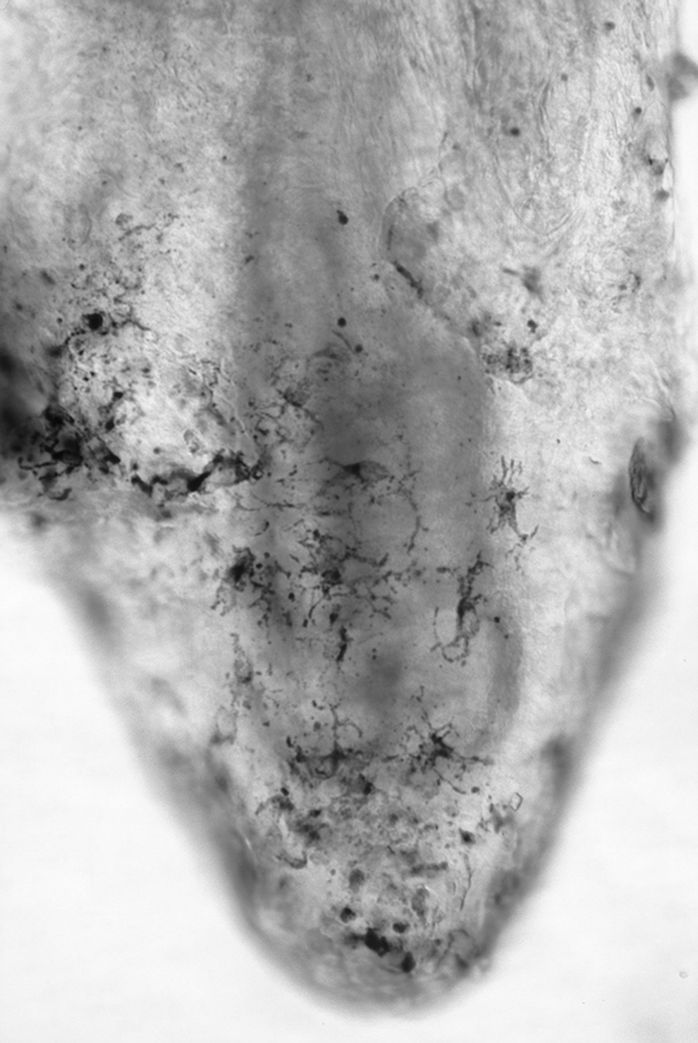
Epidermal sheet at the QRL-treated sites of a hairless dog 1 wk after QRL irradiation. DOPA-positive melanocytes with stout dendrites in the bottom of the hair follicle are seen. DOPA reaction, × 350 magnification.
The degree of pigmentation: negligible (−), very slight (±), slight (+), moderate (++), and marked (+++).
At the QRL + UVA–treated sites, the number of DOPA-positive melanocytes increased significantly from 2 wk after completion of the UVA irradiation program. Then, the number peaked at 4–6 wk and these melanocytes had well-developed dendrites as compared with those at the QRL-treated sites (Figs. 4, 5). Thereafter, the number of DOPA-positive melanocytes tended to decrease gradually and recovered to the number observed in the QRL-treated skin by 12 wk after completion of the UVA irradiation program. The DOPA-positive melanocytes gradually lost their stout dendrites and changed into round melanocytes (Fig. 6).
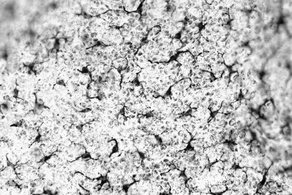
Epidermal sheet at the QRL + UVA–treated sites of a hairless dog 6 wk after completion of the UVA irradiation program. A large number of DOPA-positive melanocytes with well-developed dendrites are seen. DOPA reaction, × 350 magnification.
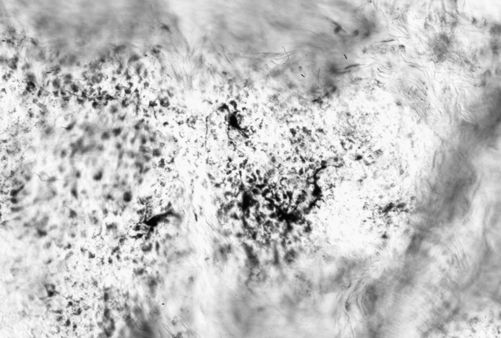
Epidermal sheet at the QRL-treated sites of a hairless dog 6 wk after QRL irradiation. A small number of DOPA-positive melanocytes are seen. DOPA reaction, × 350 magnification.
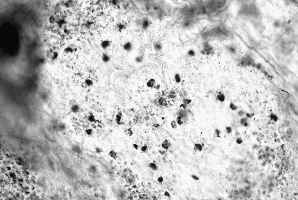
Epidermal sheet at the QRL + UVA–treated sites of a hairless dog 12 wk after completion of the UVA irradiation program. Round-shaped melanocytes without dendrites are seen. DOPA reaction, × 350 magnification.
At the QRL-treated sites, the DOPA-positive melanocytes with stout dendrites migrated from the surroundings of the depigmented sites 2 wk after completion of UVA irradiation. From 6 wk after completion of the program, there was a return to a normal distribution of the DOPA-positive melanocytes.
Histopathological findings
One day before QRL irradiation, the epidermis of the hairless dog was flat and thin, and consisted of two to three cell layers. The skin had a heavy deposition of melanin granules in the stratum basale, spinosum, and corneum. There were no melanin granules in the dermis (Fig. 7). Histopathologically, the spotty hyperpigmentation of the skin in the hairless dog seemed to be equivalent to a flat type of senile lentigo in humans.
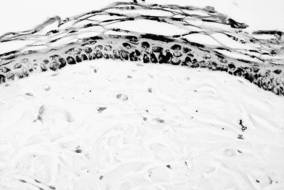
Skin with spotty pigmentation of a hairless dog 1 d before QRL irradiation. Heavy deposits of melanin granules in the stratum basale, spinosum, and corneum are seen. Fontana-Masson's (FM), × 350 magnification.
One day after QRL irradiation with 5.0 J/cm2, the depigmented sites showed epidermal excoriation and vacuolation of the pigmented cells. Disarray of keratinocytes and a separation formed at the dermoepidermal junction were also found. In both the epidermis and the dermis, a large number of erythrocytes and inflammatory cells were observed. The remaining epidermis exhibited intracellular and intercellular edema and the epidermal cells contained no melanin granules.
One week after QRL irradiation, all exposure sites were completely re-epithelialized from the surroundings of the QRL-treated sites and the surviving pilosebaceous units. The repaired skin had a thick and flat epidermis (epidermal hyperplasia) without melanin granules. In the dermis, vasodilation and inflammatory cell infiltration remained unchanged (Fig. 8).
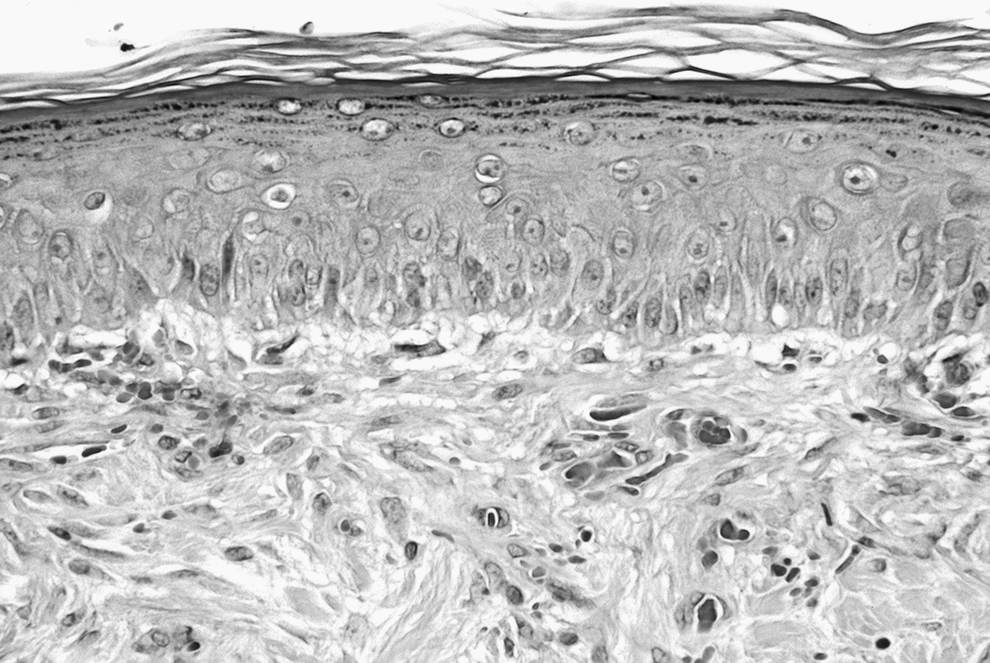
Skin of a hairless dog 1 wk after QRL irradiation. Re-epithlialized skin is seen. A thick and flat epidermis without melanin granules. Hematoxylin and eosin (HE), × 350 magnification.
Two weeks after completion of the UVA irradiation program, at the QRL + UVA–treated sites, repigmentation developed in the stratum basale, and melanin granules generally aggregated in the apical portion of the epidermal cells as nuclear caps. In the dermis, histological changes due to QRL treatment resolved and abnormal alterations such as inflammatory reactions were not observed after the UVA irradiation.
Four weeks after completion of the UVA irradiation program, in some repigmented portions of the QRL + UVA–treated sites, abundant melanin granules, released from damaged epidermal cells into the dermis, were phagocytized by dermal macrophages. The QRL-treated sites had no melanin granules in the regenerated epidermis.
From 6 weeks after completion of the UVA irradiation program, the QRL + UVA-treated sites showed a recurrence of hyperpigmentation. On the other hand, the QRL-treated sites began to show a normal redistribution of melanin granules over the stratum basale.
From 8 weeks after completion of the UVA irradiation program, in the portions with a recurrence of hyperpigmentation at the QRL + UVA–treated sites, deposits of melanin granules became more notable in the epidermis, and the pigments aggregated in the bottom portion of the nucleus of each epidermal cell (Fig. 9). Beneath the hyperpigmented epidermis, a large number of round cells producing excessive melanin granules were found in the lower portions of hair follicles. In the dermis, pigmented cells including many melanin granules were observed (Fig. 10). After QRL treatment for spotty hyperpigmentation, in the depigmented sites, follicular melanocytes seemed to produce excessive melanin granules and to transmit them to the epidermis via melanosomes and/or dermal melanocytes.
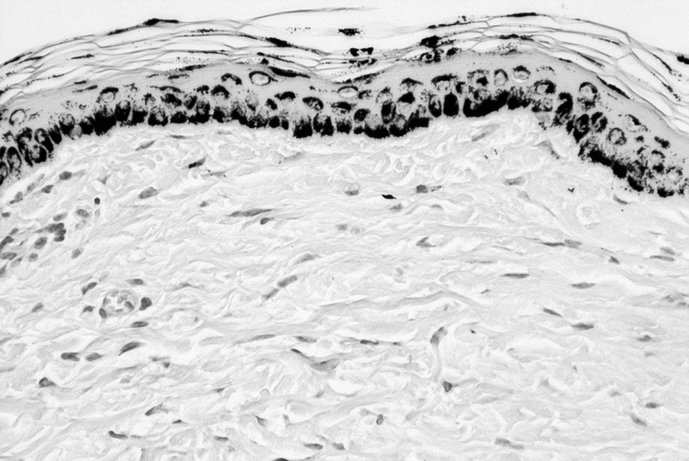
Skin of the QRL + UVA–treated sites of a hairless dog 8 wk after completion of the UVA irradiation program. This area shows a recurrence of hyperpigmentation. Melanin granules aggregate at the bottom of the nucleus of each epidermal cell. FM, × 350 magnification.
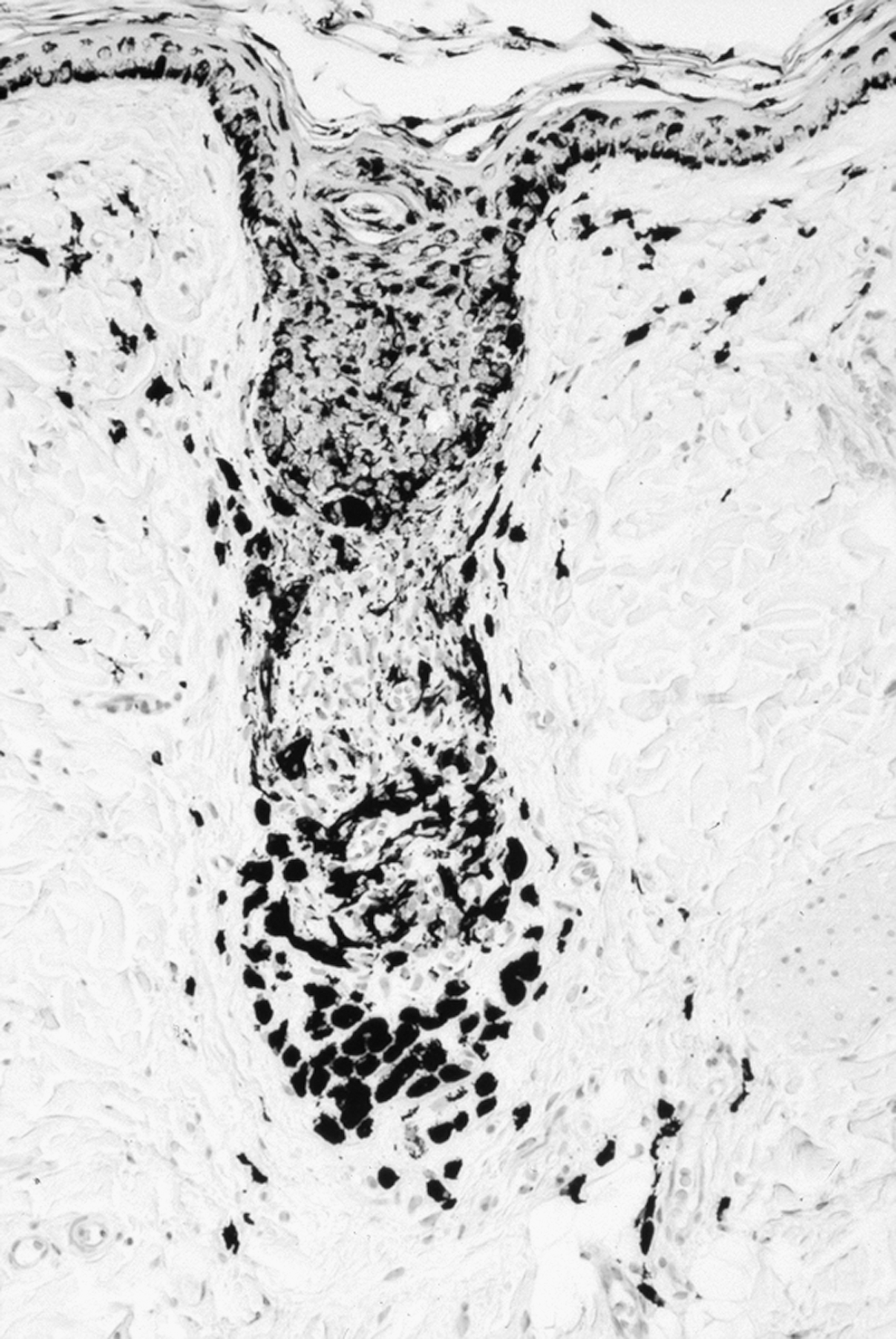
Skin of the QRL + UVA–treated sites of a hairless dog 8 wk after completion of the UVA irradiation program. A large number of round cells producing an excess of melanin granules in the lower portion of a hair follicle is seen. FM, × 170 magnification.
From 10 wk after completion of the UVA irradiation program, the QRL + UVA–treated sites showed a recurrence of spotty hyperpigmentation. There were few differences in the deposition of melanin granules between these lesions and spotty hyperpigmentation before QRL irradiation. On the other hand, the QRL-treated sites exhibited a normal skin structure, except for a small number of recurrent hyperpigmented sites.
Discussion
The present study clarifies the deleterious effects of UVA irradiation on sites treated with QRL irradiation, using UVB-induced spotty pigmentation in the skin of the hairless dog. Notably, it demonstrates that UVA irradiation causes a recurrence of spotty pigmentation with high frequency. Although the epithelialization of the hairless dog was slightly delayed, as compared with that of guinea pigs 3,20 and humans, 21 no damage from QRL irradiation remained.
Two weeks after completion of the UVA irradiation program, the QRL + UVA–treated sites had obviously developed visual pigmentation. These findings revealed that UVA irradiation produced hyperpigmentation at the depigmented sites treated with QRL irradiation.
Six weeks after completion of the UVA irradiation program, marked spotty pigmentation reappeared at the QRL + UVA-treated sites. Empirically, it has long been known that depigmented sites should be protected from solar exposure after the removal of pigmented lesions. In this study, our experimental results revealed that UVA irradiation allowing for greater dermal penetration had a deleterious influence causing a recurrence of spotty pigmentation on the QRL-treated sites. The gross observations at the QRL-treated sites were in accord with those reported in our other study. 19 The majority of depigmented lesions returned to a normal skin color in the hairless dog. These results suggested that QRL irradiation should be a safe and excellent method for the treatment of spotty pigmentation.
DOPA-positive epidermal sheets demonstrated that spotty pigmented sites of the hairless dog possessed numerous melanocytes and many clusters of melanin granules. These findings were consistent with those in senile lentigines in humans. 21
One week after QRL irradiation, visually depigmented sites had a considerably larger number of DOPA-positive melanocytes, showing that melanin granules were completely destroyed by QRL irradiation. These findings revealed that vacuolated melanocytes in the pigmented lesions survived after QRL irradiation. In addition, the melanocytes with stout dendrites at the bottom of hair follicles suggested that an insufficient dose of QRL irradiation may increase their activities.
From 2 to 6 wk after completion of the UVA irradiation program, the number of DOPA-positive melanocytes apparently increased at the QRL + UVA–treated sites and the melanocytes showed an increase in activity. These observations were similar to responses of the skin to sunlight in hairless dogs. 22,23 These findings revealed that UVA irradiation stimulated melanocytes in the depigmented skin, leading to a recurrence of spotty pigmentation.
Considering the findings at the QRL-treated sites, after UVA irradiation, the redistribution of melanocytes was speculated to occur as follows: 1) accompanied by re-epithelialization, epidermal melanocytes migrate from the surroundings of QRL-treated sites, 2) hair follicular melanocytes migrate from the follicular infundibulum into the regenerated epidermis, and 3) surviving melanocytes in the epidermis are activated and melanogenesis also recovers from degranulation. Similar to the gross observations, the epidermal split-skin preparation of the hairless dog revealed that it was important to protect the depigmented skin from solar exposure, especially for 6 wk after the treatment with QRL irradiation.
At the QRL-treated sites, an increase in the number of DOPA-positive melanocytes was noted 2 wk after QRL irradiation. Additionally, these melanocytes had a dendritic shape. DOPA-positive melanocytes that were repopulating in the hypopigmented areas seemed to be derived from surviving hair follicles as well as the periphery of the lesion areas. These findings were in agreement with an experiment performed in normal human skin, 21 and demonstrated that QRL irradiation transiently increased the activities of melanocytes and melanogenesis.
One day before QRL irradiation, the histopathological findings of spotty pigmentation of the hairless dog accorded with those reported in our other study. 18,24 Except for hairless dogs, there are no laboratory animals that have such dermatological characteristics as UVB-induced spotty pigmentation.
One day after QRL irradiation, the pigmented lesions of the hairless dog were completely devoid of melanin granules. The degeneration of the skin was severe, and these changes were in accord with those reported in our other study. 19 The effects of QRL irradiation on the skin were more severe in spotty pigmented than in normal skin of the hairless dogs.
One week after QRL irradiation, although re-epithelialization was completed, no melanin granules were observed in the epidermis or the dermis. The damage after QRL irradiation was limited to the regenerating dermis.
Two weeks after completion of the UVA irradiation program, the irradiation had apparently provoked repigmentation in the epidermis. Melanin granules deposited in the apical portion of the epidermal cells. This finding was consistent with the pigmenting responses observed in the skin of hairless dogs after solar exposure. 22,23
Four weeks after completion of the UVA irradiation program, dermal melanophages were found at the QRL + UVA–treated sites. These findings revealed that melanin granules destroyed by QRL irradiation were removed by dermal macrophages.
Six weeks after completion of the UVA irradiation program, at the QRL + UVA–treated sites, heavy deposition of melanin granules recurred in the stratum basale, spinosum, and corneum. These findings revealed that UVA irradiation promoted the deposition of melanin granules and hastened the start of hyperpigmentation as compared with the QRL- treated sites. At the QRL-treated sites, melanin granules began to be observed in the thick repaired epidermis. After QRL irradiation, there were no differences in the time of reappearance of melanin granules between the spotty pigmented and normal skin. 18,24
The aggregation of melanin in the portion of QRL + UVA–treated sites where spotty pigmentation recurred apparently differed from any melanin pigmentation in the skin of hairless dogs induced by sunlight, i.e., UVA, UVB, and UVB + UVA irradiation. The hypermelanized cells in the hair follicular infundibulum and the melanin aggregation in the bottom portion of the nucleus of each epidermal cell were characteristic findings observed in the areas where spotty pigmentation recurred. These histopathological findings may provide an important clue as to the mechanism of development of pigmented lesions such as senile lentigines.
Histopathological results revealed that UVA irradiation accelerated the reappearance and excessive deposition of melanin granules in the repaired epidermis, after the depigmentation with QRL irradiation. Therefore, after QRL irradiation, the depigmented sites should be protected with shading and/or sunscreens.
Conclusion
Animal experimentation using UVB-induced spotty pigmentation in the hairless dog demonstrated that UVA irradiation had deleterious effects on the QRL-treated sites and caused a recurrence of spotty pigmentation. Although this study was performed using a laboratory animal, hairless dogs are useful laboratory animals for evaluating the gross and histopathological changes in pigmented lesions treated with QRL irradiation. Our results showed that patients undergoing QRL irradiation should avoid exposure of their treated skin to sunlight. Because long wavelength UVA penetrates further into the skin, UVA irradiation has the potential to activate the remaining melanocytes after treatment of hyperpigmentation.
Footnotes
Disclosure Statement
No competing financial interests exist.