
Abstract
Introduction
Biochemical alterations in salivary gland composition and salivary gland hypofunction have been associated with high fasting blood glucose concentrations (hyperglycemia) and neuropathy. 4 –8 Diabetes mellitus is a group of metabolic diseases characterized by hyperglycemia resulting from defects in insulin secretion, insulin action, or both. The chronic hyperglycemia of diabetes is associated with long-term damage, dysfunction, and failure of various organs. 9 Moreover, patients with type 1 and 2 diabetes may report xerostomia and hyposalivation, along with other clinical signs and symptoms of salivary gland dysfunction. 5,6,8,10
Our research group has been studying the effects of diabetes on the salivary glands of rats. 4,11 –15 The data collected suggest that hyperglycemia increases the activity of phosphofructokinase-1 (PFK-1), 12 catalase, and peroxidase enzymes 4,15 and alters the activity of hexokinase 13 and the deposition of extracellular matrix proteins in the rat parotid gland, possibly through augmentation of transforming growth factor beta 2 expression and signaling. 14 Moreover, the diabetic state influences glycogen metabolism in the submandibular and parotid salivary glands of rats. 11
Other studies in the literature have found that hyposalivation in animals with diabetes (streptozotocin- and alloxan-induced diabetes) yields the cytoplasmic accumulation of lipid droplets in terminal secretory end pieces and intercalated duct cells of the three major salivary glands. 16 –19 It has been correlated with high blood glucose concentration. 19 In addition, ionic alterations have also been related to diabetes, for example, low magnesium and zinc concentrations. 20 –22 However, in the salivary glands, high calcium, magnesium, and potassium and low zinc concentration have been described in patients with diabetes and has been related to their oral cavity problems. 23,24
In laser phototherapy (LPT) a red or near-infrared light, in low-intensity range, is used. Our previous studies demonstrated that infrared LPT, in doses of 4 and 8 J/cm2, can stimulate salivary glands of rats 25 and increase the total protein concentration in the parotid glands. 26 Moreover, in rats with streptozotocin-induced diabetes, in the same conditions of the present study, we observed a decrease in catalase activity in the salivary glands after laser irradiation of 5 and 20 J/cm2. 15
The parotid is an exocrine gland, with structural and functional similarities to the exocrine pancreas, but an endocrine function of the salivary glands related to glucose metabolism has been reported. 27 –29 In addition, some authors consider the parotid and submandibular glands to be extrapancreatic sources of a protein-like insulin in mice. 30 Therefore, it is important to investigate not only the consequences of diabetes on salivary glands, but also the effect of LPT upon this organ, which may lead to the study of other organs directly related to the glucose metabolism, such as the pancreas and liver.
The purpose of the present study was to analyze the effect of laser irradiation on the salivary glands of rats with diabetes using histological and ionic investigations. The glycemic state of the rats was also investigated, once it had been directly associated with complications of diabetes.
Material and Methods
Experimental design, diabetes induction, and blood glucose concentration analyses
The Bioethics Committee of Animals, School of Dentistry, University of São Paulo approved the experimental protocol used in this investigation. One hundred twenty Wistar female rats weighing approximately 190 g were used. The animals were caged individually and had free access to water and solid food. The care and handling of the animals throughout the study were conducted in accordance with the principles for animal experimentation established by the Brazilian College of Animal Care.
One hundred twenty female rats were randomly divided into eight groups: four groups with diabetes and four without. Diabetes was induced using a single intraperitoneal injection of 60 mg/Kg of streptozotocin (STZ) (Sigma Chemical Co., St. Louis, MO) dissolved in 0.1 M sodium citrate buffer pH 4.5. Animals without diabetes were injected with citrate buffer alone. The development of diabetes was confirmed using blood glucose analysis using a blood glucose meter (Accu Chek Advantage, Roche Diagnostics, Mannheim, Germany) 72 h after the STZ injection after and overnight fast. Rats that had a blood glucose level higher than 14 mM (250 mg/100 mL) were considered to have diabetes.
Twenty-nine days after induction if diabetes, the animals were anesthetized by an intraperitoneal injection of chloral hydrate (400 mg/kg of body weight (bw)) and sodium diethylbarbiturate (50 mg/kg bw). The areas corresponding to the parotid and submandibular glands were shaved and demarked before irradiation or simulation. The animals included in the laser groups received laser irradiation, and those in the nonlaser groups received a simulation procedure (see below).
On the day after irradiation or simulation, the rats were euthanized. The euthanasia procedures were always performed in the morning (9:00–11:00 a.m.) to minimize the effects of circadian rhythms. The parotid and submandibular glands were then removed from 96 rats, clamped between aluminum tongs, precooled in dry ice, and stored at −80°C until being used for ionic analyses. For histological assays, the glands of 24 rats were fixed as soon being removed. The consumption of food and water was monitored during the experimental period through weekly weighing of food and diary analyses of the water bottle.
The blood glucose of fed animals was tested immediately before, and after the radiation or simulation. The animals that had fasted overnight had their blood glucose tested on the diagnosis and euthanasia days.
Laser irradiation
A semiconductor diode laser (Quantum, Ecco Fibras, São Paulo, Brazil), 660-nm wavelength and input power of 100 mW, from the Special Laboratory of Laser in Dentistry was used. The laser was applied to each parotid gland separately and both submandibular glands together; that is, three salivary gland areas of approximately 1.1 cm2 were irradiated.
For the irradiated groups, the laser irradiation was performed in continuous wave mode; the laser beam spot was 0.017 cm2; the doses were 5 (group D5 (with diabetes) and C5 (without diabetes), 10 (D10 and C10), and 20 J/cm2 (D20 and C20); and the times of laser irradiation were 1 s for groups D5 and C5, 2 s for groups D10 and C10, and 4 s for groups D20 and C20. Groups D0 and C0 received only simulated irradiation. 15
The hand-held wand was in skin contact mode, transcutaneously, and was positioned perpendicularly. Sixty-four points were necessary to evenly cover each gland area of 1.13 cm2 (1.1304 ÷ 0.01766 = 64.01 points), according to Simoes, et al. 16 A single irradiation was performed.
Because of a laser light loss of 10%, compared with the data shown on the equipment display, the actual output power was measured using a power meter (Coherent Molectron, Santa Clara, CA). To adjust the data, taking into account the above-mentioned loss, in addition to any eventual difference in fractions of seconds when performing the irradiation exposure for different groups, the calculations were made according to Simões et al. 15
Morphological analysis
The glands were fixed in Methacarn solution (60% methanol, 30% chloroform, 10% acetic acid) at 4°C for 3 h, dehydrated, and embedded in Paraffin. Sections 5 μm thick were placed on poly-y-lisine–coated slides and stained with hematoxylin and eosin. The slides were observed under an Aristoplan (Leitz, Wetzlar, Germany) microscope.
Ionic analysis
Calcium (Ca2+), magnesium (Mg2+), potassium (K+), sodium (Na+), zinc (Zn2+), and phosphorous (P−3) concentrations were analyzed using inductively coupled argon plasma with atomic emission spectrometry (Spectroflame Modula, Spectro Co., Kleve, Germany). Standard solutions were prepared from Titrisol standard solution (Merck, Darmstadt, Germany). Samples of submandibular and parotid salivary glands were treated with concentrated nitric acid and 30% hydrogen peroxide and heated until complete solubilization. The mixture was filtered, distilled water was added, and the mixture was kept at 4°C until it was used.
Statistical analysis
To perform the statistical analysis, the data were presented as means ± standard deviations. Based on the analyzed normal distribution of all parameters studied under different conditions, the analysis of variance and the Tukey test were performed, with the level of significance set at 5%.
Results
The body weight (initial and final) of the rats is shown in Table 1. During the experimental period, the groups without diabetes showed an average increase in body weight of 15% to 19%. Animals with diabetes, despite consuming more food and ingesting more water than the control group (32.51 ± 4.61 vs 13.61 ± 3.37 g/d; 171.68 ± 21.88 vs 29.23 ± 6.20 mL/d, respectively), did not show any significant increase in body weight during the experimental period (Table 1).
Different letters mean statistically different within the same column (p < 0.05).
Significantly different within the same row (p < 0.05).
After a single injection of STZ, the rats became diabetic, as evidenced by blood glucose concentration measured 72 h after induction (Fig. 1). The groups with diabetes had lower glucose concentrations on the day of sacrifice than on the day they had been diagnosed. The results were statistically significant (p < 0.05) for groups D5 and D20 (ranging from 358.97 ± 56.70 to 278.33 ± 87.98 and from 409.50 ± 124.41 to 231.80 ± 120.18 mg/100 mL, respectively) (Fig. 1). On the other hand, glycemia values obtained on the day of the sacrifice were higher for all groups without diabetes than on the day of the diagnosis (p < 0.05) (Fig. 1).

Blood glucose concentration from different groups on the days of diagnosis and sacrifice. Statistically significant difference considering the diagnosis and sacrifice glycemia in each laser dose. (p < 0.05) (10 < n < 12).
The glycemia, measured immediately before and after laser irradiation or simulation of laser irradiation, showed statistically significant variation for group D10, with the blood glucose concentration decreasing after irradiation from 527.91 ± 76.44 mg/dL to 456.82 ± 58.86 mg/100 mL (p < 0.05). Group C20 showed higher glycemia levels, ranging from 106.83 ± 19.89 mg/dL to 141.25 ± 36.48 mg/100 mL of blood, immediately after the light application (p < 0.05).
Parotid gland
For animals not receiving the irradiation, Na+ and P−3 levels were higher and Ca2+ levels lower for animals with diabetes than for those without (p < 0.05). Ca2+ concentration was also lower for D10 (p < 0.05). When 5 and 20 J/cm2 (D5 and D20) was applied, Ca2+ concentration was higher in animals with diabetes than in those without (p < 0.05) (Table 2). There are few and no conclusive studies reporting the effect of laser upon lipids.
Statistically significant considering the different conditions (with or without diabetes) and the same irradiation dose (p < 0.05).
Different letters means statistically significant difference considering the same condition (with or without diabetes) with different irradiation doses (p < 0.05).
Histological analysis showed an accumulation of lipid droplets in the cytoplasm of acinar cells of the parotid glands (Fig. 2), but when the laser was applied, this accumulation decreased for all doses (Fig. 2).
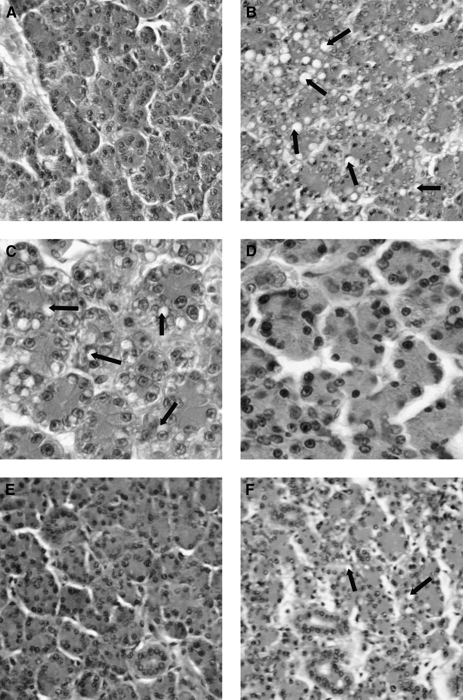
Morphology of rat parotid gland. Nonirradiated rats (
Submandibular gland
There were no differences in ionic concentrations for submandibular glands (Table 3). Moreover, no alterations were observed in the morphological analysis (data not shown).
Discussion
Our findings suggest that laser irradiation of salivary glands reduces the blood glucose concentration in rats with diabetes and the intracytoplasmic accumulation of lipid droplets in the secretory end pieces. These results amplify our previous report that showed that laser irradiation increased the salivary flow rate in rats 25 and suggested that LPT might be considered as an auxiliary therapy for salivary hypofunction. In addition, our data suggest that laser irradiation should be explored as an auxiliary therapy for control of diabetic complications because it can alter carbohydrate and lipid metabolism of rats with diabetes.
Although rats with diabetes consumed more food (polyphagia) and water (polydipsia) than the animals without, the classical symptoms of hyperglycemia, their average body weight, did not change after the experimental period (30 days). Contrarily, animals without diabetes increased their body weight by 15% to 19%. A possible explanation for the weight maintenance of rats with diabetes is insufficient insulin production due to administration of the β-cell toxin STZ and the consequent alterations in triglyceride storage in adipocytes and alteration in carbohydrate metabolism. 31
All rats injected with STZ developed diabetes, as indicated by the blood glucose analysis. On the day of sacrifice, the blood glucose concentration of the rats with diabetes was lower than the initial glycemia, measured 72 h after diabetes induction. The decrease in the final blood glucose concentration from initial levels was statistically significant for groups D5 and D20, suggesting that doses of 5 and 20 J/cm2 may alter this parameter, but no information regarding laser irradiation and blood glucose concentration has been published. The results obtained in this study led us to propose that salivary gland laser irradiation in rats with diabetes may be an auxiliary to glycemia control. Therefore, we suggest that this issue should be explored, because hyperglycemia is directly related to secondary complications of diabetes. 32 Such findings confirm some previous studies that related the endocrine function of salivary glands to glucose metabolism, 27,28,33 suggesting that parotid and submandibular glands can be extrapancreatic sources of a protein-like insulin. 30
Contrary to the results for the groups with diabetes, blood glucose concentration in animals without was higher on the day of sacrifice than initially. This increase in blood glucose concentration was expected, because the blood for the initial glycemia analysis was taken from the tail vein without anesthesia, whereas final glycemia levels were measured in animals submitted to the stress resulting from the anesthesia procedure and laser irradiation, which had been performed on the previous day. In addition, pentobarbital and chloral hydrate were related to the high blood glucose concentration because of interference in the carbohydrate metabolism. 34
Major mammalian salivary glands share many structural similarities, especially in the organization of secretory end pieces and the ductal system. The parotid gland is morphologically similar in rats and humans, containing serous secretory cells and a duct system consisting of intercalated, striated, and excretory ducts, but in the submandibular glands, male rats have a granular convoluted duct that is not present in humans and female rats, 35 complicating comparisons between them. Therefore, we chose female rats to avoid the duct variable and to allow comparisons between values obtained from both glands.
An accumulation of lipid droplets in the cytoplasm of the secretory end pieces of parotid, sublingual, and submandibular glands of animals with diabetes, as well as in the intercalary duct, have been reported in the literature 16 –19 and has been correlated with high blood glucose concentration. 19 In the present study, we observed the accumulation of lipid droplets only in the parotid gland, which decreased after laser irradiation, but ultrastructural analysis should be performed to confirm this, once few and inconclusive studies have focused this aspect. 36 –38
Chromophores in the respiratory chain of the mitochondria absorbed visible laser light, with an increase in adenosine triphosphate production and a decrease in pH; an alteration in Na+/K+, Ca2+, and cyclic adenosine monophosphate intracellular concentration; and consequently, proliferation and protein synthesis, which is related to tissue repair. 39 Moreover, LPT is able to act directly on Ca2+ channels in a plasmatic membrane, increasing its concentration in the cell. 40 –42 In this way, we performed some ionic analyses in the present study. Salivary parotid glands showed some alterations, mainly in Ca2+ concentration, which was lower in rats with diabetes than in those without, both without irradiation. However, with laser irradiation of 5 and 20 J/cm2, Ca2+ concentration increased, being similar to that of animals without diabetes. These results, together with the data from histological analysis, suggest that diabetes affect the parotid gland more than the submandibular gland, and in consequence, LPT had more effect in this gland.
Conclusion
The data from the present study suggest that red laser irradiation can be an auxiliary therapy for diabetic complications related to carbohydrate and lipid metabolisms.
Footnotes
Acknowledgments
The authors wish to thank Ms. Marinilce F. Santos from Cell and Molecular Biology Group, Laboratório Especial de Laser em Odontologia, which provided the laser equipment, and to the Brazilian National Council for Scientific and Technological Development and The State of São Paulo Research Foundation for their financial support.
Author Disclosure Statement
No competing financial interests exist.