
Abstract
Introduction
A micromechanical bond to enamel through the acid etching technique has been shown to be efficient; however, bonding to dentin has proved to be more difficult due to its higher protein and water concentration, making it much less receptive to adhesive systems. This has resulted in the development of different adhesive systems. 3
Parallel to the evolution of the adhesive systems, new technologies have been developed, such as the use of laser irradiation. Among the several types of laser, the Nd:YAG laser, with a wavelength of 1064 nm, has been shown to be effective in dentistry for dentin desensitization, 4 cleaning organic debris, sealing enamel pits and fissures, 5 removal and recontouring of soft tissue, coagulation, 6 treatment of incipient lesion, and caries removal. 7
The Er:YAG laser, with a wavelength of 2940 nm, has also been indicated for dental applications. This laser is highly absorbed by water (wavelength of 3000 nm) and by hydroxyapatite of the dental substrate (wavelength of 2900 nm) and is used for cavity preparations. 8,9 Patients readily accept this method because it is more comfortable than the use of rotary instruments for cavity preparations and is less painful, reducing the need of anesthesia. 10 Additionally, it does not cause deleterious effects to the pulp tissue. 11
With regard to the hard dental tissues, the modifications caused by the laser have a direct impact on their properties, including permeability, microhardness, and resistance to acid attack. 12 Morphological and chemical alterations caused by the application of the laser to the tooth have been widely researched. 13 –17 However, it is important to analyze the influence of laser irradiation on the hybrid layer formation, since this layer is responsible for the bonding of the adhesive to the tooth structure. 18
The objective of this study was to analyze the influence of Nd:YAG and Er:YAG irradiation on the dentin–adhesive interface with two different adhesive systems.
Materials and Methods
Twelve sound human third molars, extracted for therapeutic reasons from patients between 20 and 30 y old, were obtained from the Tooth Bank of the Pontificial Catholic University of Rio Grande do Sul Dental School. The teeth were cleaned of gross debris, disinfected with 0.5% chloramine for 24 h, and stored in distilled water at 4°C. The water was changed every week and the teeth were used within 6 months of extraction. Roots were mounted in self-cured acrylic resin, and the occlusal surface enamel was removed with a low concentration diamond disc mounted in a low speed laboratory cutting machine Labcut 1010 (Extec Corp., London, England), under cooling, to obtain a flat dentin surface. Dentin surfaces were polished with 400 and 600 grit silicon carbide abrasive paper in a polishing machine DPU-10 (Panambra, São Paulo, SP, Brazil) under water. After polishing, the teeth were randomly divided into six groups according to the adhesive system used (Table 1) and the laser applied on dentin.
Bis-GMA, bisphenol-A glycidyl methacrylate; HEMA, 2-hydroxyethyl methacrylate.
MDP, 10-methacryloyloxydecyl dihydrogen phosphate.
According to manufacturers' information.
Group 1: Adper Single Bond 2
The dentin was treated for 15 sec with 35% phosphoric acid (v/v) and rinsed for 30 sec under running tap water. The excess water was removed with a cotton pellet, leaving a moist surface. Two consecutive coats of the adhesive Single Bond (3M, St. Paul, MN, USA) were applied, using a saturated brush tip. After gently air drying for 5 sec, the material was light cured with the appliance XL 3000 (3M) for 10 sec.
Group 2: Clearfil SE Bond
A layer of primer was applied on the dentin for 20 sec, followed by a light jet of air for 5 sec. Next, the adhesive Clearfil SE Bond (Kuraray Medical, Tokyo, Japan) was applied and light cured for 20 sec.
Group 3: Nd:YAG + Adper Single Bond 2
A coat of Nanquim (Trident, Itapuí, São Paulo, Brazil) was applied over the dentin to allow better absorption of the laser on the dentin surface. The Nd:YAG Pulse Master 1000 (American Dental Technologies, Corpus Christi, TX, USA), with a wavelength of 1064 nm and 150 μm pulse duration, was applied with the parameters of 0.9 W, 15 Hz, and 60 mJ per pulse, with an optic fiber of 400 μm, generating energy of 47.70 mJ/cm2 per pulse. The optic fiber was used in a standard position, perpendicular to the dental surface and approximately 1 mm from it. The laser was applied on the entire dentinal surface for 2 min. After laser application, the rest of the Nanquim was removed with the aid of a microbrush under running water. Next the Single Bond adhesive system was applied as described for Group 1.
Group 4: Nd:YAG laser + Clearfil SE Bond
Application of laser as described for Group 3 was followed by the adhesive system as described for Group 2.
Group 5: Er:YAG laser + Adper Single Bond 2
Er:YAG laser (model KaVo KEY Laser; KaVo, Biberach, Germany) with a wavelength of 2940 nm was applied with the parameters of 200 mJ, 4 Hz, and 0.8 W, with a 2051 tip in a standardized position, perpendicular to the dentinal surface, on focus mode, at a distance of approximately 2.5 cm, with irrigation (5 mL water/min). Next, the adhesive system was applied as described for Group 1.
Group 6: Er:YAG laser + Clearfil SE Bond
Application of laser as described for Group 5 was followed by the adhesive system as described for Group 2. After the adhesive was polymerized, the surface was built up with Z250 (3M) composite resin in two layers to a height of 4 mm. Each layer was light cured for 40 sec with the appliance XL 3000. The light intensity was controlled by a radiometer model 100 (Demetron/Kerr, Danbury, CT, USA) and remained between 450 and 500 mW/cm2.
The tooth–resin composite set was stored in distilled water for 24 h at 37°C. After this period, the set was sectioned in the mesio-distal direction, in the center of the crowns, with a low concentration diamond disc mounted in a low-speed laboratory cutting machine Labcut 1010 (Extec Corp.), under water cooling.
The tooth–adhesive interfaces were polished by means of manual pressure and rotary movements with 400, 600, 1000, and 1200 grit silicone carbide abrasive papers wetted with water. After, the interfaces were polished with 6 μm, 3 μm, 1 μm, and 0.25 μm grit diamond pastes on a felt disk, also with manual pressure. All the cuts were ultrasonically cleaned in distilled water for 10 min, in order to remove the residues from polishing.
The specimens were then immersed in 6 M HCl for 2 min and washed with distilled water. Soon afterwards, they were deproteinized in a 1% NaOCl solution for 10 min and washed in distilled water. The specimens were placed on filter paper inside a closed receptacle with silicone gel and left to dry for 15 d in a suction fan. They were then mounted on stubs and covered with gold (Bal-Tec, Balzers, Liechtenstein) for observation under the scanning electron microscope (Philips XL 30; Philips Electronic Instruments Inc., Mahwah, NJ, USA). The dentin–adhesive interfaces of all the specimens were observed under × 3000 magnification.
Representative images of each group were recorded and used to qualitatively describe the topography of the dentin–adhesive interface.
Results
The Adper Single Bond 2 promoted the formation of a regular and uniform hybrid layer. There was adhesive infiltration inside the dentinal tubules, forming several resin tags with a triangular shape, as well as adhesive infiltration in the secondary ramifications of the tubules (Fig. 1).
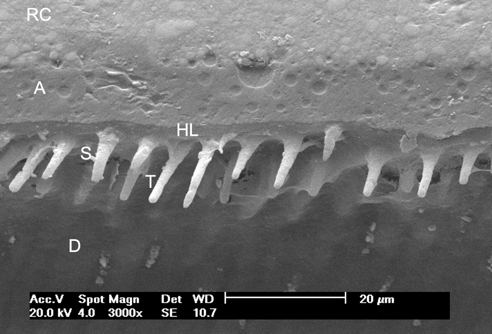
Scanning electron microscope (SEM) photomicrograph of the interface between the Adper Single Bond 2 adhesive system and dentin. RC, composite resin; A, adhesive; HL, hybrid layer; T, resin tags; S, secondary ramifications; D, dentin.
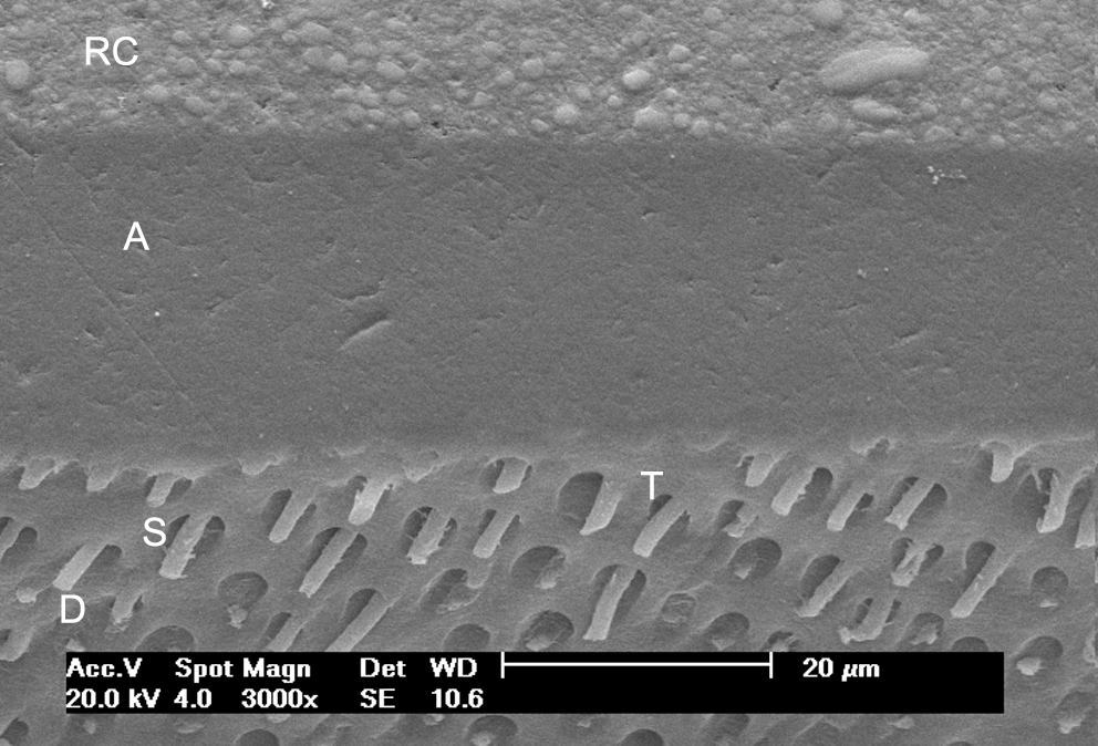
The application of the Clearfil SE Bond promoted the formation of a thinner hybrid layer when compared with Adper Single Bond 2. There was formation of shorter and fewer resin tags, with a triangular shape, without secondary ramifications (Fig. 2).

SEM photomicrograph of the interface between the Clearfil SE Bond adhesive system and dentin.
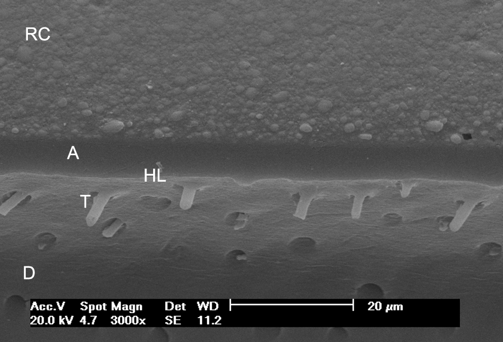
When Adper Single Bond 2 was applied on the dentin irradiated with the Nd:YAG laser, the hybrid layer that formed was thinner than in the group without the laser application. There was adhesive infiltration in the open dentinal tubules, but in fewer numbers and shorter (Fig. 3).

SEM photomicrograph of the interface between the Adper Single Bond 2 adhesive system and dentin irradiated with the Nd:YAG laser.
For Clearfil SE Bond applied on the dentin irradiated with the Nd:YAG laser, a thinner hybrid layer formed in comparison with the group without the laser application, and there were fewer and shallower resin tags (Fig. 4).
SEM photomicrograph of the interface between the Clearfil SE Bond adhesive system and dentin irradiated with the Nd:YAG laser.
When the Adper Single Bond 2 was associated with Er:YAG irradiation, a hybrid layer did not form and the typical interdiffusion zone (hybrid layer) was missing along the adhesive interface. There were more resin tags, without a triangular shape, and secondary ramifications were present (Fig. 5).
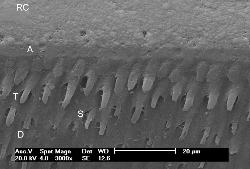
SEM photomicrograph of the interface between the Adper Single Bond 2 adhesive system and dentin irradiated with the Er:YAG laser.
For the Clearfil SE Bond applied on the dentin irradiated with the Er:YAG laser, the typical hybrid layer formation did not occur either. There were resin tags and secondary ramifications (Fig. 6).
SEM photomicrograph of the interface between the Clearfil SE Bond adhesive system and dentin irradiated with the Er:YAG laser.
Discussion
In this study two different adhesive systems were used to morphologically analyze the adhesive–dentin interface: Adper Single Bond 2, which uses an etching technique with 37% phosphoric acid as the first step, followed by the application of a primer and adhesive simultaneously; and Clearfil SE Bond, which uses a self-etching primer, followed by the application of an adhesive. These materials were applied on nonirridated human dentin and human dentin irradiated with Nd:YAG or Er:YAG laser.
For Adper Single Bond 2 on the nonirradiated dentin, a continuous and homogenous hybrid layer formed with primary and secondary resin tag formation. This resulted from the adhesive system using 37% phosphoric acid to etch the dentin, which resulted in the complete removal of the smear layer, smear plugs, and dentinal tubule openings, in addition to demineralization of the peritubular and intertubular dentin. 19,20 Thus, the hydrophilic and hydrophobic monomers in the one-bottle Adper Single Bond 2 penetrated the demineralized dentin, forming a hybrid layer and resin tags with a triangular shape. 18,21
Perdigão and Lopes 18 and Tay et al. 22 reported that the etching technique with 37% phosphoric acid may have disadvantages because it could increase permeability, possibly leading to greater pulp irritation. Furthermore, the resinous monomers cannot penetrate the dentin to the same depth as the demineralized dentin, which leaves collagen fibers exposed and subject to degradation; this is considered the weakness of the adhesive–dentin bond. 23
The Clearfil SE Bond yielded a thinner layer with shorter resin tags compared with Adper Single Bond 2. This occurred because Clearfil SE Bond uses a self-etching primer that has the acid monomer 10-methacryloyloxydecyl dihydrogen phosphate (MDP), which causes partial dissolution of the smear layer and smear plugs and more superficial decalcification of the dentin. Parallel to the demineralization procedure, infiltration of monomers occurs in this demineralized tissue and formation of a hybrid layer with an original smear layer is observed. 24,25 Yoshiyama et al. 26 and Itou et al. 27 also observed a thin hybrid layer for the adhesive systems with self-etching primers, which is around 1 μm for Clearfil Liner Bond 2V (Kuraray) and Fluoro Bond (Shofu, San Marco, CA) adhesive systems. However, Tay and Pashley 25 emphasized that the recent self-etching adhesive systems may be classified as weak, moderate, or aggressive, based on their capacity to dissolve the smear layer and demineralize the dentinal surface. Therefore, depending on the chemical composition of each material, especially the type and percentage of acid monomer, some self-etching primers may completely dissolve the smear layer and smear plugs, forming a hybrid layer with thickness between 2.5 and 5 μm. This thickness is close to the one obtained by etching of dentin with phosphoric acid.
The formation of a hybrid layer depends on the capacity of the resinous monomers to impregnate the demineralized area. Because the Adper Single Bond 2 forms a thicker hybrid layer than Clearfil SE Bond does not mean that it is clinically better, because even a thin hybrid layer with short resin tags is sufficient to promote a strong bond strength between the adhesive and dentin. 28 Furthermore, in the case of the self-etching primer, depth of demineralization is the depth of resinous monomer infiltration, avoiding collagen fiber not surrounded by monomers and their hydrolytic degradation. 29 –31
With the application of the Nd:YAG laser on the dentin surface, prior to adhesive procedures, the formation of a thinner hybrid layer and shorter and fewer resin tags occurred with both adhesive systems. These findings may be due to the Nd:YAG laser causing morphological alteration on the dentin surface.
The laser causes surface melting, followed by recrystallization, resulting in small scattered areas and some opened dentinal tubules. 32,33 The recrystallization of the dentinal apatite and formation of additional stages of calcium phosphate increase resistance of hard tissues to acid demineralization, which reduces dentin permeability. 34,35 The 37% phosphoric acid as well as the acid monomer MDP may not have had the same capacity to demineralize the dentin irradiated with the laser in comparison with the groups in which the laser was not applied. Furthermore, because some tubules were sealed, there was less formation of resin tags. Franke et al. 12 also verified less infiltration of adhesives on the dentin irradiated with the Nd:YAG laser.
When the Er:YAG laser was applied before the adhesive procedures, there was no formation of a hybrid layer, but resin tags were present, in agreement with other studies. 36,37 The phosphoric acid and the MDP probably did not cause significant morphological alterations on the dentin surface, because the laser made the dentin more acid resistant. However, the Er:YAG laser yields a dentin surface without a smear layer and smear plugs and the dentinal tubules remain open. 38,39 Because the tubules are open, the presence of a great quantity of resin tags was observed. These resin tags did not have a triangular shape, which was characteristic in the groups in which the laser was not applied, showing that intertubular dentin as well as peritubular dentin became more acid resistant.
The hybrid layer plays the major role in the mechanism of adhesion of the adhesive systems to dentin. The long-term durability of bonds between adhesive and dentin is of significant importance for the longevity of bonded restorations. 40 The Nd:YAG and Er:YAG irradiation caused different hybrid layer morphologies, which may result in differences in the quality of restorations placed on dentinal surfaces not irradiated with laser. However, clinical studies are important to determine the effect of these alterations in a restoration's longevity.
Conclusions
According to the methodology used, it was possible to conclude that (1) Adper Single Bond 2 adhesive system formed a thicker hybrid layer and longer resin tags in comparison with the Clearfil SE Bond adhesive system; (2) the Nd:YAG laser irradiation on the dentin surface, prior to adhesive procedures, caused the formation of a thinner hybrid layer and shorter resin tags; and (3) Er:YAG laser irradiation on the dentin surface, prior to adhesive procedures, did not allow the formation of a hybrid layer and there was formation of resin tags.
Footnotes
Author Disclosure Statement
The authors declare that no competing financial interests exist.