




Abstract
Introduction
Treatments that improve the activities of fibroblasts and stimulate production of new collagen should substantially improve the appearance and health of aged skin. In recent years, nonablative techniques, 5 which induce a dermal wound without epidermal ablation, have played an important role in the treatment of photoaging. One nonablative modality, intense pulsed light (IPL), also known as ‘‘photorejuvenation,’’ is a noncoherent light source from a polychromatic filtered flashlamp that emits high-intensity polychromatic light in a broad wavelength spectrum of 560–1200 nm, and has been used in the treatment of photoaging for several years. 6 IPL is frequently used to improve telangiectasias, lentigos, and skin texture. Clinical studies have demonstrated that IPL has significant photorejuvenation effects on photoaged skin. 7,8 However, the molecular mechanisms underlying the photorejuvenation of IPL treatment remain largely unknown.
The present study was designed to evaluate the mechanism and direct effect of IPL irradiation on fibroblasts in vitro by examining the expression of procollagen types I and III and ultrastructure changes on cells after IPL irradiation.
Materials and Methods
Cell culture and treatment
The designed study protocol was approved by the Ethical Committee of Shandong Provincial Hospital. Primary human skin fibroblasts were gathered from pooled adult (ages 30–50 years) skin dermis explants and cultured in high glucose Dulbecco's modified Eagle medium (DMEM) supplemented with 20% fetal bovine serum (FBS). Cells were trypsinized and expanded in the same medium. The IPLTM Quantum system (Lumenis Corp, Salt Lake City, UT) was used to irradiate the cells, and all treatments were performed using a 570 nm cutoff filter, double pulses of 4 and 6 ms with a pulse interval of 20 ms, and fluences of 18, 23, 28, and 33 J/cm2. After irradiation, medium was immediately replaced with fresh DMEM/20% FBS and left for another 24 h.
Measurement of cell activity by the methyl thiazole tetrazolium (MTT) assay
MTT assays were used to measure the number of viable cells and performed as previously described. 9 In this assay, the membrane-permeant dye is reduced by mitochondrial reductases in living cells, and spectrophotometric measurement allows for quantification of cell viability. Skin fibroblasts were plated in 96-well microtiter plates at a density of 3 × 10³ cells/well in a final volume of 100 μl of DMEM. Twenty-four hours after the initial seeding, cells were treated with various fluences of 18, 23, 28, and 33 J/cm2. Untreated cells served as a control. After treatment, the cells were incubated with fresh culture medium for 24 h at 37°C, and then a solution of methyl thiazolyl tetrazolium (MTT) at a concentration of 50 mg/100 ml was added to the culture medium. After a 4–6 h incubation period, 200 μl of dimethyl sulfoxide (DMSO) was used to replace the culture medium to dissolve formazan crystals. The results of the MTT assay were measured in terms of absorbance at 490 nm (A490). Each assay was performed in triplicate and repeated three times.
Assessment of cell cycle by flow cytometry
The skin fibroblasts were inoculated into six-well plates at a density of 105 cells/well and treated as indicated above. At the end of the incubation period, cells were harvested by trypsinization and washed twice with cold PBS. Briefly, cells were resuspended in 100 μl of 1 × annexin binding buffer, 80 μl of DNA Prep LPR, and 550 μl DNA Prep Stain were added and mixed gently. After incubation at 4°C for 30 min, the DNA contents were analyzed by flow cytometry. The results reflected the percentage of cells in each phase of the cell cycle. We used a flow cytometer (Beckman Coulter Epics XL-4, Brea, CA) for analysis of the cell cycle.
Assays of mRNA levels of procollagen I and III by quantitative real-time polymerase chain reaction (RT-PCR)
In accordance with the manufacturer's instructions (TaKaRa Corp, Shiga, Japan), total RNA was harvested with RNAisoTM Plus(TaKaRa Corp). For the reverse transcriptase polymerase chain reaction (RT-PCR), 2 μg of RNA was reverse-transcribed into complementary DNA (cDNA) by incubating with 1 μl of reverse transcriptase (TaKaRa Corp) in 20 μl of reaction buffer containing 0.1 nM of random 6 mers, 0.05 pM of Oligo dT primer, and 5 × Prime ScriptTM buffer at 37°C for 15 min and 85°C for 15 sec. The PCR reaction system (20 μl) contains 0.4 μl of cDNA, 10 μl of SYBR Premix Ex TaqTM (2X), 0.2 μl of Rox Reference Dye (2X), 0.4 μl of forward primer (10 μM) and reverse primer (10 μM) of collagen I, collagen III, or glyceraldehyde-3-phosphate dehydrogenase (GAPDH), and double-distilled water. The PCR was performed in accordance with the two-step procedure of ABI PRISM 7500 Fast Real-Time PCR System (Applied Biosystems Corp, Foster City, CA), which contains stage 1 of predegeneration, 95°C for 30 sec, and stage 2 of PCR reaction, 40 cycles of 95°C for 15 sec and 60 C for 1 min. The PCR products were scanned with an ultraviolet gel imaging system, and fluorescence intensity was analyzed by 7500 System SDS software. Compared with GAPDH product, the expression level of detected mRNA transcription was determined. The primers were as follows:
Observation of ultrastructure of fibroblasts by transmission electron microscopy
The cultured fibroblasts were trypsinized and collected into an Eppendorf tube after washing. They were fixed by 2.5% glutaraldehyde at 0°–4°C, washed by PBS, fixed by osmic acid, then washed by distilled water and dehydrated by dimethylketone. After embedment in Epon-812, the sample was cut into ultrathin sections (70 nm). The ultrathin sections were dyed with uranium acetate and plumbum citrate. They were examined with JEM-100sX electron microscopy.
Assays of protein levels of collagen I and III by enzyme-linked immunosorbent assays (ELISA)
This assay measures newly synthesized collagen. Twenty-four hours after the cells were treated as prescribed above, the protein extracts of the cell culture supernatant were prepared and assayed using a commercial enzyme-linked immunosorbent assay kit (R&D Corp, Minneapolis, MN). All reaction steps were performed according to the manufacturer's protocol, and the absorbance was read at 450 nm. The relative ratio of the treated group compared with the control group was statistically analyzed.
Statistical analysis
Data are shown as the mean ± standard deviation (SD). Statistical analyses were performed with Statistical Package for Social Sciences (SPSS) software, version 11.0, One-way analysis of variance (ANOVA) was used to determine the statistical significance of differences. The level of statistical significance was set at 0.05.
Results
Promotion of cell viability after IPL irradiation
As shown in Fig. 1, IPL irradiation significantly promoted fibroblast viability. Exposure of fibroblasts to IPL irradiation at energies of 18–33 J/cm2 induced an energy-dependent increase in cell viability, compared with the control. The 28 J/cm2 group led to a 32% promotion in cell viability and was the most obvious effect in this study.
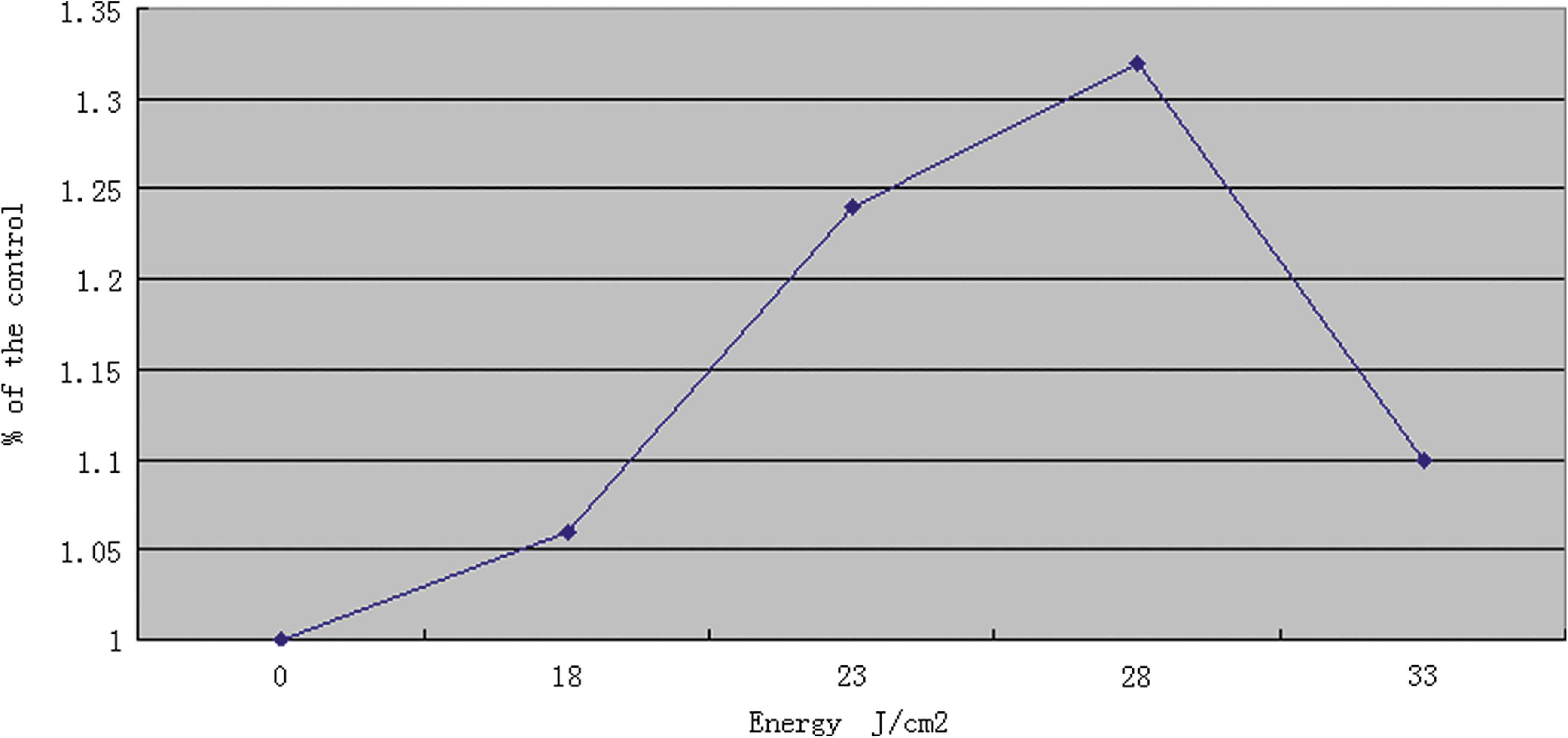
Cell viability was assessed by MTT methods 24 h after IPL irradiation. The ratio of cell viability was measured by their absorbance. Compared with the control group, the absorbance of IPL irradiation groups were significantly increased in an energy-dependent manner (p < 0.05). The most efficient effect was obtained in the 28 J/cm2 group.
Changes of cell cycle after IPL irradiation
As shown in Table 1, there was a significant change in the cell cycle of the skin fibroblasts after IPL irradiation, compared with the control group. The percentage of fibroblasts at the S stage of the cell cycle was significantly higher in samples exposed to IPL irradiation, compared with the control, with a corresponding significant decrease in the proportion of cells in the G0/G1 phase. The cell cycle changed in an energy-dependent manner.
Compared with control group, p < 0.05.
Changes of cell cycle of skin fibroblasts after IPL irradiation. Fibroblasts were irradiated using IPL as described in the Methods section. Twenty-four hours after irradiation, fibroblasts were harvested for analysis of cell cycle. The percentage of fibroblasts at the S stage of the cell cycle was significantly higher in samples exposed to IPL irradiation, compared with the control, with a corresponding significant decrease in the proportion of cells in the G0/G1 phase.
Increased mRNA levels of procollagen (types I and III) after IPL irradiation
To understand the IPL effect on collagen production, this study analyzed the mRNA expression of procollagen (types I and III) using real-time RT-PCR. Twenty-four hours after IPL irradiation, mRNA levels of procollagen (types I and III) were significantly increased. IPL irradiation increased the mRNA level of collagen I considerably, to 123%, 154%, 172%, and 141% of the control and that of collagen III to 120%, 141%, 164%, and 132%, corresponding to irradiation energies of 18, 23, 28, and 33 J/cm2. The mRNA expression of procollagen (types I and III) increased in an energy-dependent manner. Specifically, the increased level of the mRNA expression of the 33 J/cm2 group was less than that of the 28 J/cm2 group (Fig. 2).

Effect of IPL irradiation on mRNA levels of procollagen I and III. Fibroblasts were irradiated using IPL as described in the Methods section. Twenty-four hours after irradiation, fibroblasts were harvested for RNA isolation and RT-PCR. IPL irradiation energy-dependently increased the mRNA level of procollagen I and III, statistical significance was obvious compared with the control group (p < 0.05).
Ultrastructure changes of fibroblasts after IPL irradiation
The transmission electron microscope study (Fig. 3) indicated that the cells were more active after the treatment. There were more rough endoplasmic reticula, mitochondria, Golgi apparatuses, and polyribosomes in the cell matrix. Many of the organelles were dilated, and there were large nucleoli and more coarse chromosomes in the nucleus. More collagen particles were obviously seen in the cytoplasm of the cells after IPL irradiation, compared with the control group.
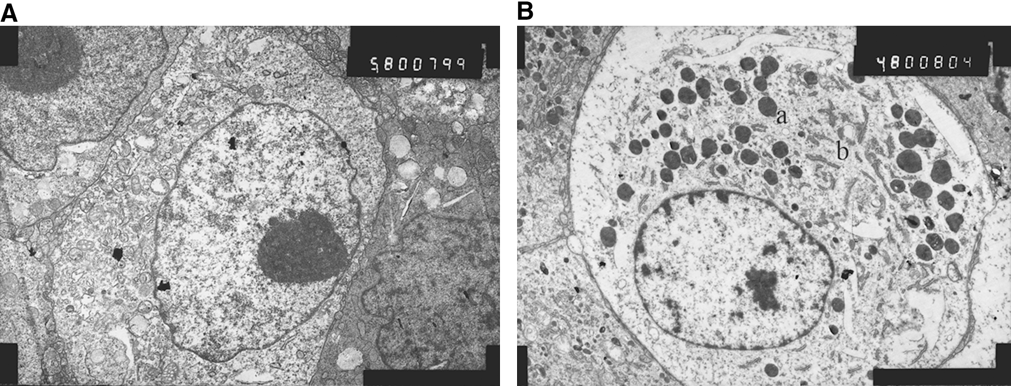
Ultrastructure of fibroblasts after IPL irradiation observed by transmission electron microscope.
Increased expression of collagen (types I and III) after IPL irradiation
The ELISA results (Table 2) showed that the relative content of collagen (types I and III) was significantly increased after IPL irradiation, and the increase was energy-dependent at the range of 18–28 J/cm2. However, when the energy reached 33 J/cm2, the expression of collagen decreased compared with the 28 J/cm2 group.
Compared with control group, p < 0.05.
Expression of collagen (types I and III) after IPL irradiation. Fibroblasts were irradiated using IPL as described in the Methods section. Twenty-four hours after irradiation, the cell culture supernatant were prepared for ELISA analysis. The relative ratio of treated group compared with control group was statistically analyzed.
Discussion
Clinical studies using objective assessments, such as spectrophotometric analysis, have demonstrated that IPL has significant photorejuvenation effects. 8,10 Fibroblasts, the major cellular components in skin dermis, are the key cells responsible for the biosynthesis of extracellular matrix (ECM) proteins. 11 The effects of IPL on skin fibroblast activity remain unknown. Some theories 12 –14 state that light energy is converted into heat in the dermis that damages the stroma in the dermis slightly and stimulates related cells to release cytokines and enzymes, which enhance fibroblast proliferation and synthesis of more collagen protein. In the present study, we found that IPL has an obvious promoting effect on fibroblast viability and expression of procollagen (type I and type III), providing experimental evidence for the direct action of IPL irradiation on the skin fibroblasts. IPL may directly promote fibroblast viability through its photothermal effect on the cells, not merely through cytokine stimulation secreted by inflammatory responses of surrounding tissues thermal injury.
In the clinical setting, an ideal balance between increasing fluences to attain optimal results and minimizing adverse effects is needed. Increasing fluences can lead to side effects such as persistent erythema, crusting, dyspigmentation, and scarring. The IPL irradiation used in this investigation was chosen according to the therapeutic mode most commonly used to treat Asian patients with photoaged skin in our hospital. This study tested a wider range of energy levels, including 18, 23, 28, and 33 J/cm2, which exerted no cytotoxic effect on the investigated condition. This range of energy levels is mostly chosen on clinical application for photoaging treatment and has been proved safe for most patients.
The present study demonstrated that IPL increased the viability of fibroblasts in an energy-dependent manner up to 28 J/cm2; as the energy level reached 33 J/cm2, the reactivity of fibroblasts decreased compared with the 28 J/cm2 group, but even increased compared with the control group. These findings suggest that IPL exerted a dual-effect on fibroblasts depending on the amount of energy, that is, low energy IPL stimulates fibroblast reactivity, whereas overhigh levels lead to cell toxicity. Moreover, after IPL irradiation, a significant percentage of fibroblasts were at the S stage of the cell cycle, whereas the proportion of cells in the G0/G1 phase was markedly less. These changes occurred in an energy-dependent manner as evidenced by flow cytometry. This further indicated that IPL stimulates the proliferation of fibroblasts in selected energy levels. IPL irradiation also increased expression of procollagen (types I and III) in an energy-dependent manner. and the most significant effect was in the 28 J/cm2 group. As the energy level rises without limitation, the toxicity of IPL is more obvious. In this study, we found that IPL had a direct promotional effect on the activity of skin fibroblasts only to a certain level. The upper threshold of fibroblast promotion was between 28–33 J/cm2 in our experimental pattern, and, importantly, exorbitant energy causes direct damage to skin fibroblasts. The present study shows that the selected energy levels have no adverse effects on fibroblasts in vitro.
It has been documented that collagen, which is responsible for the elasticity and intensity of the skin, comprises more than 90% of the total proteins in the skin. Type I collagen, which provides physical strength for the skin, is by far the most abundant. 15 Gary et al. 1 demonstrated that a major feature of aged skin is fragmentation of the dermal collagen matrix, and the levels of collagen I and III decreased in aged skin. Procollagen, a precursor of collagen, is synthesized and secreted by fibroblasts. Procollagen mRNA levels have been shown to be a reliable measure of the level of collagen production. In this study, we found that IPL irradiation produced an increase in the expression of procollagen I and mRNA in fibroblasts, which was noted after 24 hours. Our reports support previous studies demonstrating increased collagen deposition in histological specimens after IPL treatments. Feng et al. 16 described that both type I and type III collagens increased following IPL treatments, and there were more collagen fibers neatly rearranged within the stroma. Likewise, Goldman et al. demonstrated that IPL treatment may lead to increased transcription of type I collagen, and increased collagen (types I and III) and elastin in patients. 6 Negishi et al. 10 demonstrated that histological analysis of skin biopsies from IPL-treated patients showed increased staining of type I and type III collagen. The study reported herein together with the previous clinical studies demonstrating increased collagen deposition after IPL treatment provides a foundation for understanding the mechanisms that lead to clinical improvement. The increased collagen (types I and III) transcription allow us to better understand the mechanisms of photorejuvenation of IPL, and also the more viable fibroblast phenomena observed after IPL treatment.
The ultrastructural changes showed that the fibroblasts were more viable after IPL irradiation. The signs of more rough endoplasmic reticula, mitochondria, Golgi apparatuses, and polyribosomes indicated that the skin fibroblasts were activated by IPL irradiation and had laid the structural foundation for the synthesis of the collagen. IPL irradiation also induced an increase in the number of mitochondria, possibly causing an energy improvement that led to fibroblast proliferation. Rough endoplasmic reticula, polyribosomes, and Golgi of fibroblasts, which were the main sites to synthesize proteins, were abundant in the IPL irradiation group, suggesting that these organelles were well prepared for the formation of new collagen. Previous studies have demonstrated a histological increase in collagen types I and III in the dermal extracellular matrix after IPL treatment, 10,17 so this study could explain subcellular structure of the photorejuvenation effects of IPL.
Conclusion
The present study demonstrates that IPL irradiation imparts a stimulatory effect on skin fibroblasts in vitro. IPL irradiation can promote cell viability, increase the cell percentage at the S stage in the cell cycle, and improve expression of collagen (type I and type III). This provides valuable evidence regarding the mechanism of the photorejuvenation effect of IPL in vivo. Although various previous studies have demonstrated the photorejuvenation effects of IPL by providing clinical and histologic evidence, the exact molecular mechanism underlying IPL irradiation remains to be elucidated.
Footnotes
Author Disclosure Statement
No competing financial interests exist.
Yongqian Cao and Ran Huo contributed equally to this study.