




Abstract
Introduction
Such view of water solely as a ubiquitous, neutral solvent has radically changed. In 2003, Zheng and Pollack found that in the vicinity of common hydrophilic materials (including biological interfaces) water is extensively ordered. 8 It has subsequently been found that water plays a primary role in a number of mechanisms, including osmosis and diffusion, surface tension and oxygen exchange, self-assembly, light-induced effects, and vascular effects. 9 Furthermore, Del Giudice et al. 10 found that liquid water self-organizes and produces extended regions or coherence domains (CDs), which require the intervention of an EMF. Such water molecules behave in unison with the phase locked with the phase of the self-trapped EMF, in accord with the framework of quantum electrodynamics. Thus, externally applied EMFs may target the collective organization of water to influence biomolecules.
In this personal view, we propose that the modulation of the structure and function of water by light may come to embody a new mechanistic approach for the treatment of complex diseases. For example, interfacial water plays a major part in cellular recognition, especially during first contact events in which cells decide between survival and apoptosis. Light–water interactions may thus be able to steer cells toward or away from apoptosis – deregulation of which is a hallmark of cancer, neurodegenerative diseases such as age-related macular degeneration (AMD), primary open-angle glaucoma (POAG), and other complex diseases. Previously, we presented cellular and molecular bases for water-mediated, long-range energy supplementation aimed at inducing and modulating physiologically reparative processes, including apoptosis, through the mechanism termed “photo-infrared pulsed biomodulation” (PIPBM). 11 We then documented the role of water as an oscillator in near infrared (NIR) photobiomodulation, adding to a more coherent description of the central effects of NIR light on redox centers and key transmembrane enzymes such as cytochrome c oxidase (CcO). 12 Water may thus provide a novel pathway for NIR absorption and transportation, complementing and facilitating CcO energy transfer for increased efficiency in the production of adenosine triphosphate (ATP) – a vital molecule required not only as cell fuel, but also as a part of the signaling pathway connected with family-specific receptors P2 and P1, of growing importance in cancer and other complex diseases. 13,14 Here, we complement and expand on these ideas by integrating the role of the quasi-crystalline exclusion zone (EZ), part of the cell's aqueous interface which is just now beginning to be decoded.
What is the EZ?
Based on the space it occupies, water in biological systems may be classified as: (1) “bulk” water, located in intravascular, interstitial, and interfacial structures; (2) confined water, found at the intracellular level and in protein (e.g., aquaporin) channels; and (3) interfacial water, typically composed of two well-defined zones, one hydrophilic and one hydrophobic, separated by a transition phase (see top of Fig. 1). Next to the surface of the cell's hydrophilic “gel-like” area, water forms a massive “exclusion zone” (EZ).
15
The EZ is a solute-free, interfacial area that can project hundreds of microns from the cell surface. Two of the main properties of the EZ are: 1. Its viscosity is higher than that of normal water 2. It is an electron donor, or reducer, as opposed to normal water, which is a mild oxidant.
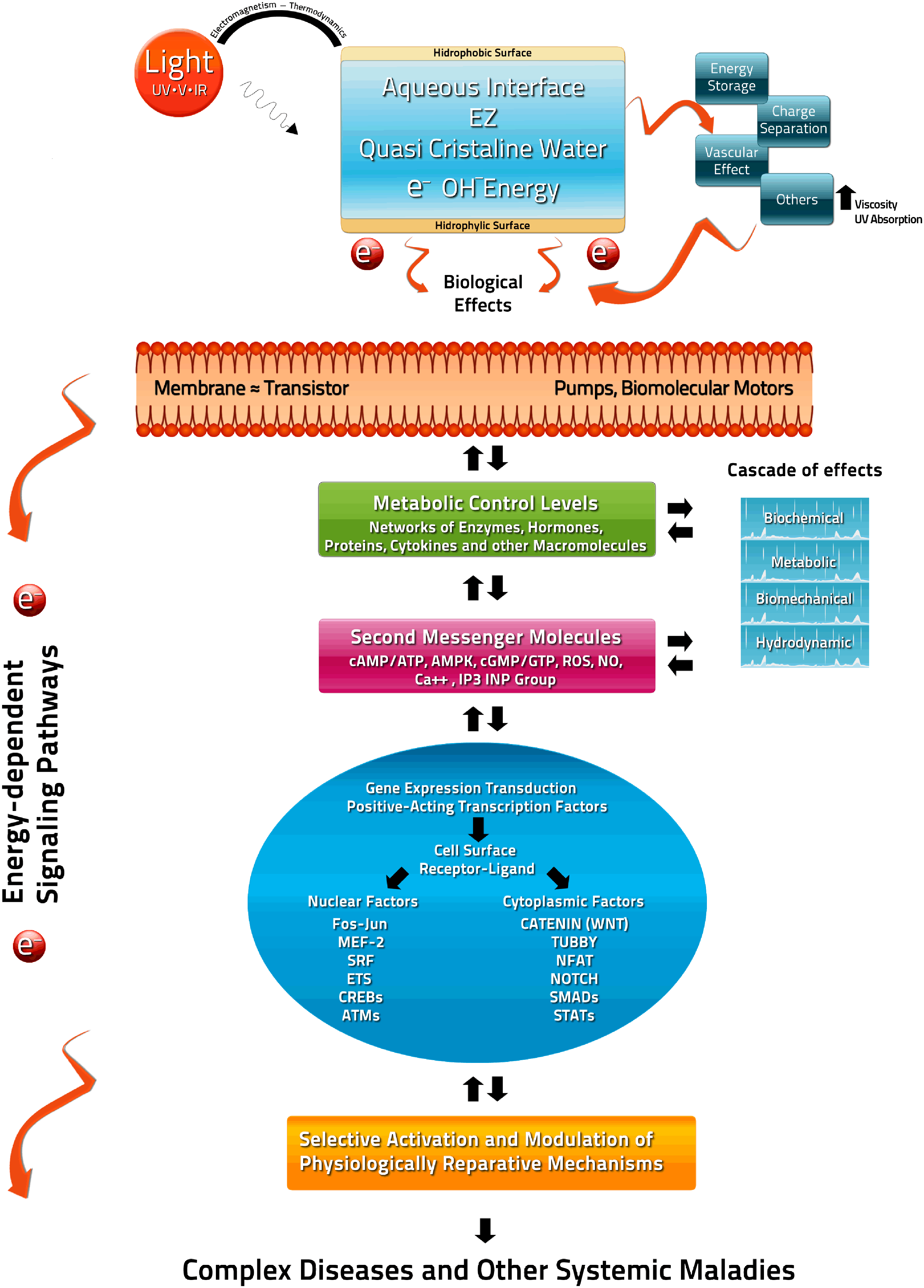
Schematic representation of the quasi-crystalline exclusion zone (EZ) as a rechargeable biological battery capable of effectively storing and providing charge to fuel cell work and power signaling pathways in the face of injury-induced redox potential, such as those caused by complex diseases. Top left, ultraviolet (UV), visible (V) and infrared (IR) light signals normally energize EZ water. Top right, energy stored in the form of charge separation leads to increased viscosity and regulates numerous effects via vascular and membrane-related pathways. Mid-center, the cell membrane is akin to a transistor capable of signal detection, switching and amplification. Center-right, in the event of energy-depleting injury potentials, EZ water can expeditiously donate electrons, triggering a cascade of biochemical, metabolic, biomechanical, and hydrodynamic effects. Lower center, effects are modulated by metabolic control levels made up of networks of enzymes, hormones, proteins, cytokines, and other macromolecules. Below, second messengers (e.g., cAMP/ATP, AMPK, cGMP) and surface receptor-ligands lead to gene expression and transduction through energy-dependent nuclear and cytoplasmic transcription factor. Bottom, it is proposed that light-induced effects can selectively activate and modulate physiologically reparative mechanisms in complex diseases and other systemic maladies.
The EZ–non-EZ water interface has been described as a widespread electrolytic pile, with redox potential jumps similar to fractions of a volt. Electron dynamics between the exited and ground state are essential to the redox properties of water in living systems. As an aside, this property was recognized by Szent-Gyorgyi, who found that different molecular species exhibited exceptionally long living states of electronic excitation with ordered water. Szent-Gyorgyi postulated that the latter was at the core of energy transfer in biological systems, and that it explained how energy from biomolecules could be translated into free energy for cell work. 2 The EZ is so large that at least hundreds of thousands of layers are involved and – if undisturbed – it can remain stable for weeks once formed. Indeed, Ling and Hu have proposed on theoretical grounds that the ordered layers could extend infinitely under ideal conditions. 16 –18 Cells and tissues are well buffered at pH values near 7.35–7.45. The EZ is highly robust under these conditions and in the presence of physiological saline solution (typically 0.9 g NaCl/L). Moreover, exclusion phenomena do not arise from electrostatic repulsion; they seem to stem from physical changes in water. Ghosh et al. have demonstrated the role of proteins (biopolymer surfaces) in the mixture to be a catalyst for formation under compression of multilayer water, which is then reintegrated into the monolayer on expansion. 19 These results suggest a very different cellular environment from what had been generally assumed. And, although similar phenomena had been found, the physicochemical properties of the EZ and their implications for cellular function are just now beginning to be understood.
The EZ as a Selective Rechargeable Biological Battery
Remarkably, EZ water can not only store charge, it can later return it in the form of current flow, with as much as 70% of the input charge being readily obtainable. 20 Although the law of electroneutrality implies that net charges should not be possible, this rule appears to be violable over restricted volumes of water. Macroscopic separation of charges can be stable for days to weeks and has unusual electric potential. Water is thus an unexpectedly effective charge separation and storage medium. Under physiological conditions, required energy for building charged EZ in biological systems appears to come from light (ultraviolet [UV], visible, and IR). Mid IR (MIR) is especially effective at 3100 nm (Fig. 2), which corresponds to the fundamental OH stretch; there is also strong resonance at the level of the fourth MIR harmonic. 21, 22 Furthermore, it seems that 270 nm absorption is a unique feature of water in contact with various charged molecules, and that this has been interpreted as a reflection of the overall quasicrystalline nature of water in the vicinity of charged or hydrophilic entities, whereas fluorescence could indicate more subtle features.
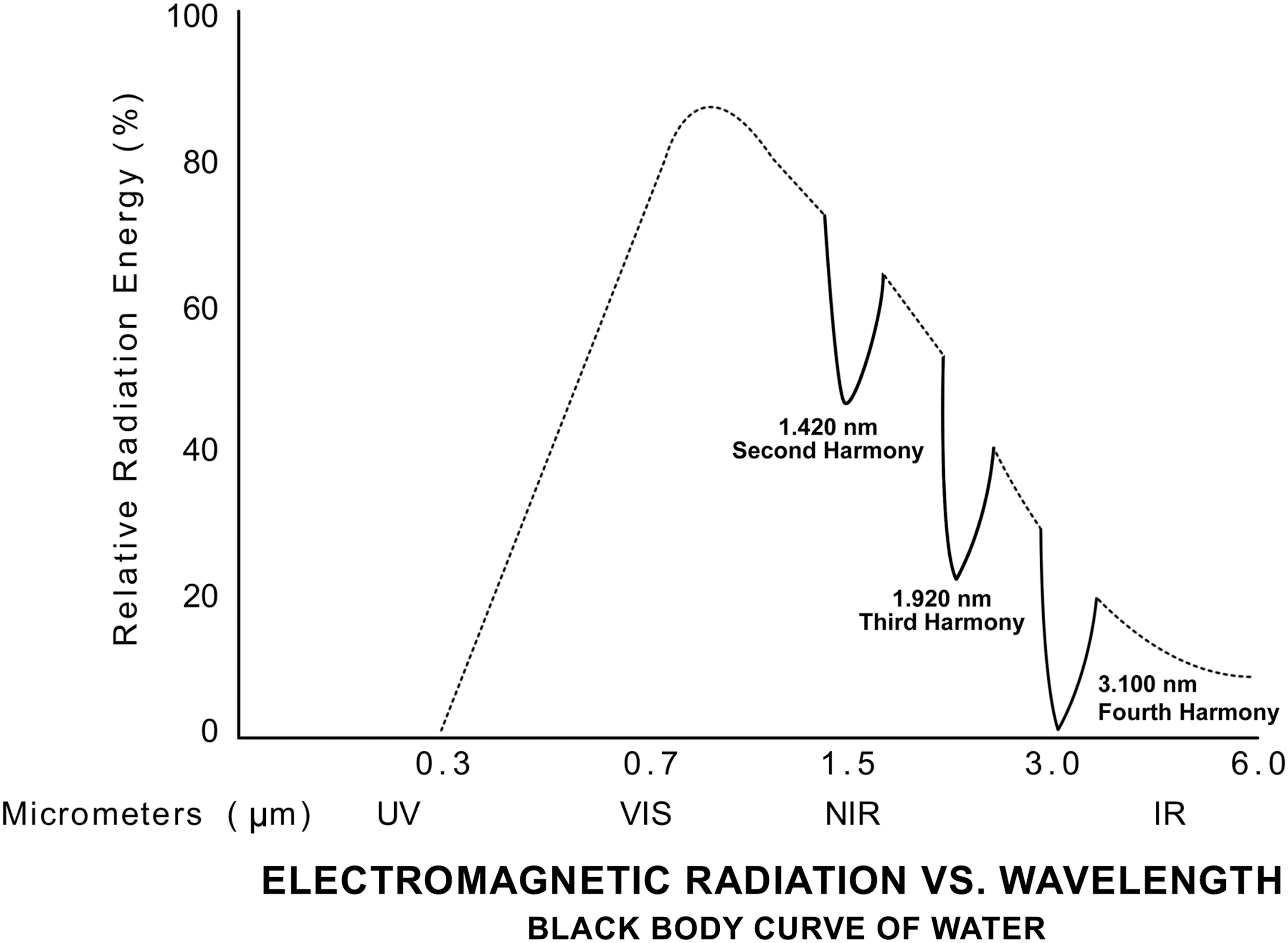
Black body curve of water. Under physiological conditions, energy required for building the charged exclusion zone (EZ) appears to come from light (UV, visible, and IR). Mid IR (MIR) is especially effective at 3100 nm, which corresponds to the fundamental OH stretch; there is also strong resonance at the level of the fourth MIR harmonic. It appears that 270-nm absorption may be unique to water in contact with various charged molecules; this may reflect the quasicrystalline nature of water in the vicinity of charged or hydrophilic entities, whereas fluorescence may indicate more subtle features.
We therefore propose that the EZ may behave as a selective rechargeable (electrolytic) bio-battery, acting as a dedicated energy reserve that selectively supplements the cell's energy demands by transferring electrons to the intracellular space when intracellular redox potentials induced by injury differ from those normally associated with metabolic activity by more than one order of magnitude (see Fig. 1). Given its ability to remain stable, and to retain and separate charges, we further propose that the EZ may be targeted as an alternate source and/or reservoir of EM energy to expeditiously cover cellular energy demands following injury-induced disturbances. This follows from the cell's resemblance to rechargeable electrolytic batteries and its capacity to transform EM energy into a high-energy compound such as ATP, needed as fuel for cellular work and for short- and long-range cellular signaling. In addition, and viewed analogously as “nanoantennas,” water molecules can be useful as key optical components for biological light harvesting.
Quasicrystaline EZ in Contact with Membrane Hydrophilic Surface
Nearly 70% of interfacial residues are hydrophilic. Hydrophobic de-wetting has been documented as a general mechanism for hydrophobic surface assembly. Previously, direct electrostatic interaction mediated by a continuous solvent had been commonly assumed. 2 Direct measurement of vibrational dynamics of tissue water interface has now been achieved 23,24 and may provide valuable insights in addition to experiments on Janus interface, and wetting/de-wetting modulation by light. 23 –27 Molecular dynamics (MD) simulations of the barnase-barstar complex have also showed that water mediates and stabilizes interactions between native contacts. 28 It has been described that “for electrostatic interactions to be important, the interfacial water's dielectric constant needs to be reduced to reduce screening. This happens as a consequence of changes in the structure of the interfacial layers (the dielectric permittivity is <50% of the bulk value for interfacial separations of <1.2 nm), and it preferentially promotes electrostatic interaction normal to the surface. 29,30 Therefore, although the association mechanism of hydrophilic interfaces remains elusive, in the words of Ball, one may say that “water does exactly what is required of it.” 31
It is worth remembering that interfacial water plays many essential roles. Water-mediated interactions drive polymers to adsorb strongly at hydrophobic interfaces, and repel them from hydrophilic ones. At hydrophilic surfaces, van der Waals interactions between the polymer and the surface mitigate water-mediated repulsion, leading to weak adsorption of the polymer. Interestingly, binding thermodynamics and kinetics correlate with macroscopic droplet contact angles that characterize the wetting properties of different interfaces, which is critical in the molecular mechanism of photobiomodulation. 12 Phase transition, a major structural change prompted by subtle environmental changes, may be another related central mechanism of interest given that it has been proposed that phase transition is responsible for much of the work done by cells 29 and may be crucial for the activation and modulation of physiological reparative mechanisms required for normal structural and functional cells and tissues.
Light, Water, Signal Transduction, and Gene Expression
Potentials associated with normal intracellular metabolic activity (∼−70 to −300 mV) are close to those associated with the EZ (∼−100 to −200 mV) 32 and are not strong enough to perturb it. 20,32 However, injury-induced potentials, such as those caused by tumors, which can exceed 1.5V, 33 can trigger electron transfer and energy supplementation from the EZ to the injured cell's intracellular space. Given the complex electrochemical nature of human eukaryotic cells, electron transfer from the EZ may activate and modulate not just bioenergetic function, but also far-reaching signaling, for example via ATP, P1/P2 and A1/A2/A3 receptors, 34,35 cyclic adenosine monophosphate (cAMP), and cyclic guanosine monophosphate (cGMP), which are prominent members of the cell's energy modulation system. As described by Steinberg et al., AMP-activated protein kinase (AMPK) acts in response to a reduction in energy charge (decrease in ATP and increase in AMP) by switching off anabolic pathways such as fatty acid, triglyceride, and cholesterol synthesis as well as protein synthesis and transcription that consume ATP, and switches on catabolic pathways that generate ATP, such as fatty acid oxidation and glycolysis. 36 The relation between decreased ATP and increased AMP could thus be modulated by electrons transferred from EZ water. It has also been argued that the AMPK pathway could be targeted to combat uncontrolled tumor growth, type II diabetes, and other conditions. 37,38 Interestingly, activation of AMPK prevents lipotoxicity in retinal microvascular pericytes, which are the first cells lost in diabetic retinopathy (DR). 39 AMPKα also contributes to the regulation of matrix metalloproteinase-9 (MMP9) expression, 40 which may accelerate the apoptosis of retinal capillary cells in the pathogenesis of DR, and plays a critical role in tissue remodeling under both physiological and pathological conditions. 40 Additionally, selective modulation of AMPKα activity may benefit patients with retinal degeneration associated with retinal pigment epithelium (RPE) cell atrophy, 41 commonly found in advanced AMD.
Experimental Evidence Relevant to LLLT
At the time of writing this article, direct experimental evidence to prove that low-level laser therapy (LLLT) will express effects via the EZ in a high-order biological system has not been attained. This may represent a significant challenge, the benefit of which could possibly be very large. Although now recognized as fundamental in biology, aqueous interfaces have been hard to investigate for years, for technical reasons. 23,24 This has also been the case with carbon nanotubules. In that case, intuited biological implications have been increasingly validated by subsequently acquired knowledge about aquasporine channels, and their deep implications in physiology and pathophysiology. 42 Nevertheless, existing experimental data already strongly support both the existence of EZ and the ability of light to modulate it.
As of today, EZ measurements have been conducted using high-resolution (750 MHz) proton nuclear magnetic resonance ( 1 H-NMR) spectroscopy in a wide range of materials, such as ion resins, polymers, functionalized monolayers, and biological tissues. 43 One of the most notable properties found is that suspended particles (monodisperse colloide) are excluded from studied surfaces by several hundred micrometers; this particle-free area is precisely the region termed the EZ. 43 Moreover, in prebiotic bacterial cells, it has been shown that the IR region of the spectrum, of particular importance in LLLT, produces an EZ with induced charge differential of −100 to −200 mV. 44,45
Using a 90 MHz (1.5 Tesla) H-NMR spectrometer, the authors showed that an LLLT-relevant 904 nm laser pulsed at 3MH (fluence 0.11 J/cm2, peak power 35 mW) induced significant short-term changes in T2 and correlation times (τc) in rat soft tissue. 46,47 A clinical (1 Tesla) MRI microdensitometry study using the same laser device (fluence 45 J/cm2) further showed changes in solid tumor T2-weighted heterogeneities, attributable to increased water diffusion associated to selective photo-induced tumor cell death. 48 Although conducted with lower resolution equipment insufficient to observe the EZ, these results highlight the broad potential of water dynamics in LLLT. Future experimental and clinical research must determine optimal light parameters and their interaction with the EZ in particular and water dynamics in general. Relevant results may, nonetheless, be found with multiple light parameters in part because of the low activation energy of water, which is slightly lower than that of UV, IR, or visible light. Potential target applications and their potential clinical application are discussed in the following section.
Potential Target Applications
Light–water interactions can activate numerous intracellular signaling pathways and regulate nucleic acid synthesis, enzyme activation, and cell cycle progression. One such mechanism lies in the photobiomodulation of cellular oxidation through reactive oxygen species (ROS). ROS are generated as byproducts of cellular metabolism, primarily in mitochondria. Studies implicate ROS generated by specialized plasma membrane oxidizes in normal physiological signaling by growth factors and cytokines. Although certain nuclear factor-κB (NF-κB)-regulated genes play a major role in regulating the amount of ROS in cells, ROS can have various inhibitory or stimulatory roles in NF-κB signaling. 49 This is significant as cancer and other complex diseases (e.g., primary open-angle glaucoma [POAG], DR, and AMD) share age-related and metabolic disorder-dependent damage of cellular energy and transport processes caused by ROS stress. 50
Studies also show that low-intensity light can catalyze nitrite-dependent nitric oxide (NO) synthesis (Cco/NO). It has been argued that this explains the increased bioavailability of NO experienced by tissues exposed to light 51 and confirms that light may modulate cell signaling. NO is a key intra- and intercellular signalling molecule. Martinez-Ruiz et al. suggest that NO signaling comprises at least three possible mechanisms: classical, less-classical, and non-classical. Classical signaling involves cGMP generation and cGMP-dependent protein kinases. Non-classical signaling is related to formation of NO-induced post-translational modification (e.g., S-nitrosylation). 52 A recently found pathway between NO and mitochondrial CcO suggests key implications for cell respiration and metabolism. 51 Both under- and overproduction of NO can lead to disease. For example, several eye and neurodegenerative disorders such as Alzheimer's and Parkinson's diseases engender synaptic and neuronal cell damage. And whereas mild oxidative and nitrosative (NO-related) stress mediates normal neuronal signaling, excessive accumulation of these free radicals is linked to neuronal cell injury or death. 53 Therefore, light-induced modulation of protein nitration, which can lead to protein aggregation, could have profound implications for the treatment of neurodegeneration. 54
In a similar vein, studies have demonstrated that LLLT can modulate gene expression and the release of growth factors and cytokines in cell cultures. 55 LLLT significantly increases the gene or protein expressions of several growth factors, including brain-derived neurotrophic factor (BDNF) 56 and insulin-like growth factor (IGF)-I. 57 Furthermore, LLLT can activate mitogen-activated protein kinases (MAPK) and PI3K/AKT pathways for BDNF and PI3K/AKT for IGF, which promote ganglion cell (RGCs) survival. 58 RGCs transmit visual information from the retina to the brain by way of the optic nerve, and their loss is a characteristic of POAG, DR, and AMD. In addition, LLLT increases transforming growth factor β1 (TGF- β1) 59 which has been found to be neuroprotective in the stroke model, 60 to maintain RGC-5 cell survival and to promote neurite outgrowth through p38 MAPK. 61 LLLT also has immune modulation effects over transforming growth factor β2 (TGF-β2) expression at sites of wound healing 62 and may activate latent TGF-β complexes via ROS. 63 TGF-β2 induces changes in extracellular matrix (ECM) and has been proposed as a novel approach to the management of POAG. 64
Additionally, LLLT stimulates cGMP synthesis
65
and modulates vascular endothelial growth factor (VEGF).
66
Decreased levels of cGMP and NO have been demonstrated in the plasma and aqueous humor of POAG patients.
67
NO can further control irradiation-activated reactions that increase cell attachment.
68
NO contributes to physiologically regulate ocular hemodynamics and cell viability, and protects vascular endothelial cells and nerve cells or fibers against pathological stressors implicated in POAG, ischemia, and diabetes.
69
In accord with this, EMFs have been found to modulate production of endothelin
Finally, we list some specific signal-dependent, positive-acting (regulatory) energy-dependent transcription factors on cell surface/receptor ligands of potential interest for EM modulation via EZ water. One potential target is the class of resident nuclear factors that includes ETS, CREB, ATMs, SRF, Fos-Jun, and Mef2. In Alzheimer's disease (AD), loss of presenilin (PS) reduces the level of cAMP response element-binding protein (CREB)-binding protein (CBP) and transcription of CREB/CBP target genes, even though CREB-mediated transcription is regulated indirectly by PS. 72 SRF acts as a master regulator of a hyperproliferative, inflammatory phenotype accompanied by neovascularization, 73 and Mef2 has been suggested to serve as an important mediator of VEGF in endothelial cells. 74 Another class of relevant latent, energy-dependent, cytoplasmic factors includes JAK-STAT, SMADs, NFkB/RelA, Notch, catenins (Wnt), tubby, and NFAT. Of special relevance is the JAK-STAT signaling pathway, which transmits information from chemical signals outside the cell, through the cell membrane, and into gene promoters on the DNA, causing DNA transcription. Among other functions, JAK/STAT mediates RGC survival following intra-ocular pressure (IOP) elevation. 75 It has also been found that pigment epithelium–derived factor (PEDF) can decrease mitochondria-derived ROS generation and downregulate VEGF expression, possibly by inhibiting high glucose-induced JAK2/STAT3 activation, which may offer a promising strategy against diabetes complications. 76 It must finally be stressed that many of the second messengers confirmed to be activated at the cell surface/interface by light/water include inositol trisphosphate (IP3), diacylglycerol (DAG), and calcium (Ca2+//CaATPase), which are all fundamental in complex diseases.
Conclusions
Given existing data, it is not possible to prove the role of EZ water in photobiomodulation at this time. This personal view is not intended to provide such proof. It presents the current state of research in this area, the technical challenges involved in obtaining direct evidence in biological systems, and some of the potential uses and implications of the EZ in medicine. Its objective is to draw attention to major recent laboratory and experimental findings in systems and conditions relevant to photobiomodulation with the goal of stimulating academic discussion, as is customary for this type of article in leading academic journals. 77 However, whereas one must highlight the novel nature of the research findings herein discussed, it is clear that light-EZ phenomena do exist and represent an area of great potential interest. Research will need to ascertain if, in the presence of injury-induced redox potentials, properly tailored external EM energy (light) may substitute and/or complement metabolic energy pathways and activate signaling pathways through electron transfer from EZ water in the cell membrane interface, as proposed in this article. Such effects may modulate cell signaling and power extensive complex networks, which lie behind the structure and functioning of metabolic control levels. In our view, involved energy (light)–matter interactions appear to satisfy the demands of quantum electrodynamics and the laws of thermodynamics, and may lead to novel deterministic and not-fully deterministic effects. Although not a panacea, this view suggests a potentially universal methodological approach to the therapeutic modulation, alone or with other therapeutic agents, of reparative physiologic processes capable of promoting positive clinical results and improving quality of life with minimal, if any, adverse effects. Because of its inherently basic bioenergetic roots, applications may extend to multiple complex diseases, some of which have few or no currently viable effective treatment alternatives.
Footnotes
Acknowledgments
This article was supported by Fundalas, an independent nonprofit foundation. The authors thank Jesús Santana-Rodríguez and Luis R. Santana-Rodríguez for editorial and technical support.
Author Disclosure Statement
No conflicting financial interests exist.