




Abstract
Introduction
Collagen, a major structural protein in the skin, gives skin strength and durability and is responsible for the smooth, plump appearance of young, healthy skin. Skin collagen decreases with age and is thought to be the main reason for skin wrinkles. The decrease of collagen content in skin could be caused by two complex processes. One results from the decreased mRNA expression of type I and III procollagen. The other is the elevated expression of matrix metalloproteinases (MMPs). As expected, collagen synthesis was found to decrease while MMP expression increased in photoaged skin compared with younger skin. 5 MMP activity can be inhibited by tissue inhibitors of metalloproteinases (TIMPs). The balance between MMP and TIMP activities is involved in both normal and pathological events, such as wound healing and tissue remodeling. 6 Therefore, procollagen, MMPs, and TIMPs are important markers for studying laser nonablative rejuvenation.
Mitogen-activated protein kinases (MAPKs) are a family of serine/threonine kinases. There are several reports showing that MAPKs play a role in regulating skin aging. Extracellular signal-related kinase (Erk) and JNK MAPK can be activated by a variety of stress signals including UV irradiation, advanced glycation end products, intense pulse light, and chemotherapeutic drugs, 7 –9 and they may be involved in the reduction of skin fibroblast proliferation, collagen synthesis inhibition, and well as the blockage of transforming growth factor-beta (TGF-β)/SMAD. However, it is still unclear whether MAPK participates in the regulation of laser on skin collagen synthesis and degradation.
In this study, to better understand the molecular activities in the skin associated with the 1064 nm Q-switched Nd:YAG laser, we investigated if the 1064 nm laser irradiation altered the mRNA expression of types I and III procollagen, MMP2, MMP3, TIMP1, and TIMP2 in an animal model. In addition, the roles of MAPK family members on the changes of collagen production induced by the laser were detected.
Materials and Methods
Animals
Six female Sprague–Dawley (SD) rats, weighing between 200 and 250 g, were purchased from the animal center at Fudan University (Shanghai, China). Animals were provided with food and water ad libitum and housed in our laboratory at a constant temperature with a 12 h light/dark cycle. Animals were acclimatized for 1 week prior to this experiment. The study was approved by the institutional review board at East China Normal University.
Laser irradiation
Experimental animals were anesthetized with 1% chloral hydrate at a dosage of 1 mL/kg intraperitoneally. The backs of the rats were shaved and then denuded with the depilatory cream ([HSCH2COO]2Ca, α-(-)-bisabolol , monoglyceryl ester, SIMP hair depressant). After 24 h, the dorsal area was divided into four 2×2 cm grids by a marker pen. One grid was shielded as the control; the other three grids were irradiated by the 1064-nm Q-switched Nd:YAG laser (Medlite IV, Conbio, CA) with a pulse width of 6 ns and a spot size of 6 mm. Three fluences were used: 0.6, 1.5, and 2.5 J/cm2, respectively. Energy was delivered with ∼10% overlap in the skin. Treatments were conducted four times at 1 day intervals.
Hydroxyproline assay
Rats were killed at 30 days, and skin samples were taken for hydroxyproline analysis. The samples were delipidized with a blade, 100 mg was weighed out, and 10% skin tissue homogenate was prepared. Then, hydroxyproline content was determined by using chloramine T oxidation and 4-dimethylaminobenzaldehyde colorimetric assay read at 550 nm absorbance according to kit instructions (Nanjing Jiancheng Bioengineering Institute, Nanjing, China).
RNA isolation and reverse transcription-polymerase chain reaction (RT-PCR)
Skin samples were obtained at the end of the experiments and weighed 100 mg. Then, all tissues were powdered in liquid nitrogen, lysed with 1mL of TRIZOL reagent (Invitrogen, USA) for 5 min, and then extracted at room temperature using 200 μL chloroform for 2 min. The total RNA was precipitated from the aqueous phase by addition of isopropanol and centrifugation. The pellet was washed with 75% ethanol and resuspended in 20 μL of diethylpyrocarbonate (DEPC)-treated water. RNA was quantified by spectrophotometry, measuring absorbance at 260/280 nm, and 1 μg total RNA was reverse transcribed into cDNA in 20μL reaction volume containing 2 μL of random oligonucleotide primers, AMV reverse transcriptase (Invitrogen, USA), and 2 mM dNTP mix. Reverse transcription was performed using a thermal program of 30°C for 10 min, 42°C for 20 min, 99°C for 5 min and 4°C for 5 min. Then, 2μL cDNA was used as the templates for PCR amplification in a thermal cycler (Bio-Rad, USA). The primers were designed by Primer 5.0 and synthesized by Invitrogen Inc. (Shanghai, China). The specific primer sequences with the size of amplified fragments for rat collagen I /III, MMP2, MMP3, TIMP1, TIMP2 and the glyceraldehyde-3-phosphate dehydrogenase (GAPDH) are provided in Table 1. The amplification condition was: 95°C for 10 min followed by 35 cycles of 95°C for 30 sec, 58°C for 30 sec, and 72°C for 60 sec. Reaction products were subjected to electrophoresis on 1% agarose gel and visualized with Tanon gel image system (Shanghai, China). GAPDH was used as internal control. Expression values of collagen, MMP. and TIMP were normalized to the GAPDH expression level.
Western blotting
Skin samples were crushed in liquid nitrogen and then lysed in NP 40 lysis buffer (50 mM Tris-Cl, pH 8.0, 100 mM NaCl, 5 mM MgCl2, 0.5% [v/v] Nonidet P-40) on ice for 30 min. The protein concentration was determined by using a BCA Protein Assay Kit (Thermo, USA) according to the manufacturer's procedure. Protein samples (30 μg) were run on a 10% sodium dodecyl sulfate (SDS)-polyacrylamide gel, transferred to a nitrocellulose membrane (Millipore) and immunoblotted with anti-Erk, anti-p38, anti-JNK, anti-phospho-Erk, anti-phospho-p38, anti-phospho-JNK (Santa Cruz, CA) at 1:1000 and anti-β-actin (Santa Cruz, CA) at 1:5000. After incubation with the fluorescently labeled secondary antibodies for 1 h, the antigen-antibody complexes were visualized with fluorescent Western blot imaging systems (Odyssey).
Statistical analysis
Statistical analyses were performed with SPSS 15.0 software. Differences among different groups were evaluated with the student's t-test, and p<0.05 was considered to be statistically significant.
Results
Effect of the Q-switched 1064 nm laser irradiation on skin collagen content
Hydroxyproline is a major component of collagen and is rarely found outside collagen. Therefore, hydroxyproline is used to determine the total collagen content. Compared with the nonirradiated control, collagen content increased markedly in the skin treated with the 1064 nm laser (p<0.05) (Table 2). Interestingly, hydroxyproline content in the skin treated with 1.5 J/cm2 was higher than treated with 0.6 or 2.5 J/cm2 (p<0.05) (Table 2). However, irradiation with 2.5 J/cm2 resulted in the lowest increase rate of hydroxyproline content.
Data were expressed as mean±SD (n=6). a p<0.05, 1.5 versus 0.6 and 2.5; b p<0.05, laser versus control.
Effect of the Q-switched 1064nm laser irradiation on procollagen mRNA expression
As shown in Fig. 1A, laser irradiation significantly increased gene expression of procollagen type I and type III on day 30 as compared with the nonirradiated control. The mRNA expression of type I procollagen after laser irradiation with 1.5 J/cm2 was elevated by 69% (p<0.05). In contrast, the level of type III procollagen induced by the same fluence is twofold higher than that of control (p<0.05)(Fig. 1B). The increase rate of procollagen expression was dependent upon the laser fluence. Laser irradiation with 1.5 J/cm2 induced new procollagen formation to a greater extent than the other energy levels (p<0.05). It was of note that the lowest increase rate of procollagen type I mRNA expression was seen in the skin treated with the highest fluence of 2.5 J/cm2 (Fig.1B).

The 1064 nm laser irradiation increased gene expression of types I and III procollagen in the rat skin at day 30 post-irradiation.
Effect of the Q-switched 1064 nm laser irradiation on the mRNA expression of MMP and its inhibitors
To investigate if gelatinases and stromelysins are involved in the changes of collagen steady-state levels induced by the Nd:YAG laser, we assessed the expression of MMP2 and MMP3 by RT-PCR. As shown in Fig. 2A, the skin treated with the Nd:YAG laser showed lower expressions of MMP2 and MMP3 when compared with the control. With irradiation of 1.5 J/cm2, MMP2 mRNA was reduced to 65.4% of the control and MMP3 was dropped to 71.1% of the control (Fig. 2B).
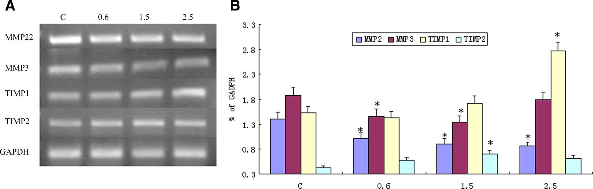
The 1064 nm laser irradiation decreased MMP2 and MMP3 levels and upregulated gene expression of TIMP1 and TIMP2 in rat skin at day 30 post-irradiation.
In contrast to MMPs, the mRNA expression of the inhibitors of MMPs was significantly elevated by the Nd:YAG laser treatments. TIMP1 showed a dose-dependent increase in mRNA levels (Fig. 2A). The amount of TIMP-1 mRNA in the treated skin with 2.5 J/cm2 was increased by an average of 81.6% when compared with the control. However, the highest TIMP2 mRNA level was seen at the skin irradiated with 1.5J/cm2, which was increased by 63.2% as compared with the control (Fig. 2B).
Activation of MAPK signal pathway by the Q-switched 1064nm laser irradiation
The anti-phospho-Erk1/2, anti-phospho-JNK and anti-phospho-p38 MAPK antibodies detected specifically the phosphorylated forms of Erk1/2, JNK, and p38 MAPK, respectively, whereas the anti-Erk1/2, anti-JNK, and anti-p38 MAPK antibodies detected total p38, JNK, or Erk1/2 proteins, respectively (Fig. 3). The Nd:YAG laser irradiation significantly increased p-Erk1/2 and p-JNK in the rat skin compared with the controls. In contrast, p38 phosphorylation did not alter in the skin treated with the laser. Moreover, laser irradiation with 1.5 J/cm2 was more effective than 0.6 and 2.5 J/cm2 at increasing the levels of p-Erk1/2 and p-JNK (Fig. 3).

The changes of mitogen-activated protein kinase (MAPK) signaling pathway in the rat skin following the 1064 nm laser treatments at 24 h. Western blot analysis showed that phosphorylation of Erk1/2 and JNK was augmented after laser irradiation at 1.5J/cm2. The total Erk, JNK, p38 and p-p38 were consistent with or without laser treatments.
Discussion
The 1064 nm Q-switched Nd:YAG laser are widely used in cosmetic surgery including treatments for wrinkles, tattoos, hair removal, and vascular lesions. Several authors have reported that the 1064 nm Q-switched Nd:YAG laser can increase dermal thickness and cause new collagen formation at the histological levels. 1 –4 Nevertheless, no study has been reported, to date, on the molecular changes in the dermis induced by the 1064 nm Q-switched Nd:YAG laser. The results of this study demonstrated for the first time that the 1064 nm Q-switched Nd:YAG laser could increase types I and III procollagen expression, decrease MMP2 and MMP3 mRNA levels, and activate Erk1/2 and JNK MAPK signal pathways in the rat skin.
Currently, clinical effectiveness of laser skin resurfacing is mainly based on the induction of collagen synthesis. In this study, hydroxyproline assay revealed the marked increase of total collagen content in the skin treated with the 1064 nm Q-switched Nd:YAG laser, indicating that the Nd:YAG laser is effective in increasing collagen production. To further assess the effects of laser on new collagen synthesis at the molecular level, RT-PCR assay was used to detect the expression of collagen. At day 30, we noted the marked upregulation of types I and III procollagen expression in the skin treated with the laser. It is therefore concluded that 1064 nm laser irradiation can increase new collagen synthesis in the rat skin.
Interestingly, we found that the mRNA level of type III procollagen was elevated to a greater extent than that of type I procollagen, which was consistent with the previous studies. 10 However, the reason is still unclear. A previous report has shown that type III collagen is common in fast-growing tissue, particularly at the early stages of wound repair, which will be replaced later by the stronger and tougher type I collagen. 11 We therefore assumed that the increase of type III collagen expression induced by the 1064 nm laser was likely the result of the short term (30 days) used in the experiments, which eventually accelerated type I collagen production. Therefore, long-term experiments will be needed to evaluate the trend of types I and III procollagen expression after Nd:YAG laser treatments.
MMPs have been reported to play an important role in the degradation of extracelluar matrix components during photoaging. 12 In the study, the 1064 nm Q-switched Nd:YAG laser was noted to reduce significantly the expression of MMP2 and MMP3, which suggested that the 1064 nm laser can effectively inhibit collagen degradation in addition to increasing collagen synthesis. TIMPs are specific inhibitors that bind in MMPs and control their activities in tissues. As expected, the 1064 nm laser markedly enhanced the expression of TIMP1 and TIMP2 as compared with the nonirradiated control. Obviously, the increases of TIMP levels after the 1064 nm laser irradiation resulted in the inhibition of MMP activities. Therefore, not only the upregulation of collagen expression but also the inhibition of collagen degradation contributed to the increase of collagen content in the skin irradiated by the laser.
We observed that the MMP2 levels in the 2.5 J/cm2-treated group were higher than those in groups treated with 0.6 and 1.5 J/cm2. As MMPs play important roles in inflammatory responses and can be usually expressed during tissue injury, we therefore assumed that the relatively higher MMP2 levels in the 2.5 J/cm2-treated group were caused by skin injury after laser irradiation of high intensity. Orringer et al. reported that 585 and 1320 nm laser irradiation caused an increase of MMP expression in the human forearm. 13 It seemed that the upregulation of MMP expression induced by 585 nm and 1320 nm laser was the result of the high fluences that they used.
The three major groups of the mammalian MAPK family have been demonstrated in collagen gene regulation in various experimental systems. 14 –16 To our knowledge, the role of Erk and JNK pathways in the 1064 nm laser nonablative resurfacing has not been reported. We observed high levels of activated Erk1/2 and JNK in the skin irradiated with the 1064 nm laser. Therefore, it seemed that the Erk1/2 and JNK pathways played an important role in the upregulated procollagen expression following the 1064 nm laser treatments. Interestingly, although p38 MAPK is one of the three major MAPK signaling cascades, it is unchanged after the 1064 nm laser treatments. A previous study demonstrated that p38 played a role in relaying the TGF-β2 signal to induce type I collagen production in the retinal pigment epithelium. 17 p38 was also reported to play a role in the regulation of inflammation after skin injury. 18 It is possible that the laser fluences used in the study were moderate, and therefore did not cause marked skin lesion, inflammatory reactions, and subsequent p38 activation.
Peplow et al. reported that laser irradiation at green, red, or infrared wavelengths at a range of dosage parameters can cause significant release of cytokines. 19 In addition, c-Jun and c-Fos, the primary effectors of the Erk 1/2 and JNK signaling pathway, are essential for regulation of human collagen, type I, alpha 2 (COL1A2) promoter activity by TGF-β. 20 Further studies are needed to determine if the cytokines related to collagen metabolism, the transcription factors such as c-Jun and c-Fos, and the TGF-β/SMAD pathways are involved in the regulation of collagen production induced by the 1064 nm laser.
In this study, laser irradiation was performed at fluences of 0.6, 1.5, or 2.5 J/cm2. The dose of 1.5 J/cm2 caused a greater increase in types I and III procollagen expression, TIMP2 expression, and MAPK activities than did the other doses. Therefore, a proper selection of the appropriate laser parameters is critical for producing the greatest clinical improvement. It is worth emphasizing that rat skin is much thinner than human skin and that therefore laser settings are difficult to translate. Further experiments in the clinic will be needed to determine the appropriate laser parameters for 1064 nm Q-switched Nd:YAG laser nonablative resurfacing.
Conclusions
Taken together, our data demonstrated that collagen increase in the rat skin irradiated with the 1064 nm Q-switched Nd:YAG laser was driven by increased collagen synthesis and decreased collagen degradation. Our study also demonstrated that both Erk1/2 and JNK MAPK signal pathways seemed to play a role in regulating skin collagen expression and steady state after 1064 nm laser treatments.
Footnotes
Author Disclosure Statement
No competing financial interests exist