
Abstract
Introduction
To treat skin aging, many researchers have attempted to evaluate the treatment effects for the different approaches, such as dermabrasion, chemical peels, and noninvasive rejuvenation methods. The noninvasive skin rejuvenation method, as one of the most normalized methods, was mostly employed to optimize the therapeutic effects by quantifying the application conditions. Dayan et al. 7 reported that the 1064 nm laser shows the overall recovery improvement for fine and coarse wrinkles, skin roughness, and uneven pigmentation. DeHoratius et al. 8 showed that the treatment effects for nonablative tissue remodeling and photo rejuvenation can be altered according to the different light sources and laser types. In addition, Alam et al. 9 performed histologic analyses to evaluate the influences of thermotherapy. On the other hand, Chen et al. 10 analyzed heat distribution according to the different laser conditions using a gel phantom. Jaunich et al. 11 attempted to find optimum laser conditions for minimizing skin damages using a tissue- mimicking phantom. Schaaf et al. 12 compared internal skin temperatures on pig skin samples according to the different penetration depths with the 2000 nm laser. Moreover, Chen et al. 13 predicted maximum thresholds of tissue damage on pig skin while transmitting the 2000 nm laser.
Despite these efforts, current approaches suffer from two main limitations. First, most existing methods did not compare the experimental results among simulations, actual experiments, and histologic approaches. Second, the previous studies evaluated the therapeutic effects subjectively through the self-assessment questionnaires, and thereby produced low reliability for predicting tissue damage. In addition, treatment effects were altered by the patients' condition, because of the absence of the quantitative evaluation criteria. It is, therefore, necessary to perform complex studies with the different laser treatment conditions to optimize therapeutic effects for skin rejuvenation. Particularly, it is necessary to perform quantitative analysis and histologic analysis simultaneously for predicting internal-external heat distributions.
In this study, we estimate heat distributions and evaluate degrees of tissue damages histologically after transmitting therapeutic lasers to find optimum ranges for skin rejuvenation. For this purpose, we transmit the 1064 nm Nd:YAG laser into a skin-mimicking phantom and pig skin samples according to the different fluences and spot diameters, and analyze its internal-external temperatures. In addition, we stain pig skin samples with hematoxylin and eosin (H&E) for evaluation of degrees of tissue damages.
Materials and Methods
Skin-mimicking phantom
The tissue-mimicking phantom was constructed by three main steps as follows. First, we produced acrylic molds that contain three measuring slots to provide the same tissue phantoms for the different laser conditions (Fig. 1A). In addition, we dried the acrylic molds at room temperature after spreading the releasing agents on the inner wall of the acrylic mold in order to separate the phantom more conveniently. Second, we composed a total of three thermocouple slots (depth of 2.5, 5.0, and 7.5 mm) on the completely dried molds using metal rods with a diameter of 2 mm. We then secured thermocouple sensors onto phantom slots by using a fixing frame, which contains the same slot conditions with acrylic mold (Fig. 1A). Finally, we injected an epoxy mixture composed of an epoxy (EP30, Master Bond, Inc., Hackenscak, NJ) and a hardener with a 3:1 ratio into the acrylic mold, and stiffened them for ∼24 h at room temperature. 14,15 Based on these procedures, we obtained a skin-mimicking phantom with a diameter of 50 mm and a height of 50 mm as depicted in Fig. 1B. The employed phantom revealed a thermal diffusivity of 0.70×10⁻⁷ m2/sec, which is within 58–85% of that for human skin, 0.82×10⁻⁷ − 1.2×10⁻⁷ m2/sec. 16 The main properties of the employed skin phantom are listed in Table 1.
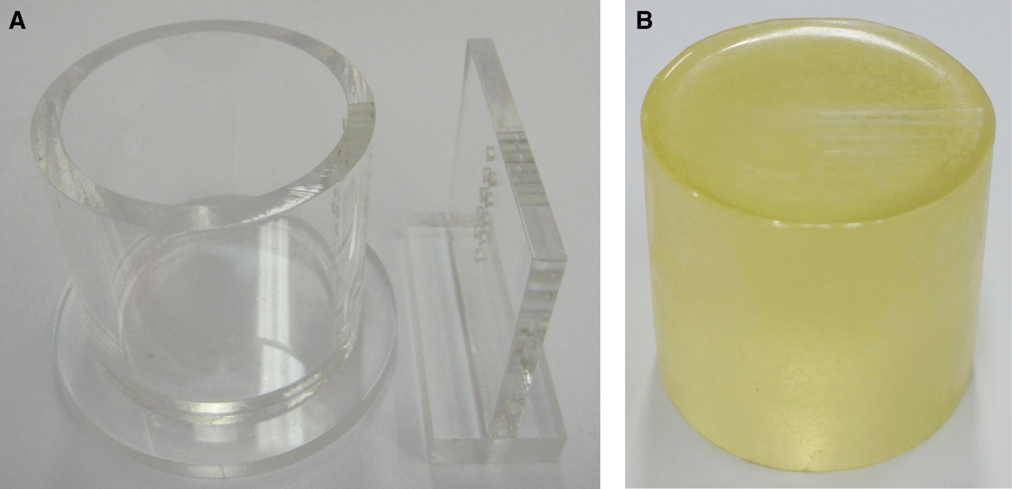
The employed skin-mimicking phantom.
Pig skin sample
Skin specimens were collected from the rear flank of Yucatan mini pigs (Optifarm Solution, Korea), which shows properties most similar to human skin, within 24 h after animal euthanasia. 12,17 The size of skin specimens was set to 50 mm (W)×50 mm (H)×30 mm (D). Additionally, the hair was removed with electric clippers before the laser transmission, for more effective heat transfer.
Additionally, in order to measure internal skin temperatures, we also composed a total of three thermocouple slots at depths of 2.5, 5.0, and 7.5 mm, respectively. For this purpose, a 21 gauge, 30 mm in, hypodermic needle (KOREA VACCINE CO., LTD., Seoul, Korea) was used to pierce the dermis of the skin and was directed parallel to the pig skin. The thermocouple needle was immediately inserted through the 21 gauge needle until it extended 10 mm past the tip. 12 Thermal diffusivity of the pig skin had a value of 0.82×10⁻⁷ − 0.86×10⁻⁷ m2/sec, within 71–100% of that for human skin (0.82×10⁻⁷−1.2×10⁻⁷ m2/sec). 16
Temperature measurement
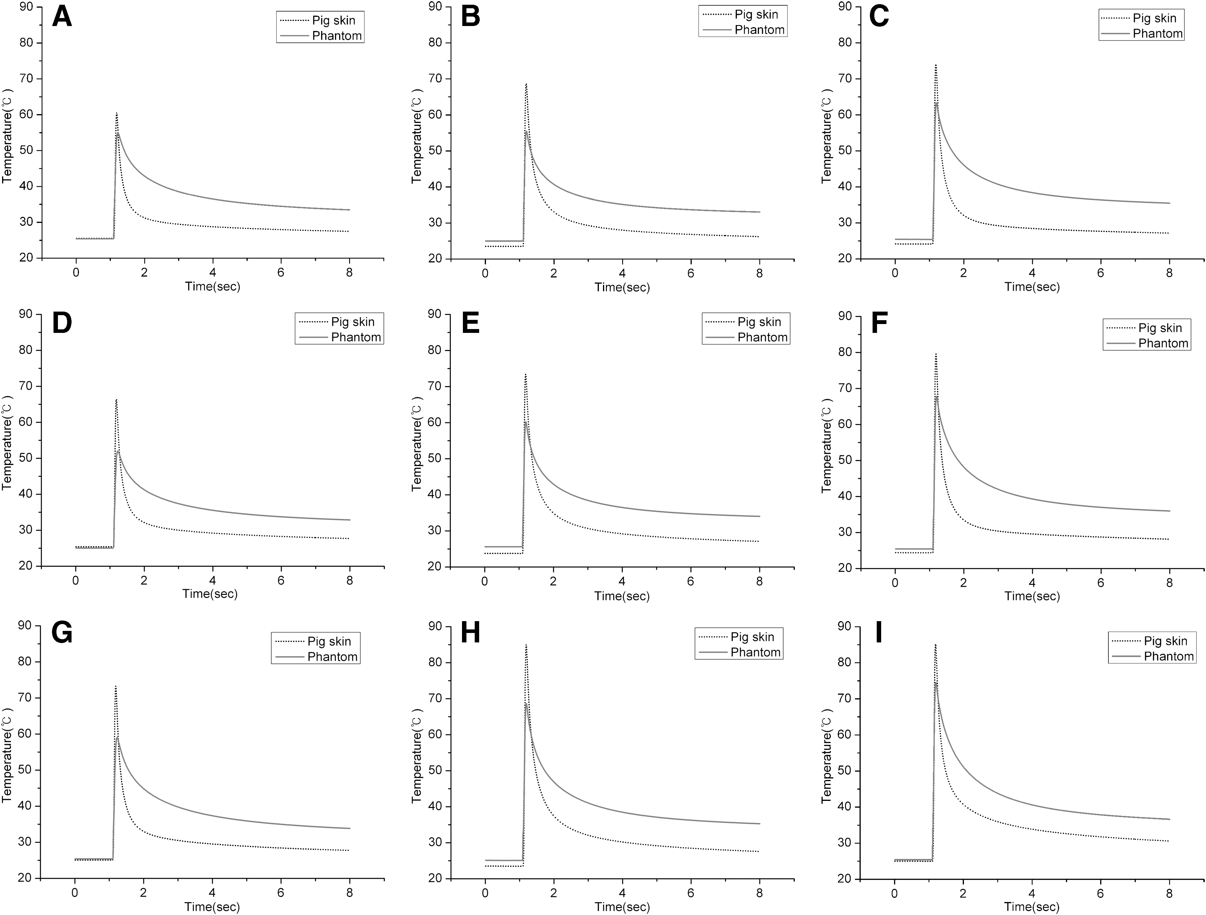
The surface temperatures of the skin mimicking phantom and pig skin were measured using the infrared (IR) camera (Testo 876, Germany) (Fig. 2A). In particular, we placed priority on the maximum temperatures of internal-external skins to examine the thermal thresholds of the skin damages. The employed camera was able to continuously measure the maximum and the minimum temperatures simultaneously (Fig. 2B). The resolution of the thermal image was 160×120 pixels, and the temperature measuring ranges were within 20°C to ∼100°C. In our experiments, the thermal images were acquired every 0.01 sec, and analyzed using AnalyzIR+1.2 software.

Measurement procedure of internal-external temperatures.
On the other hand, the internal temperatures were measured at depths of 2.5, 5.0, and 7.5 mm by using the K-type thermocouple after injecting thermocouple sensors into the composed thermocouple slots beforehand (Fig. 1A). The data acquisition was performed using a NI PCI-611 data acquisition card (DAQ) (National Instruments, Austin, TX), in cooperation with a Labview (National Instruments, Austin, TX). The radius of the K-type thermocouple was 1.57 mm, and the temperature measuring ranges were within −200°C ∼1200°C. Within the temperature ranges of interest, the error ranges were±0.75%.
To locate optimum treatment ranges, we transmitted the 1064 nm Nd:YAG laser with spot diameter conditions of 5, 8, and 10 mm, and fluence conditions of 26, 30, and 36 J/cm2. In addition, the pulse duration was set to 30 ms. Based on these measuring procedures, we evaluated treatment effects according to a total of nine different treatment conditions.
Histologic analysis
Biopsies with a thickness of 8 mm were obtained immediately using biopsy punch (BP-80F, Kai Medical, Japan) after transmission of the 1064 nm Nd:YAG laser three iterative times to conduct histologic examinations. 18 All biopsy specimens were fixed in formalin of 10%, embedded in paraffin, sectioned, and mounted on standard microscope glasses. According to the laboratory standard procedures, these samples were stained with H&E for optical microscopic examination. Biopsies were evaluated under a calibrated microscope (CH-2, Olympus, Japan).
Data analysis
For statistical analyses, data were analyzed by Pearson correlation analysis using SPSS (Ver. 12.0 for Windows, Chicago, IL). A p value of <0.01 was considered significant.
Results
Comparison of maximum temperatures between the skin-mimicking phantom and pig skin sample according to the different laser conditions
The maximum internal temperatures were revealed in the pig skin group compared with those of the skin-mimicking phantom group for all experimental conditions (Table 2). Regarding the penetration depths, the maximum internal temperatures were shown at a depth of 2.5 mm, and the temperatures were decreased as the penetration depths were deepened. In addition, the internal temperatures continuously increased as the spot diameter and the fluence increased for most experimental conditions. In contrast, the internal temperatures of pig skins for the depth of 5 and 7.5 mm with a spot diameter of 10 mm were significantly decreased as the fluences increased, and suddenly increased at a certain condition. The shorter returning time to the initial temperature was taken in the pig skin group, although the internal temperatures were higher in the pig skin group by ∼11.56±1.97°C compared with the phantom group (Fig. 3). Figure 3 demonstrates the maximum temperatures at a depth of 2.5 mm between the pig skin and skin-mimicking phantom groups for the different laser treatment conditions.
Comparison of the maximum temperatures between the skin- mimicking phantom and pig skin at a depth of 2.5 mm.
Correlations of internal temperatures between the skin-mimicking phantom and pig skin groups for the different penetration depths
We found strong correlations for internal temperatures between the pig skin and skin- mimicking phantom groups for most experimental conditions. In case of the conditions with spot diameters of 5 and 8 mm, the correlations were found relatively high at a depth of 2.5 mm, irrespective of the fluence conditions. The largest correlation coefficient of 0.965 was shown for the condition with a spot diameter of 8 mm and a fluence of 30 and 36 J/cm2. In contrast, the correlations improved as the penetration depths were deepened with a spot diameter of 10 mm. Consequently, we found the employed heat prediction method to be strongly reliable. Table 3 presents the correlation coefficients of internal maximum temperatures between the pig skin and skin-mimicking phantom groups.
Comparison of the surface temperatures between the skin-mimicking phantom and pig skin groups according to the different laser conditions
On the skin surfaces, the maximum temperatures were shown to be higher in the pig skin group than those of the skin mimicking phantom group for all laser conditions (Fig. 4). For the pig skin group, the lowest surface maximum temperature of 34.9°C was found in the condition of a spot diameter of 5 mm with a fluence of 26 J/cm2, whereas the highest temperature of 44.8°C was yielded in the condition of a spot diameter of 10 mm with a fluence of 36 J/cm2. In contrast, the lowest temperature was shown as 33°C for the condition of a spot diameter of 5 mm with a fluence of 26 J/cm2, whereas the highest value was found to be 38.4°C for the condition of a spot diameter of 8 mm with a fluence of 36 J/cm2 for the phantom group.
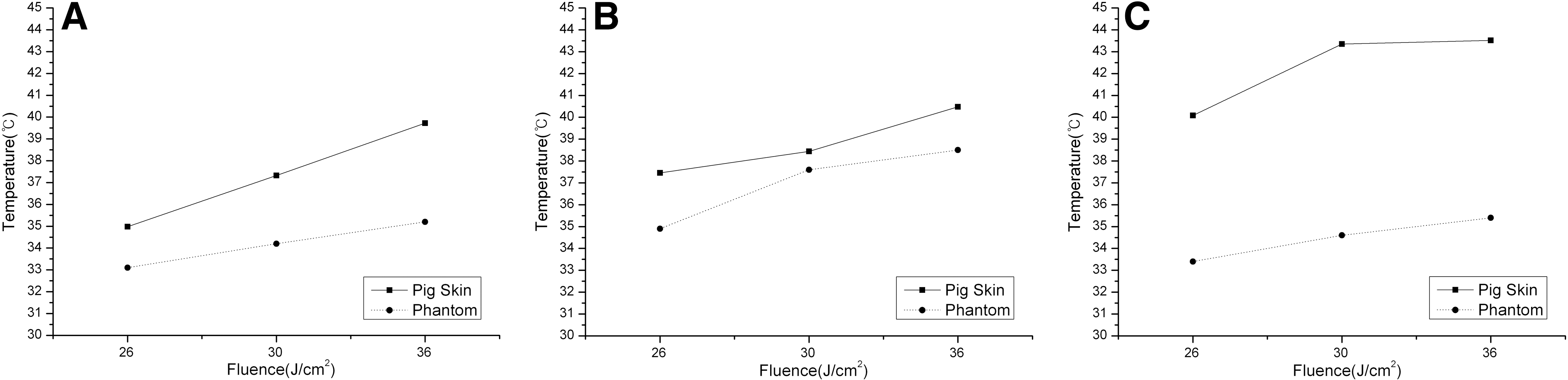
Changes of the maximum surface temperatures between the skin- mimicking phantom and pig skin according to the different laser conditions.
The differences between the pig skin and phantom groups were shown most greatly in the condition of a spot diameter of 10 mm with a fluence of 30 J/cm2, whereas the smallest differences were found in the condition of a spot diameter of 8 mm with a fluence of 30 J/cm2. In other words, the incremental tendencies of the surface temperatures varied for each experimental condition. However, internal maximum temperatures were continuously increased as the spot diameter and fluence increased. Figure 4 depicts the maximum temperatures on the surfaces of the pig skin and phantom groups according to the different laser conditions.
Histology
Figure 5A shows histologic sections of non-radiated pig skin tissue (control). Control specimen consisted of the epidermis, dermis, and dermal appendages, and did not reveal any thermal damages on the skin surface. In the condition with a spot diameter of 5 mm, slight deformations of the fibers in the dermis were revealed. Meanwhile, no serious tissue damage appeared for any of the fluence conditions (Fig. 5B–D). The epidermolysis was partially shown at the fluence condition of 30 J/cm2 (Fig. 5C). In the condition with a spot diameter of 8 mm, more fibrous tissues were formed in the dermis compared with the condition of a spot diameter of 5 mm (Fig. 5E–G). The cornification as well as epidermolysis occurred at the fluence condition of 36 J/cm2 (Fig. 5G). These tissue damages were worsened at the condition with a spot diameter of 10 mm (Fig. 5H–J). There was no serious tissue damage, except for the formation of fibrous tissues, for the fluence condition of 26 J/cm2 (Fig. 5H). However, the partial epidermolysis and the damages on the stratum spinosum occurred in the fluence condition of 30 J/cm2 (Fig. 5I). Moreover, the collapse of basal layers, epidermolysis, and epidermal necrosis appeared in the fluence condition of 36 J/cm2 (Fig. 5J). Consequently, the treatment conditions of a spot diameter of 8 mm with a fluence of 36 J/cm2 and a spot diameter of 10 mm with fluences of 30 and 36 J/cm2 caused irreversible tissue damage on the epidermal and dermal layers.

Hematoxylin and eosin (H&E) stained slides of pig skin according to the different laser treatment conditions:
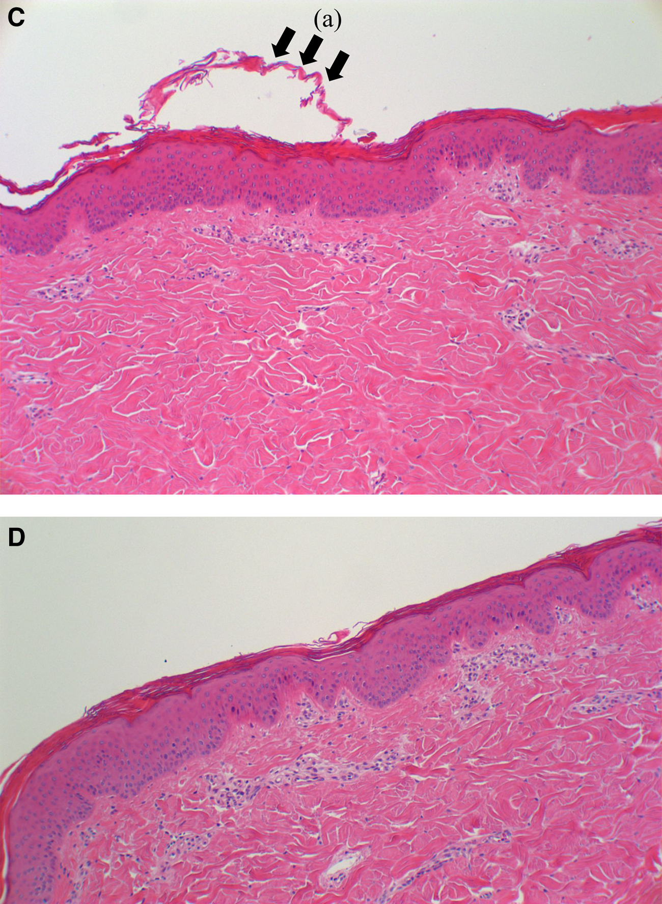
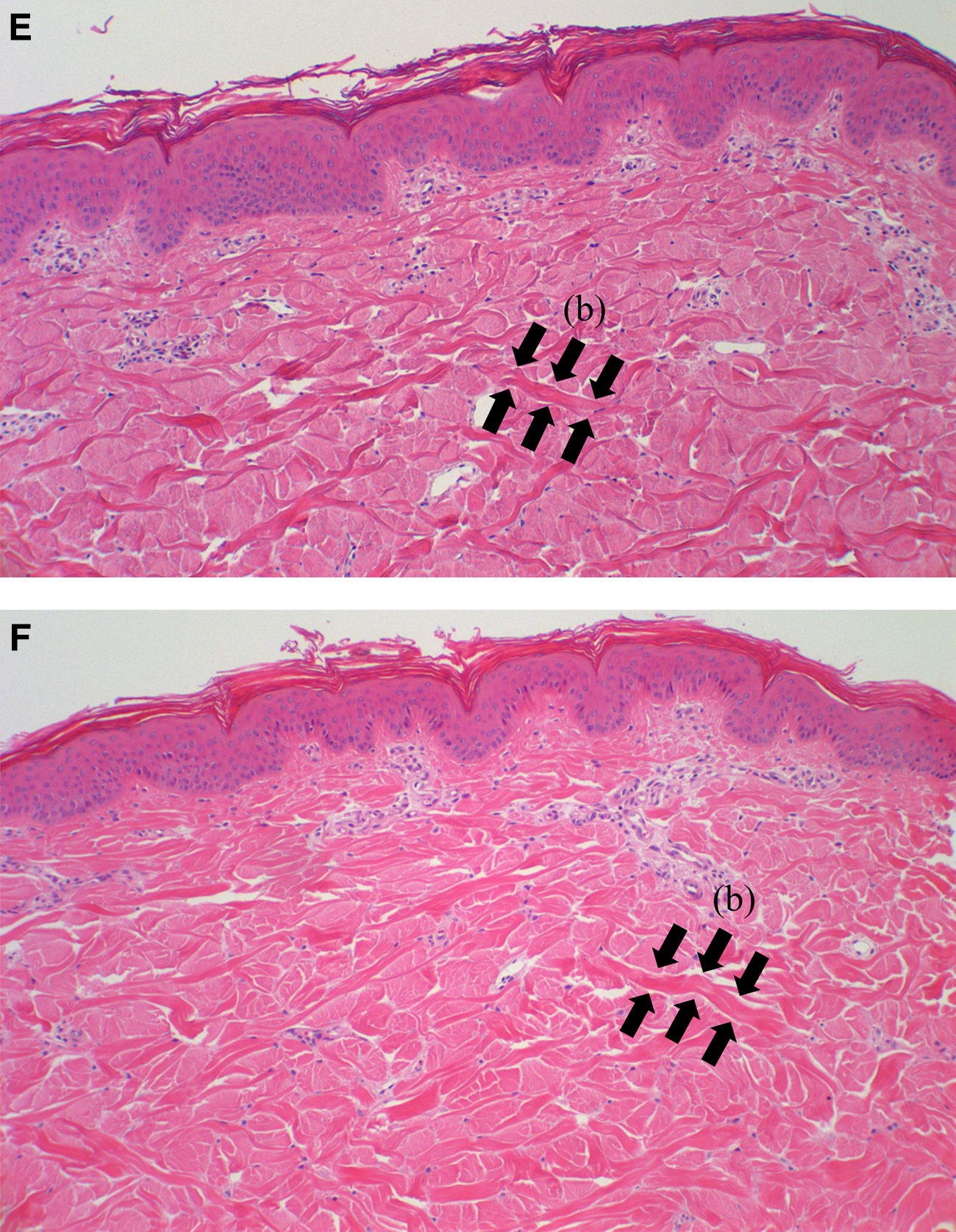
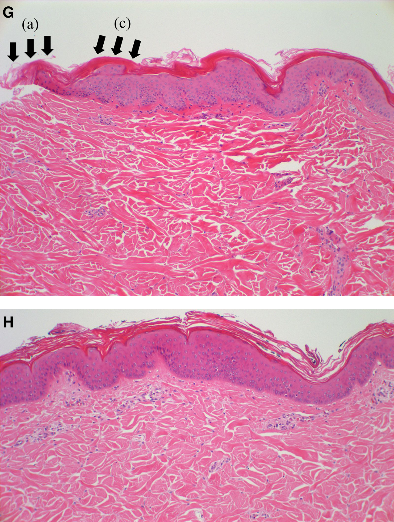
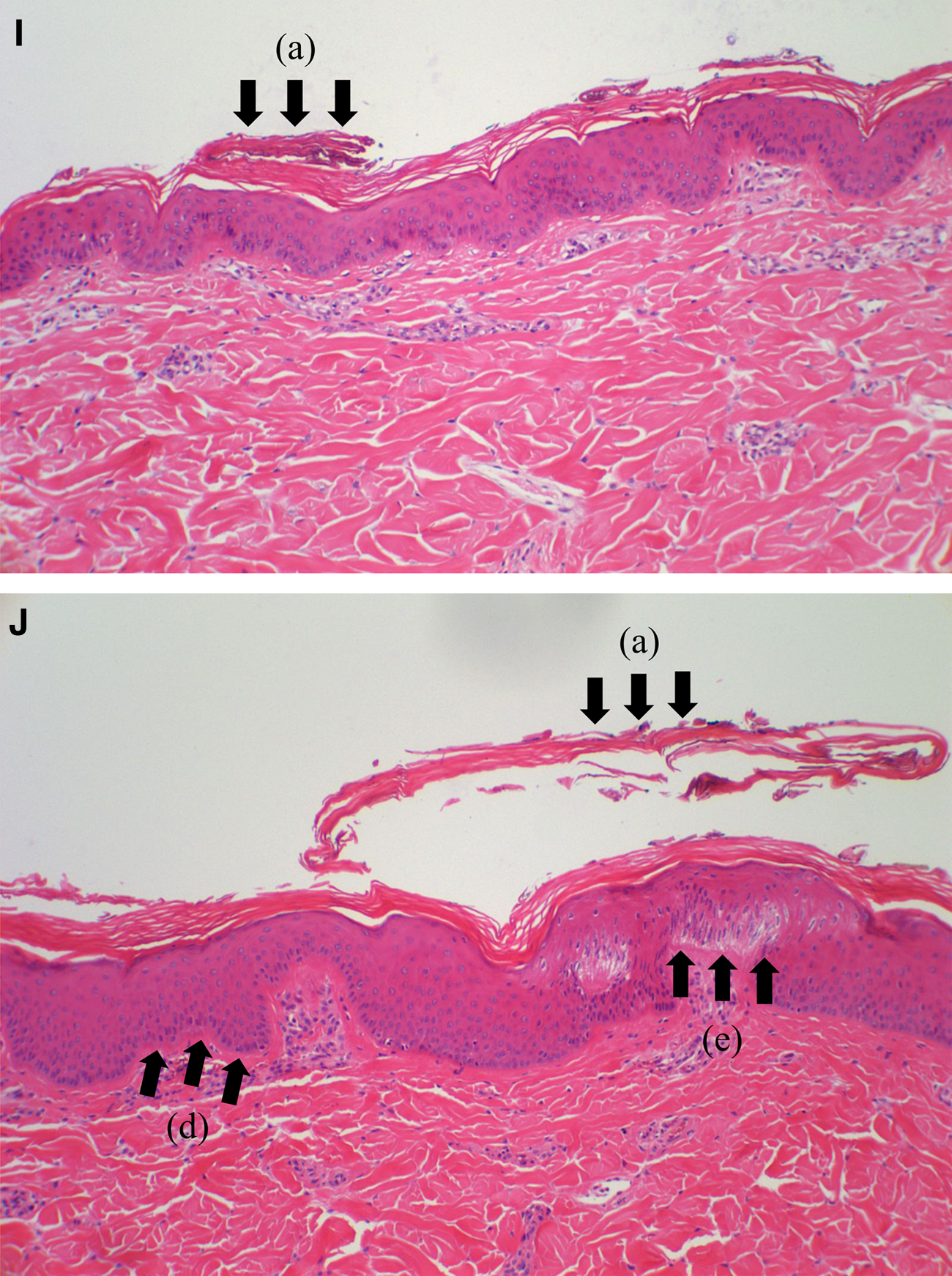
Regarding degrees of tissue damage for the different laser conditions, we found that tissue damages are influenced more seriously by spot diameters than by fluences. In addition, tissue damages occurred only up to below the epidermal layers. Figure 5 demonstrates the histologic analyses with regard to the degrees of tissue damage for the different laser conditions.
Discussion
The ideal treatment for skin rejuvenation causes tissue damage on the dermis without any influence on the epidermis. 19 For this reason, it is essential to transmit therapeutic lasers on the targeted lesions selectively, by proper combination of wavelengths, pulse durations, and fluences. 20 –22 In our study, we gave priority to internal-external maximum temperatures according to the different laser conditions, as the maximum temperature is one of the most important factors for evaluating degrees of tissue damage.
In order to evaluate the heat distribution quantitatively, we estimated internal-external temperatures for the various transmitting conditions of the therapeutic laser. As a result, we found that the heat is transmitted at the depth of 7.5 mm for all test conditions. The maximum surface temperatures were revealed>40°C only under certain limited conditions for the pig skin group, although the surface temperatures of the pig skin group were much higher than those seen in the phantom group. In addition, the maximum internal temperatures were shown mostly at a depth of 2.5 mm, and were <80°C for most experimental conditions. In general, the depth of 2.5 mm indicates the dermal layers, and the dermal temperature is known to be the most reliable factor for evaluating the treatment effects for skin rejuvenation. The existing studies reported that the thermal injuries occurred when the dermal temperature increased to>65–70°C. 23,24 Furthermore, it is also known that these thermal injuries produce smooth skins by accelerating collagen synthesis. 25 –27 Similarly, noninvasive studies also revealed that heating the dermis through the epidermis, and limiting surface temperature to ∼40°C associated with a dermal temperature of 60–70°C, provides skin texture improvements. 9,28,29 In contrast, irreversible injuries can occur when the dermal temperature increased to>80°C. 30 In our experiments, the conditions of a spot diameter of 5 mm with a fluence of 36 J/cm2 and a spot diameter of 10 mm with a fluence of 26 J/cm2 yielded the maximum surface temperatures>40°C. In addition, the maximum temperatures at the depth of 2.5 mm were within the range of 60–70°C. Therefore, these conditions only corresponded to the abovementioned ideal treatment conditions. However, the condition of a spot diameter of 10 mm with a fluence of 36 J/cm2 was unsuitable for skin rejuvenation, as internal temperatures increased to >80°C.
Regarding histologic evaluations, we found that the degrees of internal thermal injuries worsened as spot diameters and fluences increased. The conditions of a spot diameter of 8 mm with a fluence of 36 J/cm2 and a spot diameter of 10 mm with fluences of 30 and 36 J/cm2 revealed irreversible injuries on the dermis layer. However, conditions with a spot diameter of 5 mm produced significant therapeutic effects, irrespective of fluence conditions. Moreover, the conditions of a spot diameter of 8 mm with fluences of 26 and 30 J/cm2 as well as a spot diameter of 10 mm with a fluence of 26 J/cm2 also induced reliable thermal damages. Based on these experimental results, we selected the optimum treatment conditions for skin rejuvenation as being the laser condition of a spot diameter of 5 mm with a fluence of 36 J/cm2 and a spot diameter of 10 mm with a fluence of 26 J/cm2.
Despite these efforts, our study has two main limitations. First, the surface temperatures were shown to be relatively high, as the cooling procedures were not employed during laser transmission. The cooling process can maximize the therapeutic effects by minimizing the heat transfer to the surrounding tissues of the targeted areas. It is, therefore, expected that the optimum treatment ranges will be expanded, when the cooling procedures are employed during laser transmission. Second, the employed skin-mimicking phantom and pig skin samples presented relatively different properties compared with human skin. To improve the reliability of our results, additional experiments with more reliable testing materials will be required.
Conclusions
In this study, we analyzed internal-external heat distributions after transmitting therapeutic lasers to find optimum ranges for skin rejuvenation. We employed the 1064 nm Nd:YAG laser in the skin-mimicking phantom and pig skin samples according to the different fluences and spot diameters, and evaluated its internal-external temperatures. For histologic analyses, we also stained biopsy specimens with H&E and compared degrees of tissue damage for each experimental condition. Consequently, we selected the optimum treatment conditions for skin rejuvenation as the condition of a spot diameter of 5 mm with a fluence of 36 J/cm2 and that of a spot diameter of 10 mm with a fluence of 26 J/cm2. In our future study, we will focus on additional experiments involving in vivo environments using hairless mice. Moreover, we will evaluate the degrees of wrinkle reduction and collagen expression quantitatively.
Footnotes
Acknowledgments
This work was supported by the Dongguk University-Seoul Research Fund.
Author Disclosure Statement
No competing financial interests exist