




Abstract
Introduction
Z
It is important for a restorative material to have favorable mechanical properties; however, long-term retention of the restoration primarily depends upon the bond strength and durability of resin cement to the tooth and ceramic. In order to obtain adhesion between a luting agent and a restoration, pretreatment of the ceramic surface is required. 2,3 A strong resin bond depends upon micromechanical interlocking and chemical bonding, which requires surface roughening for adequate surface activation. 4 Surface treatments increase the microporosities on the ceramic, enhancing the mechanical retention. Treatment alternatives consist of coarse grinding, using diamond burs, air-particle abrasion with Al2O3 particles, silica coating, acid-etching, and any combination of these techniques. 3
The lasers have been widely used in medicine and dentistry since the first applications of ruby laser were introduced by Maiman in 1960. 4 The investigations of laser treatment for dental materials are more recent, especially for ceramics. Erbium: yttrium-aluminum-garnet (Er:YAG) and Neodmium: yttrium-aluminum-garnet (Nd:YAG) lasers are used for this purpose; 5 however, there is little available information regarding the effects of laser irradiation on the surface properties of high-strength zirconia ceramics. 6 –9
A clinical problem with the use of zirconia ceramics is the difficulty in achieving a suitable adhesion with resin cements. 10 Zirconia ceramics are not silica based; therefore, standard application of hydrofluoric acid will not create a sufficient surface roughness. Although the effects of surface treatments on glass ceramics are well documented, the best method for zirconia ceramics is still a subject of controversy. 11 According to most studies in the field, airborne particle abrasion with Al2O3 particles coupled with silica coating was reported to be one of the most effective methods for obtaining higher bond strengths for oxide-ceramics such as zirconia and alumina. 12 Furthermore, although the effects of laser on hard and soft tissue surfaces have been investigated, 13,14 studies evaluating its effects on the surface topography of zirconia are scarce.
Therefore, the aim of this in vitro study is to evaluate the surface roughness of zirconia ceramic surfaces after laser etching by Er:YAG laser at different pulse lengths, and airborne particle abrasion. The null hypothesis was that laser treatment would not increase the surface roughness compared with airborne particle abrasion.
Materials and Methods
Specimen preparation
One hundred and forty zirconia discs were prepared using the Cercon system (DeguDent, Hanau, Germany). The specimens were prepared 10 mm in diameter and 1.2 mm in thickness by an authorized dental laboratory, according to the manufacturer's instructions. A 1.0 mm deep stainless steel mold and a digital caliper (Neiko 01407A, Neiko Tools, Montevideo, Uruguay) was used to standardize the thickness of the specimens. The surfaces of the specimens were polished using 600, 800, and 1200 grit silicone carbide papers (English Abrasives, Stafford, UK) under water cooling using a special polishing machine (LaboPol-5, Struer, Denmark). Polishing to the final thickness of 1.00 (±0.13) mm was done using a special brush and polishing paste designed for clinical applications of zirconia ceramics (Wheel Brush Rodeo and Zi-Polish, Bredent UK, Derbyshire, UK).
Surface treatment groups
The specimens were randomly divided into 14 groups (n=10). One group was left as polished as a control, one group was airborne particle abraded, and 12 groups received laser treatment (Table 1).
SiC, silicone carbide; SSP, super short pulse; MSP, medium short pulse; SP, short pulse; LP, long pulse.
For the air-particle abrasion group, the specimens were abraded with 110 μm Al2O3 particles (EasyBlast, Bego, Bremen, Germany) at a pressure of 3 bars, from a distance of 10 mm perpendicular to the treated surface, for 15 sec.
For the laser treatment groups, an Er:YAG laser (Fotona; AT Fidelis, Ljubljana, Slovenia) with 2940 nm wavelength was used. Laser etching was applied at three different pulse energy levels (100, 200, and 300 mJ) with a pulse rate of 10 Hz at 10 W power. For each pulse energy level, four different pulse lengths; super short pulse (SSP, 50 μs), medium short pulse (MSP, 100 μs), short pulse (SP, 300 μs) and long pulse (LP, 600 μs) were tested. A contact hand piece (R14) was placed perpendicular to the specimen using a cylindrical sapphire optical fiber tip (1.3 mm in diameter and 8 mm in length). The laser was applied from a 1 mm distance (noncontact mode) using a specially prepared two part apparatus; the fixed upper part connected to the handpiece and mobile lower part where specimen was fixated with screws. The entire surface area was manually irradiated with sweeping movements with water cooling for 20 sec. The irrigation was standardized using the electronic spray control settings of the laser device (6.75 mL/min). The energy densities for 100, 200, and 300 mJ groups were 7.54, 15.08, and 22.61 J/cm2/pulse respectively.
Surface treatment evaluation
The influence of micromechanical retention produced by airborne-particle abrasion or laser treatment on the surface topography of zirconia specimens was evaluated using an optical profilometer (Wyko NT1100; Veeco Metrology, Inc., Tucson, AZ). Three dimensional images were captured at x5 magnification, covering an area of 1.2×0.9 mm. Software of the equipment with a measurement array of 768 x 480 (Wyko Vision 32) was used for evaluation. Three measurements were made for each specimen, and average roughness (Ra) values in micrometers were calculated.
Scanning electron microscopy (SEM) evaluation
One specimen from each group, with an Ra value closest to the mean value of the group, was selected for qualitative microscope analysis. The specimens were sputter coated with a thin layer of platinum, and evaluated using SEM (JSM 7000F, JEOL, Japan) at 2000x magnification.
Statistical analysis
Mean Ra values of laser etched and airborne particle abraded surfaces were compared with one way ANOVA and Tukey multiple comparison tests at a significance level of α=0.05.
Results
The surface roughness results of the 14 groups can be observed in Table 2.
Means followed by different capital letters were statistically different at p<0.05.
SSP, super short pulse; MSP, medium short pulse; SP, short pulse; LP, long pulse.
Control group observed to have the lowest surface roughness value in all groups (p<0.05).
For the 100 mJ laser etching groups, 50 and 100 μs laser duration resulted in significantly higher surface roughness than air-particle abrasion (p<0.05). For the 300 μs group, the Ra value was significantly higher than for the air-particle abrasion group, but significantly lower than for 50 and 100 μs groups (p<0.05). The difference between the Ra values of the 600 μs and air-particle abrasion groups was not statistically significant (p>0.05).
For the 200 mJ laser etching groups, 100 mJ 50 μs and 100 μs laser duration resulted in significantly higher surface roughness than air-particle abrasion (p<0.05). The difference between the Ra values of the 300 μs, 600 μs, and air-particle abrasion groups was not statistically significant (p>0.05).
For the 300 mJ laser etching groups, there was no statistically significant difference in the Ra values between them and the 50 μs, 100 μs, 300 μs, 600 μs, and air-particle abrasion groups (p>0.05).
In SEM imagery and optical profilometry, the increase in the roughness of the Er:YAG treated specimens can be observed in accordance with increase in the Ra values. The surface of the specimen from the control group displayed low surface roughness; however, the SEM imagery displayed sharper particle edges; this appearance was not present in specimens with surface treatments (Fig. 1). The SEM image of the air-particle abraded sample did not display the “sharp and grainy” look of the control specimen; the surface was observed to be smoother (Fig. 2). In the laser-treated specimens, profilometry and SEM images displayed an even smoother look, and blister-like globules. At higher energy levels and longer pulse durations, smoother areas, fewer undercut regions, and shallow pits were observed because of melting and cooling of the surface (Figs. 2 –4).
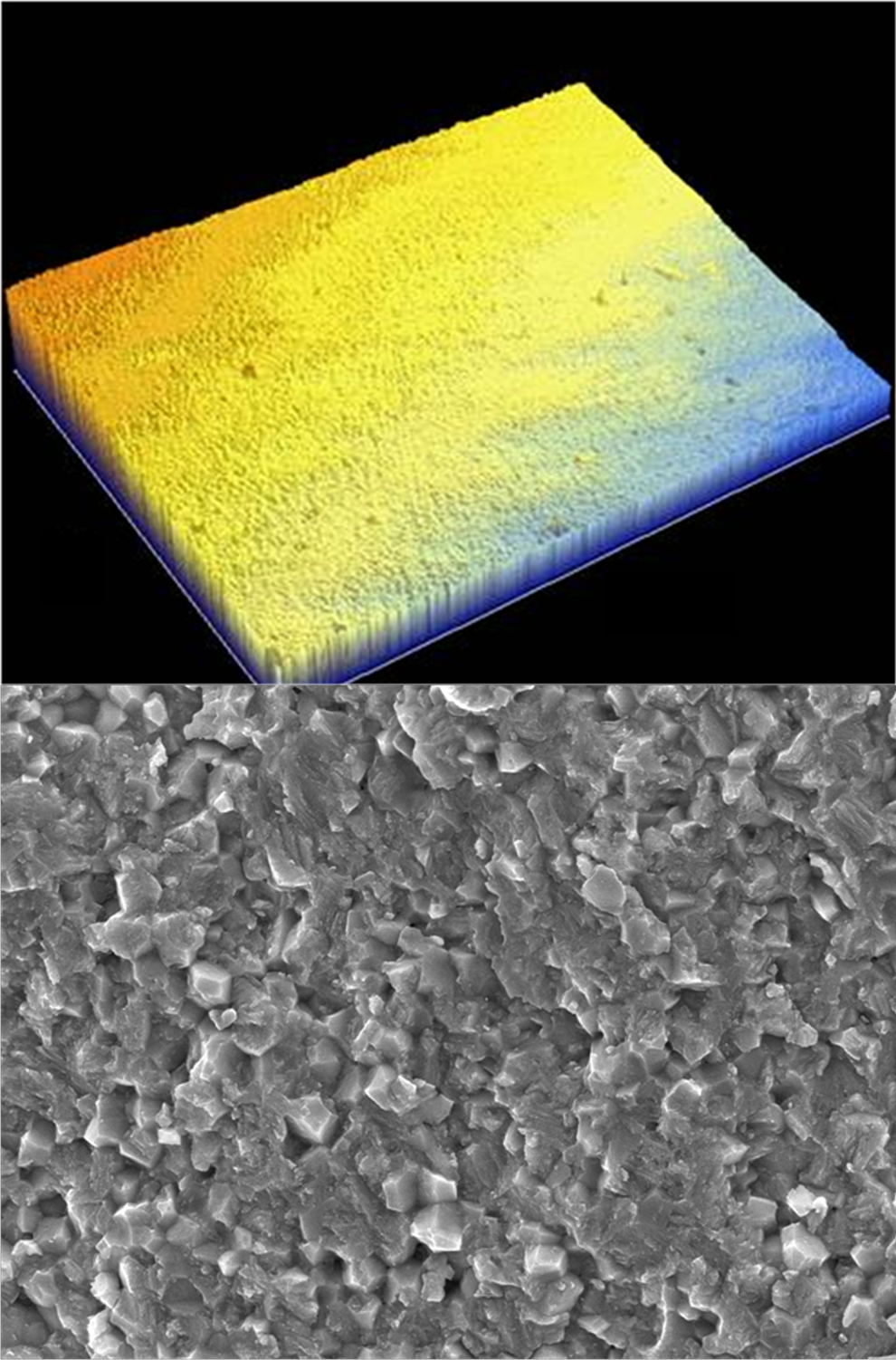
Top: Three dimensional (3D) profilometric image of a specimen from polished control group. Bottom: Scanning electron microscopic (SEM) image of a specimen from polished control group.
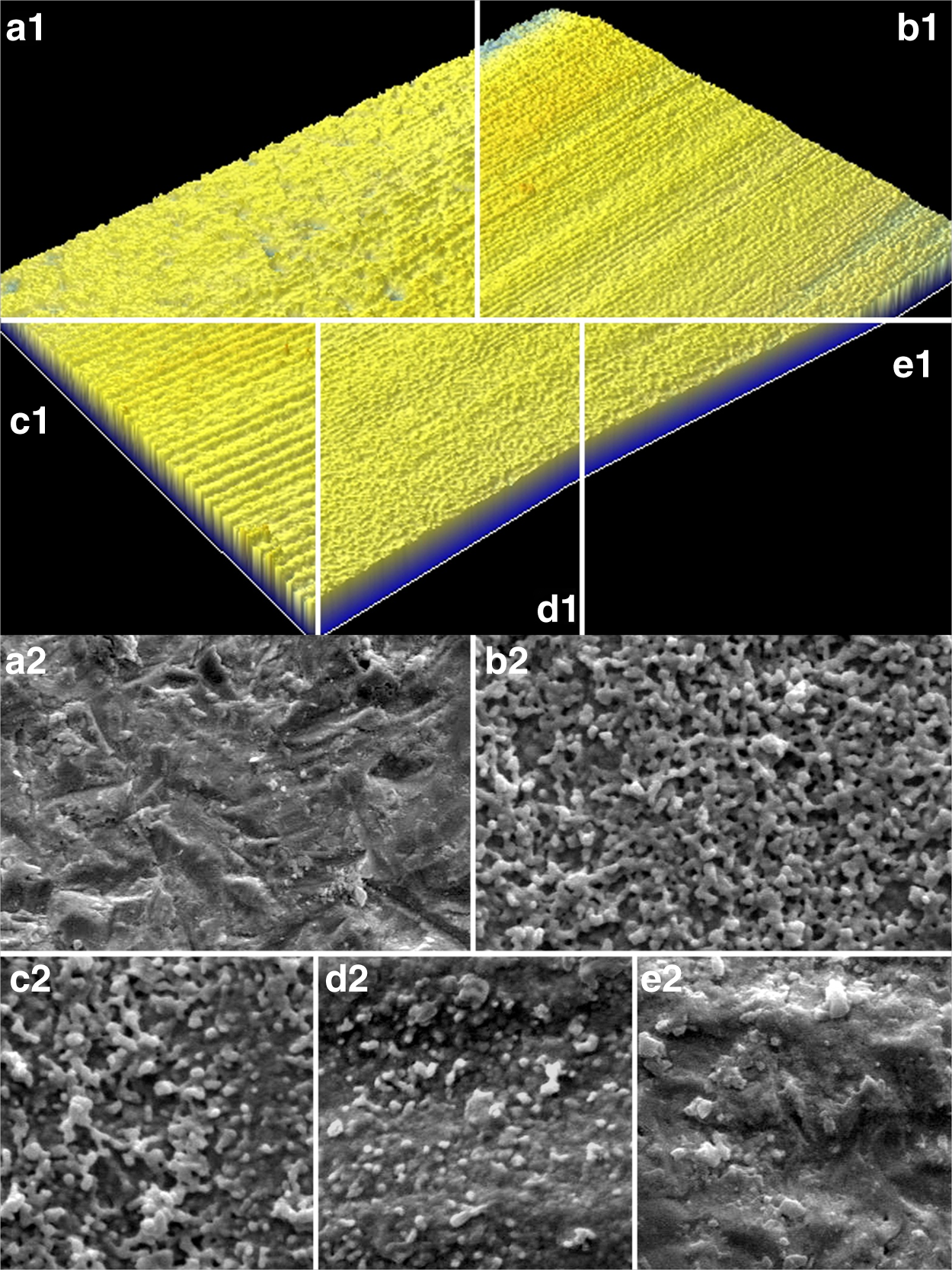
Top: Three dimensional (3D) profilometric images of air-particle air particle abrasion (a1), 100 super short pulse (SSP) (b1), 100 medium short pulse (MSP) (c1), 100 short pulse (SP) (d1), and 100 long pulse (LP) (e1) groups. (The images from different samples were arranged digitally for better visual comparison.) Bottom: SEM images of air particle abrasion (a2), 100 SSP (b2), 100 MSP (c2), 100 SP (d2), and 100 LP(e2) groups.
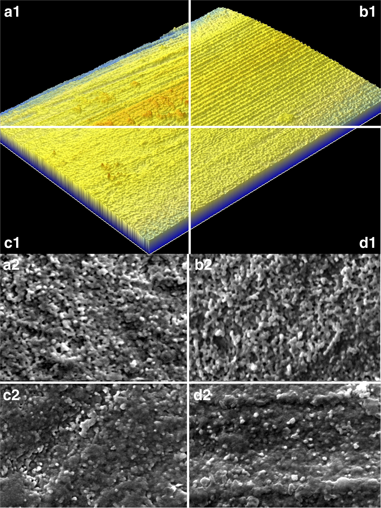
Top: Three dimensional (3D) profilometric images of 200 super short pulse (SSP) (a1), 200 medium short pulse (MSP) (b1), 200 short pulse (SP) (c1), and 200 long pulse (LP) (d1) groups. (The images from different samples were arranged digitally for better visual comparison.) Bottom: Scanninig electron microscopic (SEM) images of 200 SSP (a2), 200 MSP (b2), 200 SP (c2), and 200 LP (d2) groups.

Top: Three dimensional (3D) profilometric images of 300 super short pulse (SSP) (a1), 300 medium short pulse (MSP) (b1), 300 short pulse (SP) (c1), and 300 long pulse (LP) (d1) groups.(The images from different samples were arranged digitally for better visual comparison.) Bottom : Scanninig electron microscopic (SEM) images of 300 SSP (a1), 300 MSP (b1), 300 SP (c1), and 300 LP (d1) groups.
Discussion
The results of the current study support the partial rejection of the hypothesis; treating the zirconia surface with Er:YAG laser increases surface roughness and microirregularities. At lower energy levels and low duration, this increase in surface roughness is greater than with air-particle abrasion. However, as the energy and duration increase, the surface roughness significantly decreases.
The changing parameters of laser treatment in the current study were pulse energy (in mJ) and pulse length (in μs). In order to evaluate the results of the study, the effective radiating area (ERA) and energy density terms need to be explained. ERA is defined as “area covered by the beam either at the tip of the applicator or any given distance beyond that point,” 15 which is the same as the spot size at the tip of the applicator, and it is a constant for all specimens in this study. Energy density is the amount of energy delivered per unit area, which is not constant for all groups. As the pulse energy and pulse length increase, the energy density increases, because the amount of energy on the ERA increases. 15 As a result, higher heat energy is produced per unit area, resulting in increased melting of the zirconia surface followed by immediate water cooling.
According to the findings of the current study, it can be speculated that there is a threshold for this melting and cooling cycle to produce a meaningful change in surface roughness. If the laser energy is delivered over a larger area with a tip with a larger diameter, the effective energy density would decrease. This aspect may be modified by the user to decrease the energy received per area. Enwemeka 16 stated that energy density may be considered as the dose of the treatment, and it is critical in determining the outcome of the application. It was also reported that as the power of the device increases, the treatment time can be decreased to achieve the same or even a better result (e.g., better tissue healing). This approach may be used when treating zirconia material, as perhaps applying the laser energy with a larger tip for a shorter time and at a lower pulse length may result in better surface properties for the clinical application of the material.
Although many studies have investigated the effects of lasers on dental hard tissues, their effect on dental ceramics still is not clear. In principle, laser energy affects the dental hard tissues by ablation. Ablation is the result of microexplosions of water molecules present in the crystalline and organic components in dentin. The surface pigmentation and water content are the main factors determining the energy absorbed by the irradiated area. 15,17 In comparison, zirconia ceramic does not contain any water molecules, and has an opaque coloration, which may influence the laser absorption negatively. 9 Also, it is suggested that the reflective surface of the material affects the laser absorption negatively, 18 and that coating the surface with graphite powder may increase the effectiveness of laser treatment. 19
The studies conducted on laser treatment of zirconia ceramic are even fewer than those on lithia-based ceramics. The effects of laser treatment on oxide ceramics are very different than those on lithia-based ceramics, because of the presence of a glass phase in the latter. 1,20 Also, there are studies evaluating the changes in pulse energy, 1 –6,9,10,18,19 but the importance of pulse length in treating zirconia is not investigated in the literature.
In the current study, changes in the pulse length displayed significant changes in the 100 and 200 mJ groups. However, in the 300 mJ group, the pulse length did not produce any significant change in the surface topography. In the profilometry images and SEM photographs, the specimens with high Ra values displayed a rough and irregular appearance, which was qualitatively different than the polished and air-particle abrasion specimens. The dark pits, globules, and smooth protrusions in the imagery may be the result of melting and immediate cooling after the laser application. However, as the pulse energy and pulse length increased, the surfaces were observed to have fewer dark areas, hence less depth and fewer shallow protrusions. The reason for the decrease in roughness may be the result of excessive melting and cooling on the surface of the material, resulting in a smoother topography.
Different laser systems with various parameters of laser energy were used by researchers on zirconia ceramics; 1 –6,9,10,18,19 however, in the current literature, there is a lack of consensus on the effectiveness of laser etching on zirconia. In accordance with the present study, Usumez et al. 1 suggested that with a strong output power (>3 W) Nd:YAG laser increases surface roughness and bonding of zirconia. Cavalcanti et al., 19 comparing the laser treatment and air abrasion with Al2O3 particles, and metal primer application of zirconia, reported that laser treatment affects the bonding positively. They used 200, 400, and 600 mJ energy intensities for two different commercial zirconia brands (Cercon and Procera), and reported that at 200 mJ, Procera specimens were rougher than air-particle abrasion specimens. However, for Cercon specimens, the result was the opposite. At 400 and 600 mJ energies, they 19 reported excessive deterioration at the surface, which is in accordance with findings of current study. Kirmali et al. 21 applied Er:YAG and Nd:YAG laser treatment to zirconia specimens, both coupled with and without air-particle abrasion, and reported that whereas Er:YAG treatment resulted in significant increase in surface roughness, Nd:YAG treatment was not effective. Akin et al. 3 also compared Er:YAG and Nd:YAG laser treatment and air-particle abrasion by evaluating the shear bond strength, and concluded that both laser treatments improved the bond strength compared with air-particle abrasion.
Several studies reported laser treatment to be inefficient for surface treatment compared with air-particle abrasion. Dilber et al. 5 evaluated only a single setting of Er:YAG laser at 500 mJ energy and 100 μs pulse length, and compared the results with air-particle abrasion. At those parameters, Er:YAG treated surfaces displayed lower surface roughness than those treated with air-particle abrasion. Demir et al. 6 treated zirconia surfaces with Er:YAG laser at 200, 300, and 400 mJ intensities at 100 μs, and reported that the laser application was not as effective as air abrasion with 110μm Al2O3 particles.
The current study compared different pulse energy and pulse length parameters, which had not been evaluated in previous studies. The results displayed that changes in the pulse energy and pulse length influenced the surface topography of zirconia significantly. The change in surface properties may affect the bonding of resin cement to the restoration, and, therefore, have a clinical significance. It has been reported that an increase in surface roughness increases the bonding of resin cement to zirconia, affecting the clinical success positively. 22 It is even suggested that micromechanical retention, which increases with the increase in surface roughness, may be the most effective mechanism for the adhesion of oxide ceramics. 23 Several studies evaluated different pulse energies, but the current study shows that pulse length should be taken into consideration as well. The ceramics in the market have differences in composition and microstructure, and these variations might affect the results of laser application on the surface properties of the material. The subject of the study can be investigated further by evaluating the bonding and surface properties in detail. Further studies are recommended that would investigate the bond strength via microtensile and microshear tests, and evaluate the surface roughness in more detail with profilometry parameters such as root mean square roughness (Rq), maximum height of the roughness (Rt) and average maximum height of the profile (Rz).
Conclusions
In order to increase surface roughness and promote better bonding to resin luting agents, Er:YAG laser etching may be an alternative to air-particle abrasion for zirconia ceramics. However, high levels of pulse energy and longer pulse length may decrease surface roughness and possibly have an adverse effect on micromechanical locking properties.
Footnotes
Author Disclosure Statement
No competing financial interests exist.