
Abstract
Introduction
U
Hard tissue laser applications in orthodontics comprise enamel etching, bracket bonding, and debonding. The bonding of brackets on the tooth surface requires penetration of the bonding material into the etched enamel. Etching by laser radiation, mainly Nd:YAG, 8 Er:YAG, 9 and Er:YSGG, 10 can produce retentive surface known as microirregularities suitable for resin penetration. The acid-resistant effect of the laser is superior to conventional acid laser conditioning for both the dentin and enamel surfaces, but it is necessary to monitor the laser energy output to avoid substructural damage. The difference between enamel (96% in weight) and dentin (about 70% in weight) is based on mineralization of tissues. 11
The last method where laser radiation is frequently used is laser debonding. The principle of safe bracket debonding is to degrade the adhesive resin strength connecting the tooth and bracket. Removal of adhesive resin from tooth surfaces without iatrogenic damage (enamel loss) is generally the main problem of this method. 12 The composite remnants after classical mechanical debonding had a mean volume of 2.48 mm3 (SD ±0.92 mm3), 13 and therefore, the following surface cleaning can destroy the layer of enamel. The occurrence of enamel fracture is also relatively higher with ceramic brackets because they are more brittle and resistant to deformation and have higher bond strengths than metal and plastic brackets. 14
According to Tocchio et al., 15 laser energy can degrade the adhesive resin by thermal softening, thermal ablation, or photoablation. 15 Thermal softening occurs if the laser heats the bonding agent until it softens, so the bracket succumbs to gravity and slides off the tooth surface. Thermal ablation occurs if heating is fast enough to raise the temperature of resin to vaporization before debonding by thermal softening. The third type of interaction, photoablation, can take place only if the adhesive material is exposed to very high energy of laser light (e.g., UV), and energetic photons can break down the molecular bonds of the target material. The adhesive resin is thus decomposed. The main prerequisites of laser photoablation are high energies and very short pulses. 16
Laser debonding is used not only for brackets but also for veneers 16,17 and all-ceramic crown removal. 18,19 For these purposes, Er:YAG, Er,Cr:YSGG, CO2, Nd:YAG, and Tm:YAG were applied. 1
Clinically, mainly Rechmann et al. 18,19 have shown the practical benefits of laser debonding, that is, mainly how to remove ceramics from the tooth without crown destruction. Of interest are also the application time, reduction of debonding force, and a possible enamel damage. An important factor is also intrapulpal temperature increase, types of lasers and brackets, and different resins with varied reactions to specific laser radiation.
The aim of this study is the evaluation of the Er:YAG laser radiation (wavelength 2.94 μm) effect on bracket debonding. The main goal of our contribution was to monitor the intrapulpal temperature increase and the interaction with various metal or ceramic brackets and adhesive materials.
Materials and Methods
Specimen preparation
The study used a set of 43rd molars of young patients (aged 15–25 years), extracted for orthodontic reasons. Patients were requested to provide consent to the experiment. Ethical approval for the study was obtained from the Faculty Hospital Motol and 2nd Medical Faculty of Charles University Ethics Committees. The extracted teeth were stored in 100% humidity at 37°C until testing started.
Ten ceramic brackets (Clarity™ Advanced; 3M Unitek, Monrovia, CA) and 10 metal brackets (Victory Series™; 3M Unitek) were bonded on extracted teeth using the adhesive system (Transbond™ XT Light Cure Adhesive; 3M Unitek). The last group of 10 ceramic brackets (Clarity Advanced; 3M Unitek) was bonded using Variolink II Professional Set adhesive system (Ivoclar Vivadent AG, Schaan, Liechtenstein).
In the first two groups of brackets (10 ceramic and 10 metal), where Transbond XT Light Cure Adhesive Paste was used as adhesive system, the enamel surface was cleaned with nonfluoride polishing paste, rinsed with a stream of water, and air-dried. Transbond Plus Self-Etching Primer (3M Unitek) was applied for 10 sec on the labial surface of extracted teeth and smoothly blown. The bases of brackets were covered with Transbond XT Light Cure Adhesive Paste, then placed on prepared labial sides of extracted teeth, and light-cured with Ortholux™ Luminous Curing Light (3M Unitek) (outputs of 1600 mW/cm2) for 10 sec.
In the series of 10 ceramic brackets, an adhesive Variolink II Professional Set system (dual-curing composite system) was used; at first, the surfaces of the extracted teeth were totally etched with phosphoric acid for 30 sec. After air-drying, the enamel showed a chalky white hue. Monobond Plus was applied with a tiny brush on the ceramic bracket base for 60 sec and then air-dried. The last step consisted in covering the prepared ceramic bracket base and the labial surface of the tooth with Heliobond. Variolink II (mix 1:1, catalyst:base) was used as the adhesive system. After the bracket was seated on the prepared labial side of the tooth, it was light-cured for 40 sec.
The control group of nine brackets was prepared, where the orthodontic plier was used for debonding (without the laser irradiation). Three samples were used for every type of brackets and adhesive system; one other tooth was only cleaned with nonfluoride polishing paste, rinsed with a stream of water, and air-dried. It was used for checking of unattached enamel. The surface was also checked in SEM.
Laser irradiation source
The experiment used a laboratory prototype of flashlamp-pumped solid-state Er:YAG laser with lasing medium—erbium-doped yttrium aluminum garnet (Er:Y3Al5O12) emitting 2.94-μm radiation (Czech Technical University in Prague, FNSPE prototype, Prague, Czech Republic) (Fig. 1). The laser parameters are shown in Table 1.
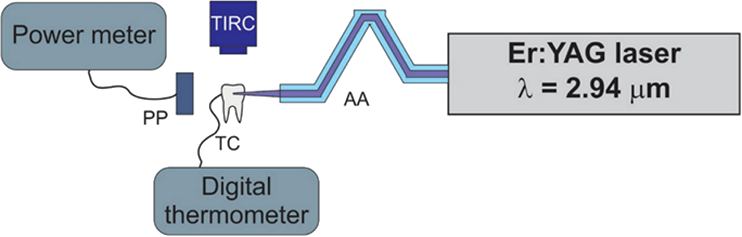
Experimental arrangement. PP, power probe; TIRC, thermo IR camera; AA, articulated arm.
During our experiments, fine water mist (water 10 mL/min) was used. The delivery system for this case was the same as in the classical high-speed drilling machine (two channels: one for water and one for air).
Analysis methods and measuring instruments
To determine the energy and time characteristics of the Er:YAG laser system, we used Coherent probes (P1) J25LP-ERBI and (P2) J-10MB-LE (Molectron-Coherent, Portland, OR) and Vigo PVI6 IR detector (Boston Electronics Corporation, Brookline, MA) (rise time 10 nsec) or InAs/InAsSbP photodiode (model PD36-05, IBSG, spectral range 0.8–3.8 μm, and rise time 150 ns) (Lambda Photometrics, Hertfordshire, United Kingdom) along with the Tektronix DPO4104B-L oscilloscope (Tektronix, Portland, OR). The laser beam spatial profile was monitored with IR-sensitive Pyrocam III pyroelectric camera (Ophir-Spiricon, North Logan, UT) (spectral range 2–8 μm).
Before the interaction experiment, all the measured teeth with brackets were photographed with a Nikon SMZ-2T (Osaka, Japan) stereomicroscope connected to a Mintron color video camera (MTV-73X11P-R; Mintron Enterprise, Fremont, CA) and a computer. The teeth were divided into three groups as follows: (1) ceramic bracket Clarity Advanced and Transbond XT Light Cure Adhesive Paste, (2) ceramic bracket Clarity Advanced and Variolink II Professional Set adhesive system, and (3) metal Victory Series and Transbond XT Light Cure Adhesive Paste. Each group had its control samples—debonded without laser irradiation.
For interaction measurements, the tooth with the bracket was fixed in a special holder, and the above-described Er:YAG laser radiation was used for debonding. The radiation energy used was 280 mJ and the repetition rate 6 Hz, and the irradiation time derived from the preliminary experiments was 140 sec. During irradiation, the tooth with the bracket was cooled. The water flow was 10 mL/min.
During irradiation, the bracket and tooth surface temperature spatial distribution were monitored with a thermal image infrared camera Optilas–Electrophysics PV320L2E (Alphen aan den Rijn, Netherlands). The temperature changes inside the tooth (in the root area) during the bracket irradiation were recorded with the NiCr-Ni thermocouple and digital thermometer GMH 3210.
After irradiation, the bracket was removed from the tooth surface by professional 3M Unitek band removing pliers as it is also in reality. After treatment, all the irradiated samples were once again photographically documented with the Nikon SMZ-2T stereomicroscope, and then, the tooth tissue surface was analyzed with a JSM 5510 LV scanning electron microscope (SEM; JEOL, Tokyo, Japan).
Statistical analysis
Statistical Student's t-test (at the significance level p = 0.01), including standard deviation, was implemented to monitor objective measurements.
Results
Debonding procedure and temperature measurement
The irradiation conditions and results obtained are summarized in Fig. 2, where the blue color shows that temperature increase is higher in the metal bracket. Overheating was limited and does not interfere surrounding tissues, mainly with enamel and dental pulp.
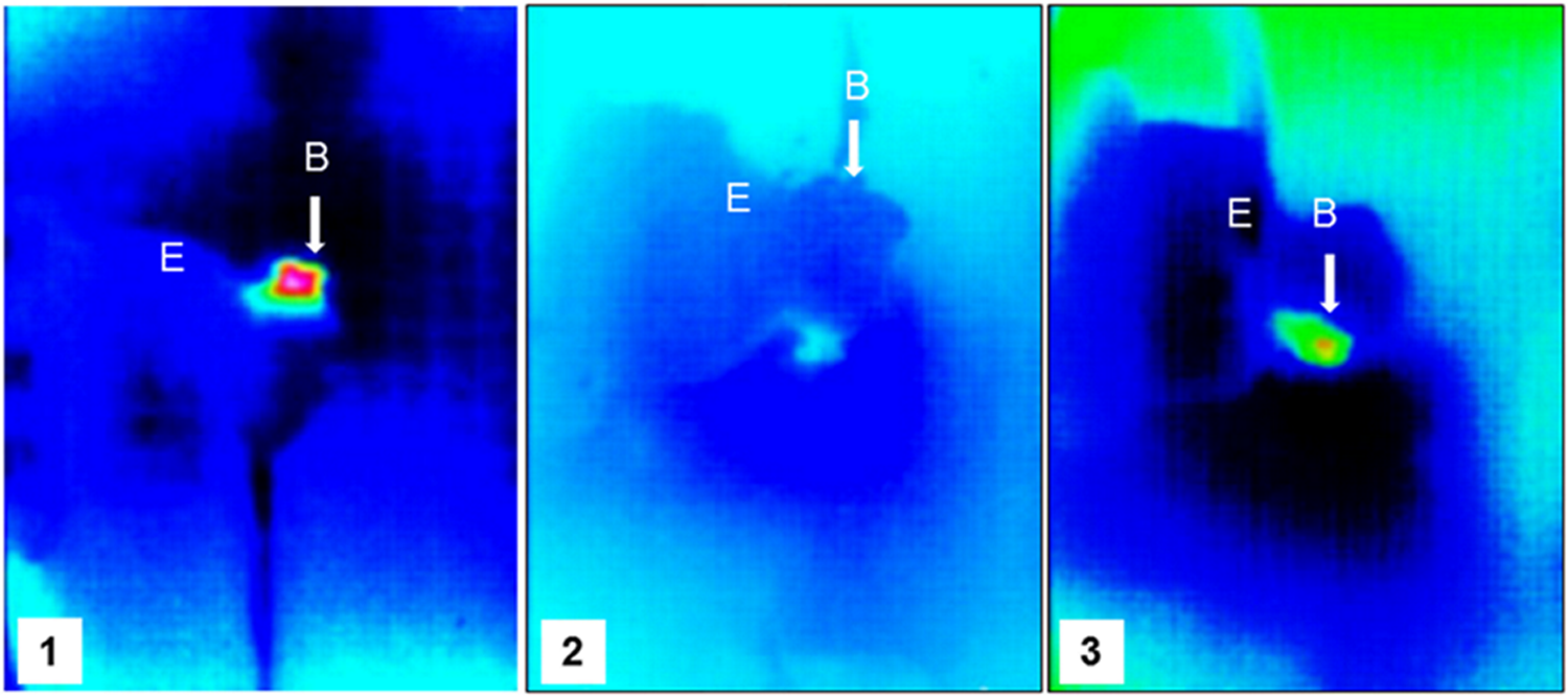
Temperature rise monitoring. B, bracket; E, enamel; 1, Victory™ Series + Transbond™ XT Light Cure Adhesive Paste; 2, Clarity™ Advanced + Transbond XT Light Cure Adhesive Paste; 3, Clarity Advanced + Variolink II Professional Set adhesive system.
The Er:YAG laser was used in conjunction with water cooling during the 140-sec bracket irradiation. A thermocouple placed inside the pulp chamber verified that water cooling protected the tooth; the heat was concentrated inside the bracket and adhesive resin only. Then it was easy to remove the bracket with special pliers.
From the temperature profile measurements, it was apparent that bracket heating due to radiation (from 2.2°C to 3.0°C for 140-sec irradiation time) was safe, and the remaining radiation transmitted through the tooth tissue did not heat the thermocouple (Fig. 3).

Temperature rise during bracket debonding 140 sec. 1, Victory Series + Transbond XT Light Cure Adhesive Paste; 2, Clarity Advanced + Transbond XT Light Cure Adhesive Paste; 3, Clarity Advanced + Variolink II Professional Set adhesive system.
Scanning electron microscopy investigation of the tooth after debonding
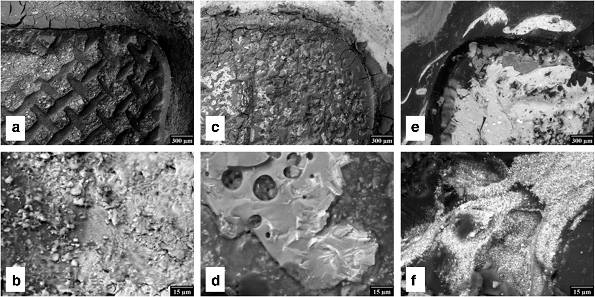
It has been found that Er:YAG laser radiation is an effective tool for safe debonding. The results for clinical practice depend on the bracket material as well as the adhesive systems. The criterion of success was the amount of clearly visible resin remnant on each tooth and its quality ascertainable in SEM. The residual adhesive can be seen in Fig. 4.

SEM evaluation—surface after laser debonding.
During laser application, the bracket metal material overheated the adhesive paste. Then it was very easy to remove the brackets; nevertheless, the rest of the gluing layer stayed strongly connected with enamel (Fig. 4a, b).
The ceramic bracket was directly connected with enamel, and so, the aesthetic quality was excellent. After laser irradiation, the bracket was removed without cracks, and the rest of Transbond XT Light Cure Adhesive Paste was visible on the enamel surface (Fig. 4c). Therefore, it was evident that after bracket debonding, some resin remnant had to be mechanically removed (Fig 4d).
Quite different results were observed after applying the Variolink II Professional Set adhesive system (Fig. 4e, f). This adhesive system was also removed with most amount of adhesive material, and only finishing and polishing of enamel had to be applied.
The control group for each type of brackets and adhesive resin was also evaluated in SEM. The amount of adhesive material is higher for all types of brackets (Fig. 5a–f). In the case of ceramic brackets also, the rest of ceramic material was found (Fig. 5c–f).
SEM evaluation—control group surface after orthodontic plier debonding.
Discussion
Experimental setup was based on standardization of treatment parameters, including a detailed description of the laser system. 20 Of considerable importance was dose limitation, 21 which is directly connected with enamel cracks presence. 22 Of importance for erbium family lasers are not only the wavelength, power, irradiation time, beam area in tooth, pulse parameters, anatomical location, and time of therapy 23,24 but also water flow during tissue water cooling. 25 For this reason, we prepared an experimental arrangement and Er:YAG laser parameters, which could be safe from the point of view of Er:YAG radiation application dose response. 26,27
We used the laser parameters, which overheat the laser adhesive system but not the dental pulp; temperature increase was safe but did not exceed 3.2°C. The system was able to remove bracket without ceramic material fracture and enamel ablation.
The disadvantages of classical debonding include enamel or bracket fractures and pain due to extensive debonding force. Laser debonding has several advantages: It saves time and reduces debonding force and enamel damage. 1 Er:YAG 28 and Er:YSGG 18,19 lasers seem to be the most effective devices for clinical practice. Our study using Er:YAG laser radiation, similar to the study by Rechmann 18,19 using Er:YSGG laser system, has confirmed that strong water cooling is necessary for protection of hard dental tissues and pulp. Zach and Cohen 29 found that the pulp tolerates an increase of 5.5°C in intrapulpal temperature. Overheating harms the pulpal tissue. During Er:YAG laser irradiation, temperature rise increase ranged from 2.0°C to 3.2°C in the dental pulp. Our temperature maps have shown that the debonding process was based on thermomechanical ablation in the superficial part of the adhesive layer. In addition, Rueggeberg 30 reported that various resins respond differently to certain types of lasers.
After classical therapy, the brackets are debonded, and resin remnant must be carefully mechanically removed. For the clinician also, the time needed for complete cleaning is essential. Several factors, such as the type of adhesive resin and debonding instruments, are related to the amount of enamel loss. 31 Enamel breakouts are frequent in debonded surface teeth, for example, 25–30% in molars. 13 The enamel lost during orthodontic procedures is not significant due to enamel thickness. 32 The thickness of adhesive paste is approximately 1500–2000 μm. For this reason, it would be optimal if laser radiation debonding softened the adhesive resin and if after laser irradiation not only the bracket but also the resin remnant could be removed. We have found that Er:YAG laser radiation used for debonding is able to remove not only the ceramic bracket Clarity Advanced but also most of the Variolink II Professional Set adhesive system (Fig. 3e, f). SEM images show an almost pure layer of enamel, which must be subject to long-term follow-up because of a possible future caries presence based on unsuccessful dynamic balance between demineralization and remineralization. 33,34 Similar findings were observed during all-ceramic crowns and dentin debonding. 18,19 A control group (orthodontic pliers bracket debonding) for every type of brackets and adhesive resin was also evaluated in SEM. It was found that the amount of adhesive material is higher for metal and ceramic brackets. In the case of ceramic brackets, the rest of ceramic material was also detected. It could be dangerous for enamel in the case of mechanical removing. It was proved that laser irradiation helps protect enamel surface.
Conclusions
In our study, it has been observed that bracket removal was easier after the Er:YAG laser irradiation. The temperature rise was limited (from 2.0°C to 3.2°C) also for metal brackets. As against the nonirradiated samples, SEM investigation has confirmed no damage of enamel.
Irradiation with Er:YAG laser before debonding of ceramic brackets significantly decreases the bonding failure and amount of remaining adhesive.
In the case of mechanical removing with orthodontic pliers, only the rest of ceramic material was regularly detected. It was proved that laser irradiation helps protect enamel surface.
Footnotes
Acknowledgments
This research has been supported by the Grant of the Czech Ministry of Education no. MSM68407700 and project no. 00064203 (FN MOTOL).
Author Disclosure Statement
No competing financial interests exist.