
Abstract
Introduction
A
The literature is prolific in demonstrating effects of PBMT at several cell functional levels, such as proliferation, migration, differentiation, and protein synthesis, as well as growth factors, all of them important for tissue regeneration that include fibroblast, osteoblast, muscle, and endothelial cell cultures, among others. 3 –6
The authors developed an innovative technique, 7 hemolasertherapy (HLT), which is described in the clinical cases. The advantage of this technique is that it is nonsurgical and simple to execute, giving it an edge over the conventional techniques. HLT is the result of hemotherapy treatments carried out with blood, wherein the patient's blood drops are used in a given area together with laser PBMT.
The authors started developing this technique in early 2010 and nowadays have several clinical cases and a wide follow-up to present. This clinical study was carried out at the Biophotonics Center of Institute Brugnera and Zanin, São Paulo, Brazil, where results of three clinical cases treated with HLT to close black spaces were evaluated. The researchers' question was how the gingiva could grow so fast, occupy the adequate space, and keep stable for long follow-up periods.
Simultaneously to the clinical study, a preliminary experiment was conducted at the School of Dentistry Laboratory of University of São Paulo, Brazil, to demonstrate what could be happening at the cellular level in clinical cases of bleeding caused in the gingival tissue when associated with the application of laser PBMT.
Many in vitro studies using cell cultures have widely contributed to the knowledge of mechanisms subjacent to the clinical effects of PBMT. 3,8 Recent publication of a systematic review on the effect of PBMT on mesenchymal stem cells (MSCs) concluded that it has a positive effect influencing proliferation of the studied stem cells. 8
This initial in vitro study demonstrated that the blood clot originated from the bleeding provoked in the gingival area is rich in MSCs. 9 However, they need more stimuli to preserve viability and additional differentiation. The authors analyzed the effects of a provoked bleeding in the marginal gingival of patients with black spaces followed by PBMT and present the outcome of a combined regenerative therapy with PMBT through provoked bleeding. A positive expression for all analyzed genes was increased after PBMT.
PBMT showed positive effects when applied in tissue engineering. 3 PBMT improves survival, proliferation, migration, and differentiation of originated dentoalveolar stem cells. 8 In addition, human dental pulp regeneration was obtained by combining PBMT with bleeding provoked in the radicular canal. 10
Based on that the authors hypothesized that gingival papilla regeneration could be obtained by combining PBMT bleeding in the gingival papilla area with loss or anatomical deformity of the papilla. The blood clot would act as a natural scaffolding containing stem cells and growth factors present in blood platelets, whereas PBMT would improve survival and differentiation of MSCs. To test this hypothesis, the authors gently provoked a bleeding on the gingival margin of patients having black spaces in anterior teeth and after that applied PBMT with adequate parameters.
Based on the previous studies, 3,7,8,10 the authors are interested in investigating the clinical cases presented, the effect of laser therapy on gingival papilla growth, and how the stem cells respond to such therapy.
Materials and Methods
This new technique (HLT) suggests the use of 660 nm diode laser associated with gingival bleeding to fill black spaces, both in the same session, after the cementation of porcelain veneers. Gingival bleeding is caused by gently stimulating the gingival sulcus with an n.5 clinical probe. This procedure aims to release mesenchymal stem cells (MSCs) along with the blood to favor neovascularization and reconstruction of the interdental papilla.
Criteria for patient's choice
Three patients (P1, P2, and P3) were chosen from Institute Brugnera and Zanin. P1, RR, is a woman, 61 years old, Caucasian; P2, HC, is a man, 60 years old, Caucasian; and P3, LC, is a woman, 42 years old, Caucasian. After cementation and correct placement of porcelain laminates, there still were black spaces of about 2–3 mm high by 1–2 mm base between the four superior incisors, totaling three black spaces. To confirm the patients' periodontal health, a prior evaluation was performed to ensure they had healthy gums, no edema, bleeding, inflammation, or gum retraction. Patients should not present gingival alterations, inflammation, dental defects, such as abfraction, irregularities, or excess on the nearby dental surface, as it would act as a guide for the growth of the gingival papilla.
In addition, oral hygiene had to be excellent and periodontal maintenance should have been done at some point in the previous 3 months. Patients were not diabetic, cardiopathic, or smokers. Control patients were treated with the conventional technique by dentists at the IBZ. Neither patients who only received gingival bleeding nor those who just received PBMT were able to fill in the black spaces.
HLT clinical protocol
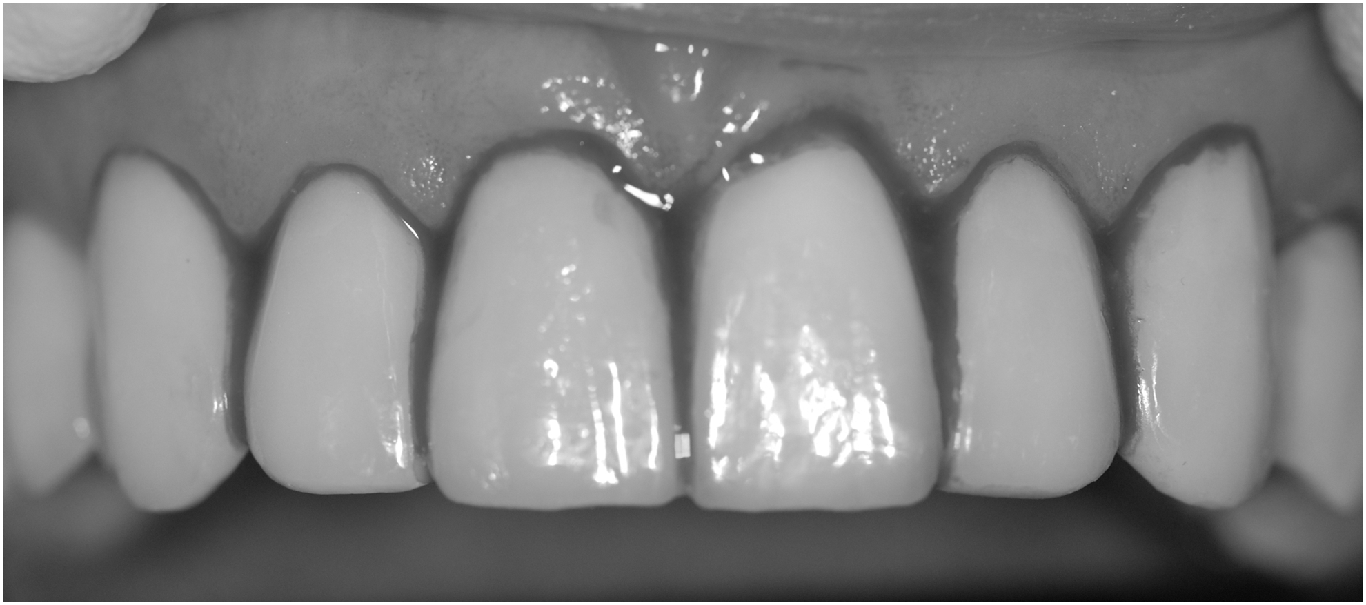
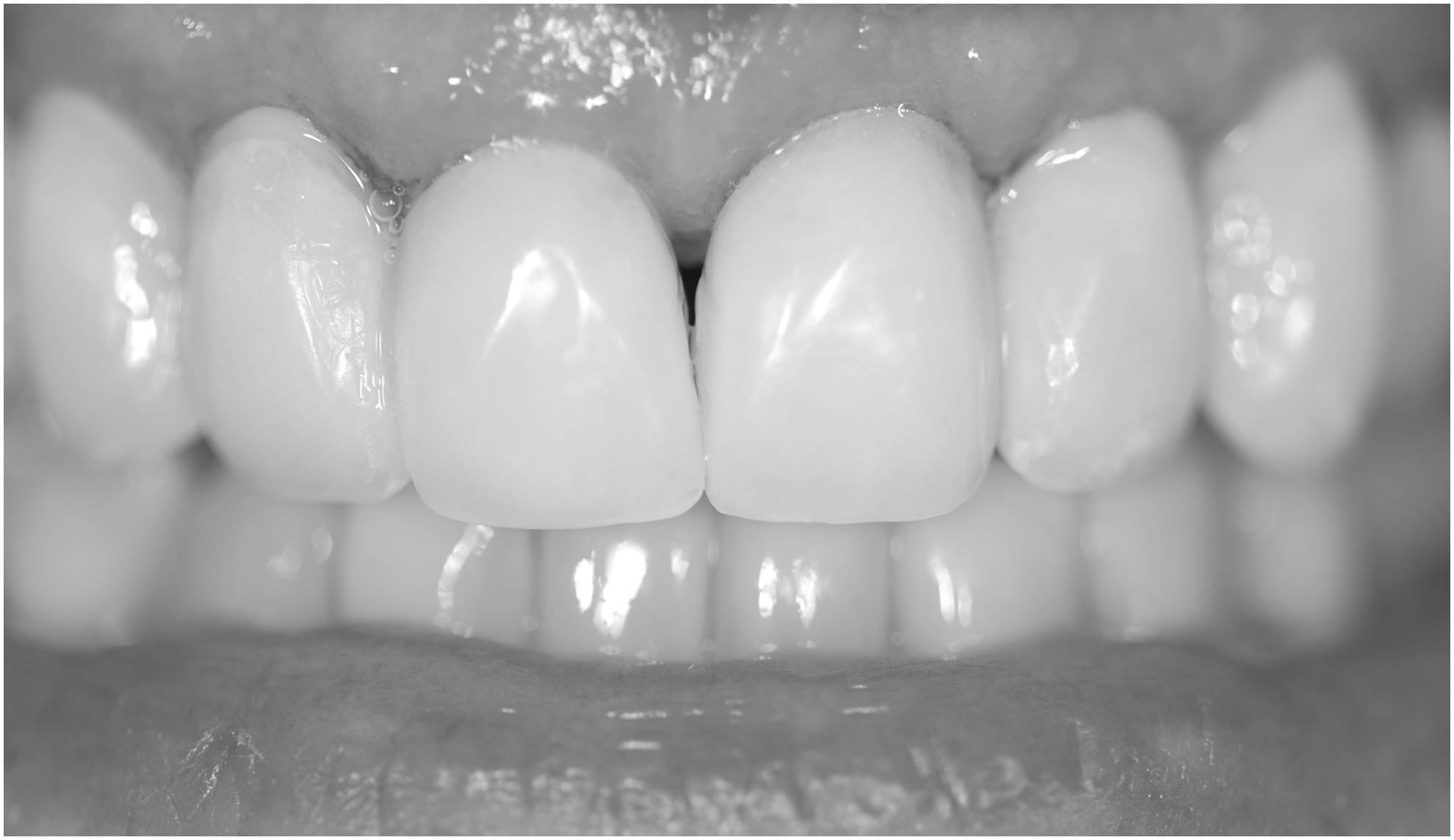
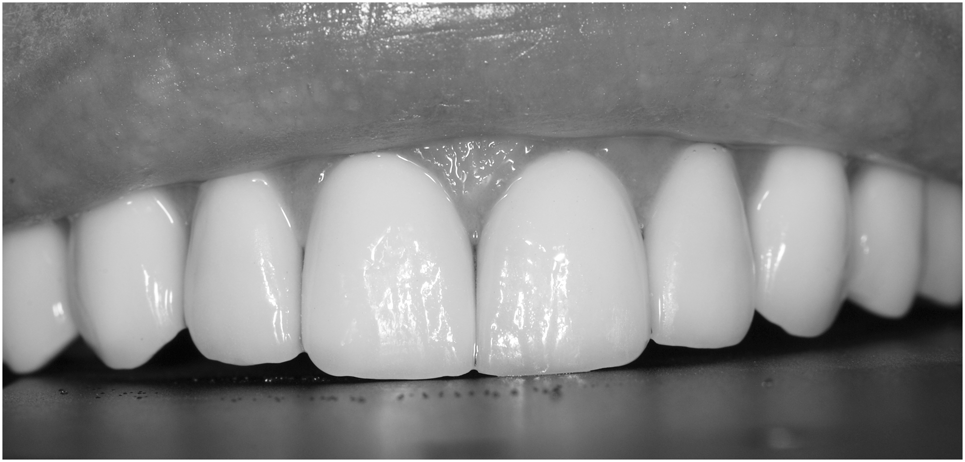
(a) After the clinical examination and the signature of the term of consent, a photographic protocol and measurement of black spaces with a pachymeter were done. Results were triangular shape 2.5 mm high by 1.5 mm base, on average, considering the three patients (Fig. 1).
(b) Local anesthesia was not necessary for this procedure.
(c) Day 1: After correct placement of porcelain laminates and laser PBM application, polishing was done with orthodontic drills (30 blades drills Jet Carbide Burns FG 9714 Canada) between the gingiva and the tooth surface to remove surface debris and smoothen the tooth–porcelain interface. These orthodontic drills cannot scratch either the tooth or the porcelain but promote bleeding of the marginal gingiva (Fig. 2).
(d) PBMT was applied with a diode laser in two stages: before bleeding (first PBMT) for analgesia and stimulation of the local microcirculation and immediately after bleeding (second PBMT) 14 points per daily session: 2 J per point, 7 points distributed in the central portion of the cervical gingiva of each one of the four superior incisors and in the center of the three faulty papilla; energy = 14 J before bleeding and 14 J after bleeding. Total of two sessions, interval of 1 week between treatment sessions (Fig. 3).
(e) The dose of each application point was chosen according to the size of the incisors so that it did not generate heat and preserved the vitality of the dental and gingival tissues 11 (Table 1).
(f) The power output of the laser was checked before each PBMT session with the Laser Check (MMOptics, São Carlos, SP, Brazil). It uses 9 V battery and it shows results of calibration of immediate form. The power of the laser equipment can be checked by selecting the wavelength of 660, 780, or 808/830 nm.
(g) Day 2: After a 1-week interval, same procedures were applied in the second session, in two stages: before bleeding and immediately after bleeding, using a clinical probe n.5 Duflex Explorer SS White, USA, to gently induce gingival bleeding in the space where the papilla should continue to grow in the internal region of the gingival sulcus, in the vestibular and interproximal areas of the teeth.
(h) Immediately after filling in the black space with blood, a new application of PBMT was done with the same parameters: 2 J in the seven points of previous application, resulting in 28 J of application on the first day.
(i) After each session, patients were oriented to not use the dental floss within the next 24 h to preserve the stimulated area.
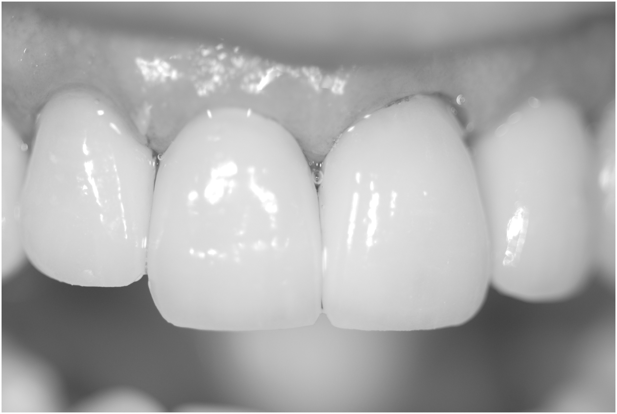
(j) After the second session, new clinical examination and pictures were taken to quantitatively and qualitatively compare the aspects of papilla before and after treatment (Fig. 4).

Black spaces. Absence of interdental papilla, after cementation of porcelain veneers before hemolasertherapy technique; P2 04 30, 2013.
Bleeding first session; P2 04 30, 2013.
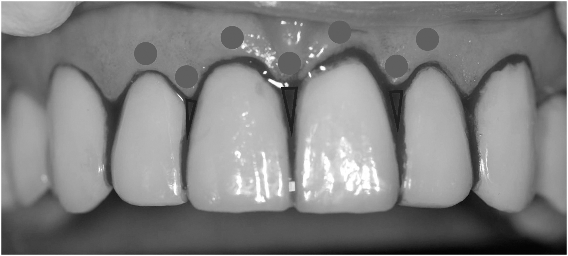
Schematic drawing of the laser application points in the three black triangles.
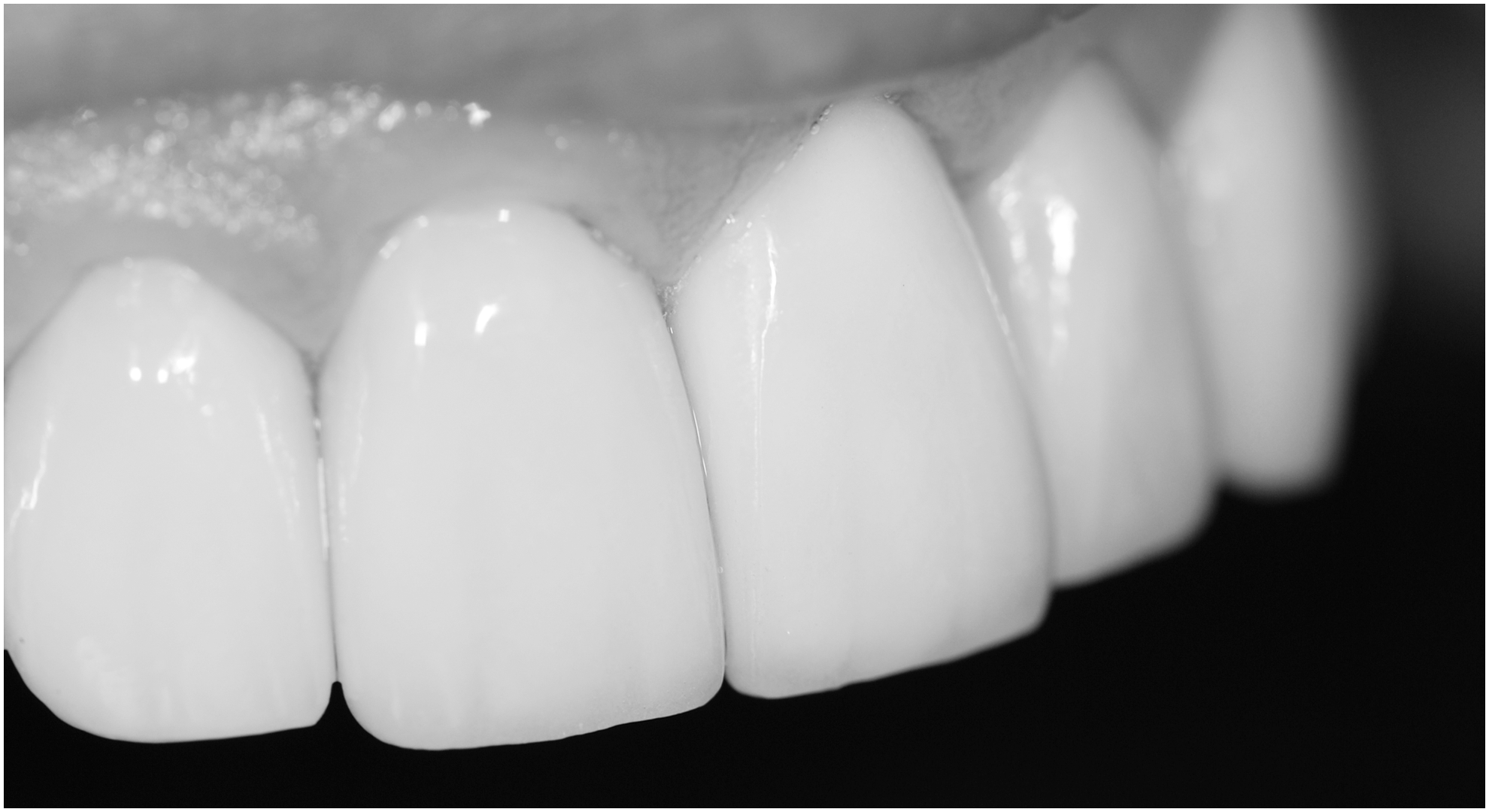
Aspects of the compete filling of gingival papilla after second session; P2 05 20, 2013.
WALT, World Association for Laser Therapy.
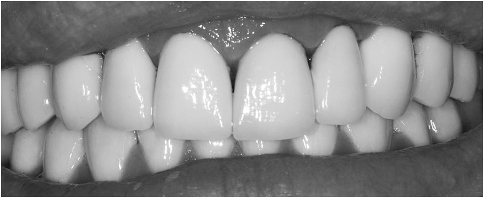
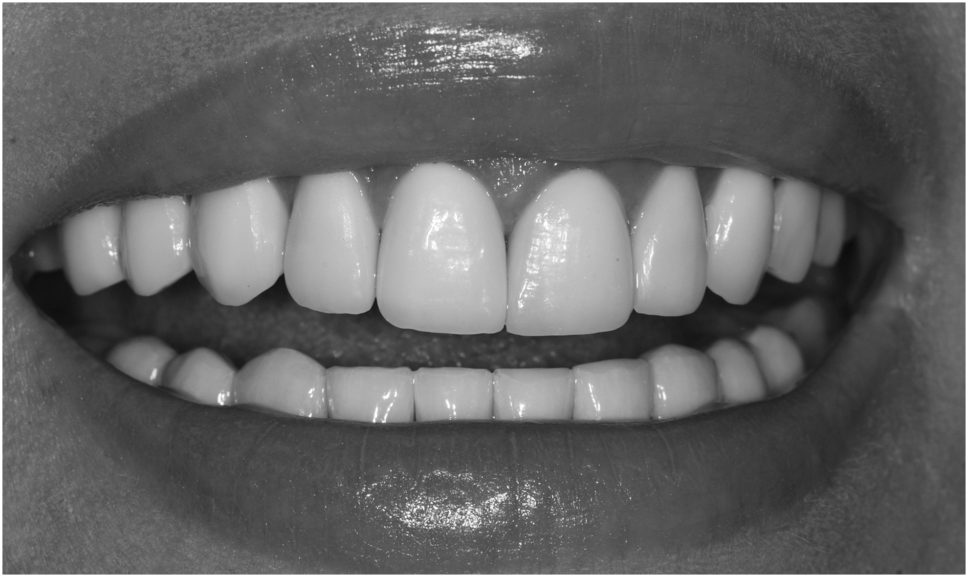
Follow-up: Clinical pictures of the three patients were taken during monitoring twice a year and showed the integrity of formed papilla and maintenance of gingival health (Fig. 5). In both cases, papilla had occupied adequate space (Figs. 6 –10). According to these case reports, PBMT showed positive clinical effects when applied in gingival tissue engineering.
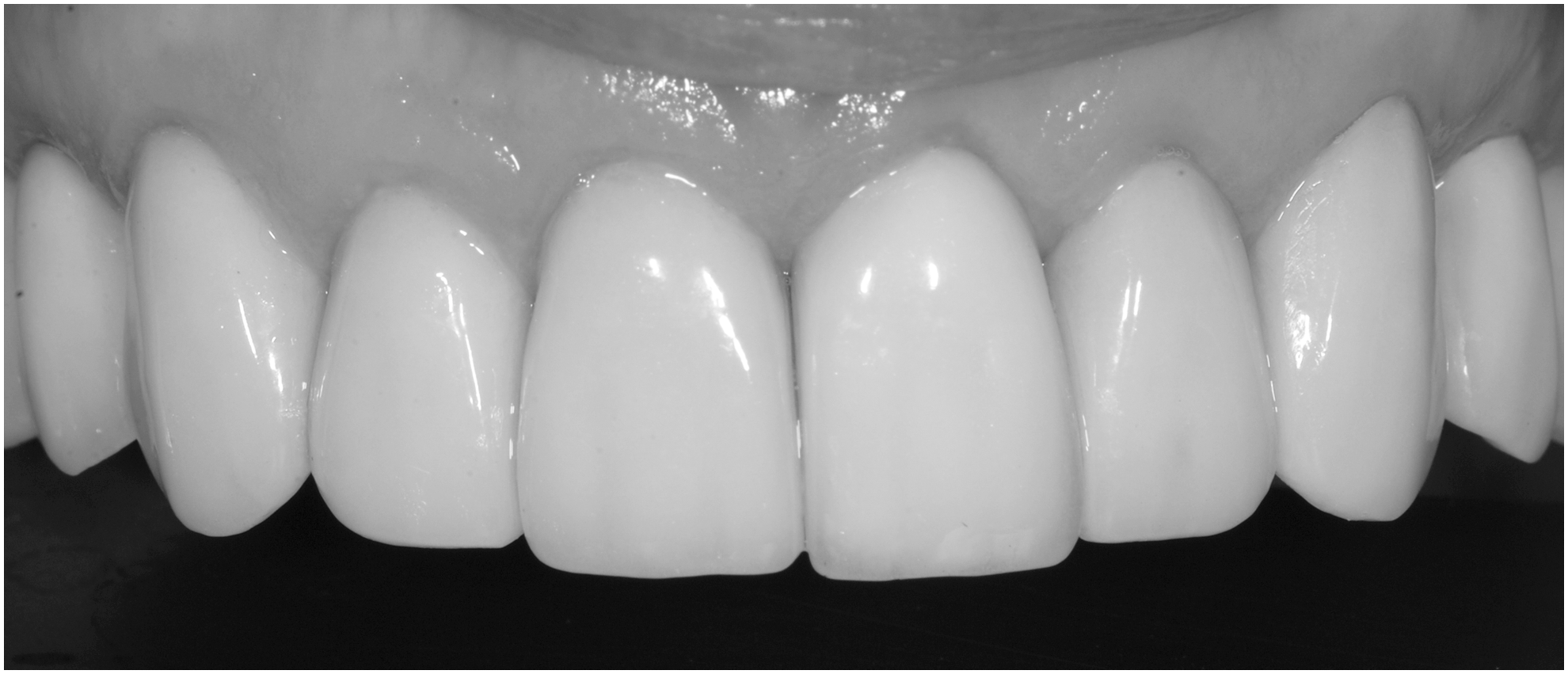
Four years follow-up; P2 10 24, 2017.
P1 02 09, 2012.
P1 second session; 02 22, 2012.
P3 11 28, 2013.
P3 second session; 12 05, 2013.
P3 final.
Laboratory protocol
To research MSCs, blood samples were collected from gingiva of a patient subjected to reverse transcription polymerase chain reaction (RT-qPCR). Blood samples were obtained three times: immediately after provoked bleeding (first sample), after second laser irradiation (second sample), and 5 min later (third sample). Sterile paper points were introduced inside gingival creviculae for 2 min to absorb the blood that was immediately placed into Eppendorf tubes containing RNAlater® solution (Invitrogen; Ambion, Austin, EUA). Total extraction of RNA was done. Evaluated genes and respective primer sequences are listed in Table 2. These were analyzed based on the Ct value (cycle threshold). Means of the Ct values of measurements in duplicate were used to calculate the target gene expression with normalization to the endogenous control (GAPDH) and compared with a reference sample (first sample) to calculate relative quantification. Values of the relative expression of each gene before and after PBMT were subjected to the t test (p < 0.05).
We searched for stem cell markers whose primers are currently available at our Laboratory at the Department of Dentistry, Dental School, University of São Paulo. They were embryonic markers (OCT-4 and Nanog) and the mesenchymal marker (CD90). Several studies have chosen only two or three markers for detecting stem cells in several different tissues. 12 We could have used other markers but our objective at that time was to preliminarily observe the presence or absence of stem cells in the blood of the irradiated gingiva.
Results
All three patients presented improvement in the size of interdental papilla. In the first week, a significant increase of the papillae was observed with the spaces almost closed, as height was reduced to 1 mm and base to 0.5 mm. In the three reported cases, interdental papillae were completely filled by the newly formed tissue in the first 14 days. Positive expressions for all analyzed genes were observed in the blood samples collected immediately after the bleeding (first sample) and after second PBMT (second sample). Third sample presented expression of only CD90 (Table 3).
Discussion
According to the clinical follow-up, PBMT suggests regeneration of gingival papilla tissue and proved feasible. We propose that therapy with PBM stimulates the return of gingival stem cells and supports their survival and differentiation in the blood clot favoring interdental papilla regeneration.
In fact, authors showed that evoked bleeding in this proposed regenerative procedures triggered accumulation of undifferentiated stem cells into the dental papilla space, as demonstrated by RT-qPCR. Qualitative and quantitative analysis of clinical cases showed successful levels of gingival papilla recuperation. In the first week, a significant increase of papilla and almost filled in spaces was observed as its height measurements reduced to nearly 1 mm and the base to 0.5 mm. After 2 weeks, all three patients presented decrease in black spaces and the filled in papilla gave a better aesthetic aspect to the gingiva. After both PMBT applications, cells in blood samples expressed undifferentiated stem cell markers (Nanog and OCT-4). Although preliminary, this result could indicate a participation of PBMT in the localization of these MSCs. Moreover, only CD90 remained expressed in the cells 5 min after the last PBMT application. CD90 is a stem cell marker also expressed by fibroblasts and myofibroblasts. Thus, it may be supposed that MSCs were subjected to some kind of commitment to differentiate into connective tissue cells (e.g., formation of the newly formed dental papilla).
This is a preliminary and innovative study, which nevertheless points to a possible participation of MSCs' homing in gingival papilla regeneration. Besides, based on the known effect of PBMT in tissue engineering wherein MSCs survived and differentiated inside the blood clot after PBMT, 10 we may suppose that this process also happened in these cases.
Conclusions
According to reports and 4- and 5-year follow-ups of the clinical cases treated with this innovative technique (HLT) proposed by the authors and the preliminary results in vitro, it can be considered that there may have been MSCs participation in the regeneration of gingival papilla at the cellular level. Since the blood clot originated by provoked bleeding in the gingival area is rich in MSCs when associated with the application of PBMT, it is able to preserve viability and further differentiation, stimulating the return of gingival stem cells, which, in turn, would support their survival and differentiation in the blood clot, favoring interdental papilla regeneration. This seems to be an innovative and noninvasive therapy to fill in black spaces, thus improving aesthetics and restoring the function of papilla in patients with black spaces.
Footnotes
Author Disclosure Statement
No competing financial interests exist.