
Abstract
Introduction
T
LIPUS is a type of mechanical energy carried by sound waves over the hearing limits of the human ear. The energy of ultrasound is delivered as acoustic pressure waves. LIPUS therapeutic mechanism of action remains elusive, but as reported in this study, the beneficial osteogenic effects observed are nonthermal and probably mechanical under suggested operating parameters. LIPUS most likely stimulates healing by key molecular and cellular pathways. It is used in clinics for diagnostic, therapeutic, and surgical purposes. 6 LIPUS forms micromechanical strains at the cellular level as they pass through the human body. Studies have shown that these stresses increase angiogenesis, vascular permeability, 7 and cellular proliferation, 8 and accelerate bone formation. 9 –11 LIPUS has shown positive effects on delayed bone fracture healing by forming micromechanical tensions on the bone similar to Wolf's law. 12,13
Another biostimulation application by direct irradiation without affecting surface temperature is LLLT. It has a photochemical effect and low level of this energy lead cellular stress. However, biomedical mechanisms underlying the beneficiary effects are not thoroughly understood.
LLLT has low-level energy output and classified as soft laser. Its analgesic and anti-inflammatory effects have been reported in clinical practice. 14 It has been reported that LLLT treatment improves cellular proliferation in the early phase of healing 15,16 and accelerates bone regeneration. 17,18
Although the positive effects of LIPUS and LLLT applications have been demonstrated in laboratory and animal studies and in clinical practice, there are doubts about their use due to the side effects that arise due to parameters such as intensity, duration of application, frequency, and power to be applied.
Studies conducted with LIPUS applications were mainly performed using a specific device (Exogen). This system uses a frequency of 1.5 MHz and uses a spatial average-temporal average (SATA) intensity of 30 mW/cm2, a duty cycle of 1:4, and a pulse frequency of 1 kHz. 19 Acar et al. 20 used Exogen's parameters for LIPUS in their study of bone regeneration with gallium–aluminum–arsenide (GaAlAs) diode laser for LLLT at 810 nm continuous wave length and 0.1 W power output for 120 sec at 4 J/cm2 dose. Babuccu et al. 21 employed LLLT and LIPUS on bone regeneration in rats and in ultrasound application, a 1:4 pulse interval at 1 MHz, 0.5 W, and 16 J/cm2 as SATA intensity preferred, while in the LLLT application, diode laser was applied at 820 nm, 0.5 W power output, and 16 J/cm2. Nakamura et al. 22 investigated the effects of LIPUS on synovitis, they preferred a frequency of 3 MHz, intensity of 30 mW/cm2, and pulse interval 1:4. In addition to all these applied parameter changes, some studies have shown that LLLT has a stimulative effect on fibroblast cells at low dose of 2–4 J/cm2 and high dose of 16 J/cm2. 23
Reported parameters of the studies determined by LLLT and LIPUS in the literature vary. For this reason, in this study, we aimed to compare the effects of all the parameters that can be used in vitro on both fibroblast and osteoblast cell cultures to determine safe cell survival and optimal cellular induction conditions.
Materials and Methods
Cell culture
L929 fibroblast cells (mouse embryo tissue, CRL-9642; ATCC) and osteoblast cells (human bone tissue, CRL-11372; ATCC) incubated in 10% Dulbecco's Modified Eagle Medium (DMEM) (Invitrogen, Carlsbad, CA) were supplemented with 1% fetal bovine serum (FBS) and 10 mg/mL of penicillin/streptomycin (Invitrogen). The cells were incubated at 37°C in a 5% CO2 atmosphere for 24 h before being subjected to ultrasound and laser treatment (Table 1). Cells were seeded and incubated overnight to attach them on the plate and then, laser and ultrasound were applied without changing the medium to prevent handling dependent cellular stress. Plates were incubated for 24 and 48 h. After the treatment, the medium was replaced for XTT experiments. Briefly, the mitochondrial dehydrogenase enzyme activity turns XTT salt (2,3-bis (2-methoxy-4-nitro-5-sulfophenyl)-5-[(phenylamino) carbonyl]-2H-tetrazolium hydroxide) color from yellow to orange. This is used as a measure of cell viability and absorption at 450 nm provides a valuable tool to determine cell survival rate.
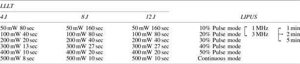
LIPUS, low-intensity pulsed ultrasound; LLLT, low-level laser therapy.
Phenol-free medium was used in all phases of the experiments to avoid any light-dependent undesired side reactions. FBS was added to irradiation/sound shock medium since serum contains growth, adhesion factors, lipids, and minerals and further, it is essential for cell proliferation and the attachment process. Phenol red-free medium was used in cell culture to avoid XTT absorbance reading interferences. The medium exchange was performed before XTT treatment so that the cells were not exposed to any stress conditions. Similar conditions were applied over control cells to eliminate any possible effect of dead cell-originated interference on calculations.
Ultrasound and laser application
Before ultrasound application, fibroblast and osteoblast cell cultures were subcultivated in six-well plates and 1 × 10⁶ cells were allocated to each well. The pulse duration (10–50%) and continuous mode, application time (1, 3, and 5 min), and frequency (1 and 3 MHz) variations were tested with a 30 mW/cm2 intensity (n:9). LIPUS (Gymna Uniphy, Pulson 200, Belgium) was applied after a 24-h incubation when the cells adhered to the plates. The probe was immersed to the culture medium around 3–4 mm above the cell layer. Laser-applied cells were subcultivated in 96-well plates with the energy (4, 8, and 16 J) at 50, 100, 200, 300, 400, and 500 mV settings at 820 nm (n:9). Laser (Doris CTL1106 MX, Poland) was gallium–aluminum–arsenide (GaAlAs) and the optic fiber was 1 cm2. Cell viability and proliferation were analyzed using the XTT assay.
Statistical analysis
Mean values were compared by one-way analysis of variance using SPSS 15.0 software package, and the post hoc Tukey Honestly Significant Difference (HSD) test was also performed to justify differences among the groups. T-test was used to distinguish paired and independent samples. Values of p < 0.05 were considered statistically significant.
Results
LIPUS treatment
Pulse mode at 10% (1:9 duty cycle) at 1–3 MHz for 1–5 min promotes cell proliferation both for osteoblast and fibroblast cells. In 10% pulsed mode osteoblast groups, the differences between 1, 3, and 5 min were significant and the difference between 1 and 3 MHz was not significant, only in the 3-min application group (p > 0.05) (Figs. 1 and 2). In 10% pulsed mode fibroblast groups, no significant difference was observed between 1, 3, and 5 min. However, cell survival percentage showed significant difference between 1 and 3 MHz in all time groups. Osteoblast cells cannot maintain cell viability after 24 h in contrast to that of fibroblast cells (Tables 2 and 3). Cell proliferation at other tested parameters indicated loss of cell viability. This loss is mainly caused by heat generated from LIPUS probe as the pulse rate increased to 20% continuous mode. Since pulse rate above 40% caused overheating of cell media, the experiment stopped at this value.
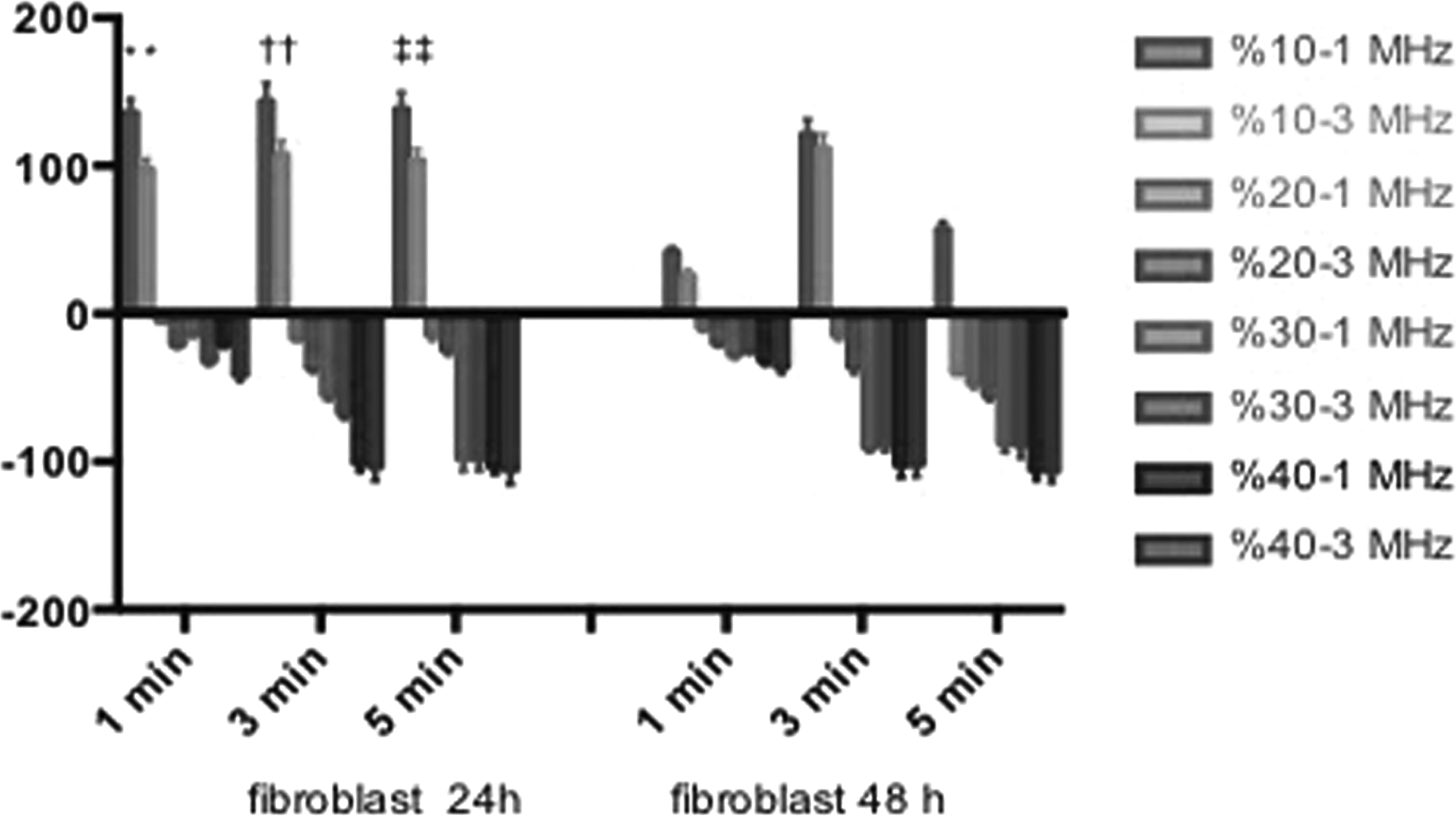
LIPUS-treated fibroblast cell survival percentage over untreated cells after 24 and 48 h. *,†,‡ p < 0.05, significant difference. LIPUS, low-ıntensity pulsed ultrasound.
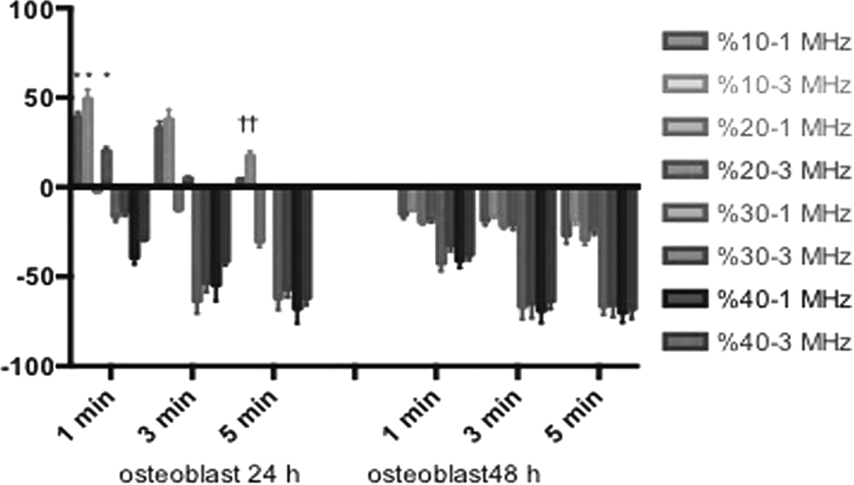
LIPUS-treated osteoblast cell survival percentage over untreated cells after 24 and 48 h. *,† p < 0.05, significant difference.
p < 0.05, significant difference.
Bold values indicate safe limit intervals for LIPUS treatment and its clinical benefits for osteoblast cells.
p < 0.05, significant difference.
Bold values indicate safe limit intervals for LIPUS treatment and its clinical benefits for fibroblast cells.
LLLT treatment
Laser treatment of fibroblast and osteoblast cells induced cell proliferation for both 24- and 48-h period at 4, 8, and 16 J and the differences between output levels at all energy levels (Tables 4 and 5) were statistically significant (p < 0.05). The range of 50–500 mV induced cell proliferation, but 200–400 mV range gave the best results. Due to nature of laser light, the heat is not generated in the wells. This advantageous provide LLLT-treated cell amplification (Fig. 3). LLLT does not propagate its energy to certain part of the cell, but rather disseminates it to the whole cell. Thus, energy quants may not lead vibration of the molecular energy levels and may not have adverse thermal outcome.
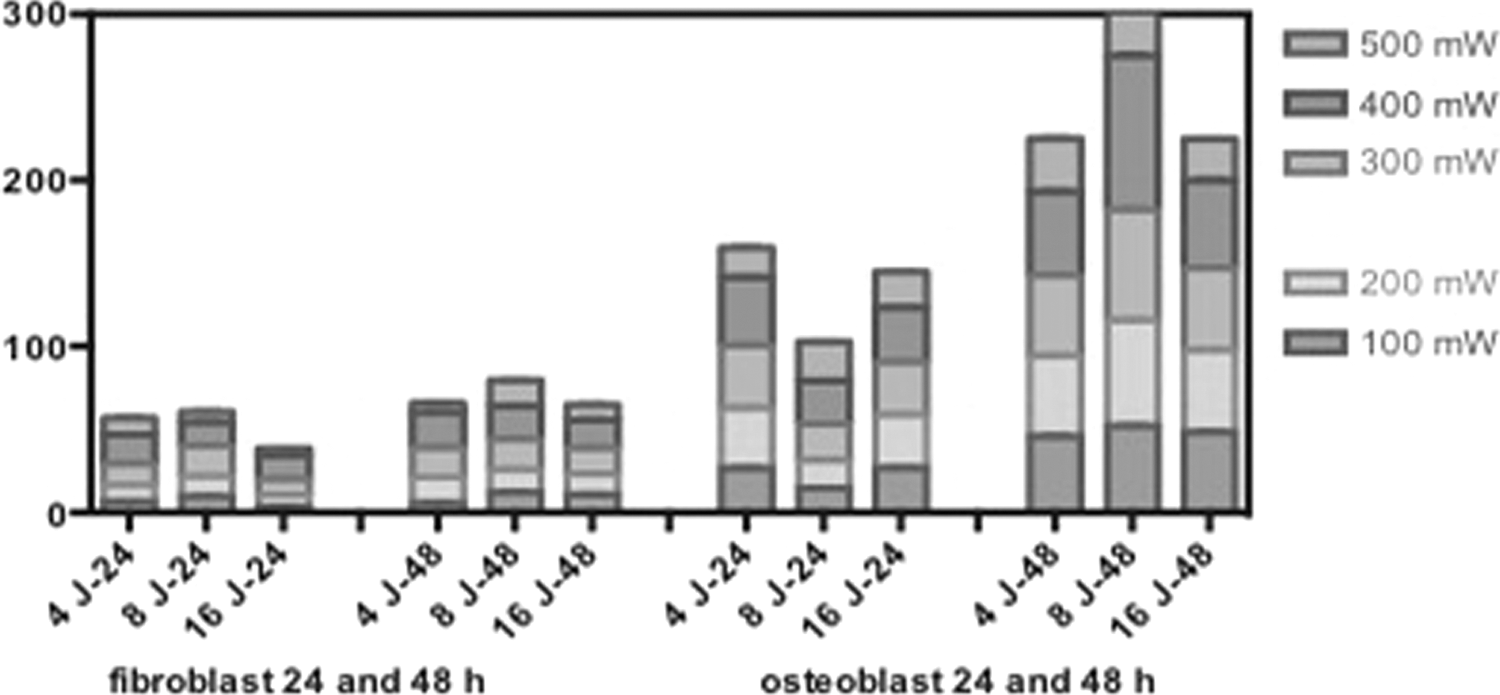
Laser-treated fibroblast and osteoblast cell survival percentage over untreated cells after 24 and 48 h.
p < 0.05, significant difference.
Discussion
Ultrasound is mechanical energy transmitted by high-frequency acoustic waves and when passes through a tissue ultrasound heat up the medium up. 24 The heat can be dissipated into surrounding tissues, but heat concentrating locally causes disruption of consequences rather than stimulating proliferation during repair phase of the cell.
Local heat may cause a damaging effect on culturing conditions, but in vivo it may lead to beneficiary results due to heat dispensing over matrix. Heat in the culture medium may not dissipate, but scaffolds (extracellular matrix) may disperse the heat in the pulp. This may lead osteoblast cell induction for its regeneration or may affect dental pulp stem cell differentiation. Dental pulp stem cells differentiate toward osteoblast and several studies showed that mimicking extracellular matrix helps this transformation. 25,26
Therefore, the heat to a certain extent may activate heat shock response and may induce cell regeneration and/or induce cell proliferation. Matrix dissipates heat over cells evenly, but excessive heat may cause apoptosis. Therefore, response at cell culture and responsive cell behavior on the pulp may show differences, that is, optimal parameters for LLLT and LIPUS on the tooth environment may slightly vary. Despite these, the parameters determined in the study trigger cellular activity and increasing the dose of optimal parameters may cause adverse molecular events in cells. Further, animal experiments can be performed to determine upper limits of these variables.
The results of this study suggest that LIPUS is not effective as LLLT. LIPUS does not promote cell proliferation, except at 1–3 MHz-10% pulse mode both in osteoblast and fibroblast cells. However, after 24 h, LIPUS-treated cell viability decreases significantly (Tables 1 and 3). This scenario is different in LLLT-treated cells and after 24 h, both cell type proliferation increases significantly (Tables 2 and 4). During experiments, LIPUS treatment heats the cells up and the generated heat caused cell death. The nature of ultrasound generates heat, but in soft gingiva and hard tooth tissues, dissipation of local heat is not effective. As results indicate, the LIPUS-treated fibroblast and osteoblast cell maintenance and cell survival after 24 h are lower and ineffective. However, same experiments with LLLT are much more effective since laser does not affect surface temperature. This property makes laser application superior over ultrasound application. Further, increasing LLLT energy from 4 to 16 J does not affect cell number and stability, but shortens laser exposure time. Heat generation and prolonged treatment of LIPUS-LLLT boost cancer cell proliferation and may cause several undesired side effects (unpublished results). Therefore, it is advantageous and safe to employ LLLT for healing procedures. This safely triggers cellular response and minimizes cancer cell augmentation.
Laser affect may be modified by matrix or functional biomaterials as tested in the literature over stem cells by employing silicon scaffolds and biomaterial, which consist of demineralized bone matrix, a chitosan hydrogel, and mesenchymal stem cell affinity peptide, respectively. The matrix may also provide promising results for LIPUS at less harsh conditions. 27,28
Literature reports that comparison of both techniques found similar outcomes to our results. The study determined that, while LIPUS enhances bone repair by promoting bone resorption, LLLT accelerates healing process by promoting new bone formation. 15 Another study on new bone formation compared the two techniques in rat tibial defects and interestingly, LLLT, but not LIPUS, displayed positive effect in this study. 29 LLLT biomodulatory positive effect was also reported by Dr. Danillo Barbosa group. 30 Further, LIPUS limited the effect reported by Claes and Willie. The study showed that LIPUS is effective in soft tissues, but not in bone tissues. 31 Molecular level understanding of the mechanism has been investigated for years. Omics technologies provide an overall look to comparative gene expression pattern and pathway analysis. Thus, LLLT efficiency on accelerating newly formed bone at gene level was studied by microarray technology and modulating factors were determined. These factors include inflammatory and angiogenic agents and these factors accelerate early stage of bone healing. 32 This study initiates determination of a detailed molecular mechanism.
LIPUS energy is transmitted to heat and bone absorbs more heat compared to soft tissues. High frequencies generate short waves and this limits LIPUS penetration. On the other hand, low frequencies generate long waves with deeper penetration; therefore, higher frequencies (3 MHz) are employed in superficial wounds, but lower frequencies are used for deeper wounds. 33 LIPUS results of this work show problematic energy dissemination of LIPUS-treated cells. LIPUS is effective in cell proliferation only at 10% pulse-24-h condition. Thus, a caution must be taken in LIPUS-based treatments in adjusting the dose response.
The outcomes are consistent with our study since LIPUS-treated cell proliferation decreases after 24 h and is not effective as LLLT treatment. While ultrasound exposure perturbed cell maintenance, the laser irradiation did not affect cell viability during the experiment.
Conclusions
Comparing LLLT and LIPUS over two key cell lines showed that laser treatment was found to be more effective in promoting cell proliferation and shortening healing period effectively. The study determined optimal dose and duration parameters for both techniques. Clinical treatments may be adopted to the determined doses given in this work for efficient and safe treatment.
Footnotes
Acknowledgments
This study was funded by Scientific Research Projects Unit of Cumhuriyet University, Sivas, Turkey (Project No. DIS-130).
Author Disclosure Statement
No competing financial interests exist.