
Abstract
Objective:
To determine the optimal parameters of power, energy, and time for the application of a carbon dioxide laser for Tribal Black ink tattoo removal.
Background data:
The use of antiquated techniques to remove tattoos demonstrates the difficulty of making advances in this field. Studies by the American Society for Dermatologic Surgery have shown that 5% of the global population has at least one tattoo on the body, with 10% of them wanting a tattoo to be removed. Laser removal has been studied and improved as a less invasive and safer method of surgical removal; however, the ideal dosimetry is not yet established.
Materials and methods:
Thirty-three male Wistar rats were anesthetized and tattooed in the dorsal region in a quadrangular manner. The rats were distributed under low/null luminosity for 4 months into three equal and random groups for the application of the laser, namely, G1 (P = 0.6 W, Et = 0.9 J), G2 (P = 0.8 W, Et = 1, 2 J), and G3 (P = 1 W, Et = 1.5 J), with the application time standardized to 0.15 sec with 10 passes per application. The procedure was repeated at intervals of 4 weeks until 10 cycles of laser application were completed. The images were studied using the ImageJ program and histological analysis and subjected to the one-way ANOVA test for Tukey's multiple comparison post-test.
Results:
We observed a significant difference between groups 1 and 3 and between groups 2 and 3.
Conclusions:
The laser with the parameters of P = 1 W, Et = 1.5 J, and t = 0.15 sec yields better Tribal Black ink removal results.
Introduction
T
A 2008 study showed that 14% of the New York population had tattoos, 3 and in 2013, the first tattoo census in Brazil revealed that 51% of the women in the country (59.9% of the total population) were tattooed, along with 9% of the Brazilian population between 19 and 25 years old (48.2% of the total population) and 14.8% of those educated or who received higher education (61.2% of the population). In 2006, the Journal of the American Academy of Dermatology published an article that sought to determine the prevalence of tattooed people in the United States and revealed that 24% of the population had at least one artistic tattoo, and 21% of the nontattooed considered getting one. This study also revealed that 17% of the tattooed people (practically one in five) contemplated the removal of an existing tattoo. 4
A study by the American Society for Dermatologic Procedure (ASDP) revealed that in 2013, 2.25 million laser/light/energy procedures were performed (a 34% increase from 2012), and one of the procedures in greatest demand has been tattoo removal, along with resurfacing and scar removal. 5 Due to the absence of techniques that have been proven safe, those who seek such treatments are exposing themselves to the risks of the current procedures, which usually do not culminate in the expected result and may lead to the adverse events that have already been described. Thus, the study of tattoo removal must be expanded. This study aims to determine the carbon dioxide (CO2) laser optimal parameters of power, energy, and time for Tribal Black ink tattoo removal.
Materials and Methods
Thirty-three male Wistar rats of the Rattus norvegicus species with a mean age of 12 weeks and a mean weight of 300 g underwent antisepsis of the back with nonalcoholic Chlorotex® solution and trichotomy. General anesthesia was administered with intraperitoneal ketamine 80 mg/kg and xylazine 10 mg/kg with a 30G needle, and a 2.2 × 2.2 cm full square tattoo 6 of Tribal Starbrite Colors 30 mL (recognized by ANVISA in accordance with the standard established in December 2009 and sold by Starbrite Colors—Amazon Indústria, Comércio, Exportação e Importação de Produtos Especializados) with Black Line Machine BK 210 drawn using a Black Line 07 MG needle on the back. At the end, the tattoo was covered with PVC film for 1 day, and the animals were kept in an environment with low/null solar incidence for 4 months (Fig. 1).
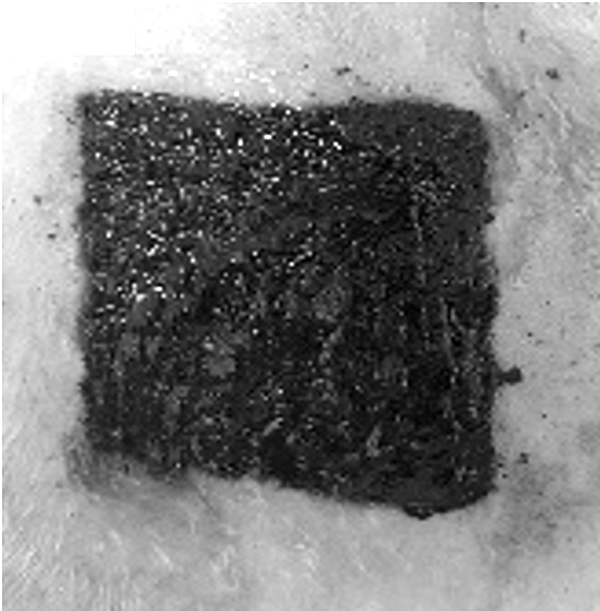
Example of a finished tattoo on the back of an animal.
After 4 months, the rats were randomly assigned into three groups (G1, G2, and G3) of 11 animals each. All animals underwent a new dorsal trichotomy and were anesthetized with an inhalation application of 1.5 mL of 30% v/v isoflurane solution in propylene glycol. In G1, the CO2 laser (AcuPulse CO2 model; Lumenis brand) was applied with 0.6 W of power, 0.15-sec pulse duration time, and total energy of 0.9 J. In G2, a power of 0.8 W, time of 0.15 sec, and energy of 1.2 J were used. In G3, a power of 1 W, time of 0.15 sec, and energy of 1.5 J were used. The laser was pointed perpendicular to the tattooed field and at the focal length and was operated after the pedal was activated. There were 10 laser passes per application in each animal, and the procedure was repeated at 4-week intervals for a total of 10 applications (Fig. 2).
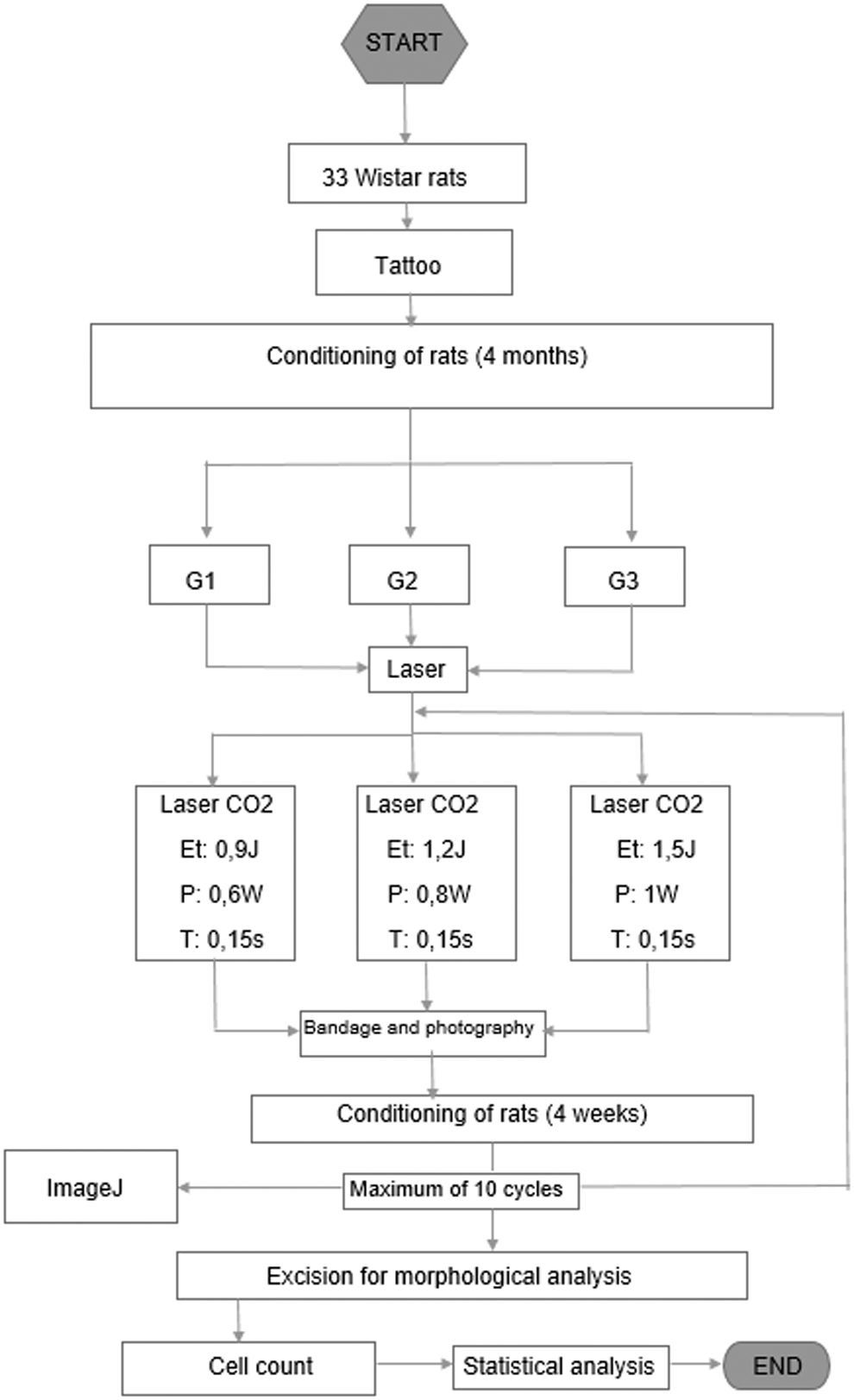
Flowchart of the study design.
In all applications, the studied region was photographed before and after the use of the laser with a 12 MP instrument and a resolution of 4290 × 2800 pixels to be studied in the ImageJ program with the use of the color threshold adjustment tool to create comparative images of the black color of the tattoo and the white color, representing the skin of the animal without ink, for subsequent analysis of particles for counting dots that are filled by black color.
At the end of the experiment, a 0.5 × 2.0 cm rectangular excision was performed from the center of the previously tattooed area for analysis of results in a histological study after euthanasia of the animals using general anesthesia of intraperitoneal ketamine 80 mg/kg and xylazine 10 mg/kg, followed by exsanguination.
The excised samples were prepared on histological slides that were stained with hematoxylin and eosin and analyzed under an optical microscope with a photographic record through the AxioVision program for cell counting.
All procedures were carried out in the experimental laboratory of the Centro de Desenvolvimento de Modelos Experimentais (CEDEME) of the Escola Paulista de Medicina (EPM/UNIFESP) and the Laboratory of Microscopy of the Escola Paulista de Medicina (EPM/UNIFESP).
The inclusion criteria were male animals, 12 weeks of age, and an average weight of 300 g. The animals 1, 2, 4, 8, and 11 of G1; 12, 13, and 16 of G2; and 24, 27, 31, 32, and 33 of G3 died and were excluded from the study.
Results
For comparison between groups (group 1 with 2; 1 with 3, and 2 with 3), the one-way ANOVA technique was used for Tukey's multiple comparison post-test.
First, a comparison was performed before the laser was applied to analyze possible significant differences in the application of the tattoo that could influence the result. The analysis was based on counting prelaser black points using the ImageJ® tool (Table 1).
A second analysis was performed on the postlaser results by counting black points with the ImageJ tool (Table 1).
Finally, an analysis of the postlaser results was performed by counting black points through histological analysis (Table 2).
All applied tests were of one-tailed hypotheses, with a p-value <0.05 defining statistical significance.
The results should be analyzed in three scopes: visual, ImageJ, and histological.
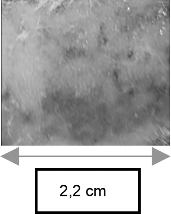
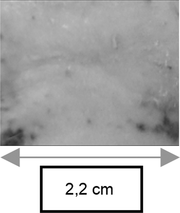
Regarding the visual scope, the best results of each group were from animals 5 (G1), 15 (G2), and 25 (G3). A direct comparison among these three animals indicates a better result with the application of the laser with the parameters of G3 (Et: 1.5 J; P: 1 W; t: 0.15 sec) since, visually, the tattoo was removed almost completely, leaving only a few points of greater concentration on the periphery of the tattoo. This outcome differs from the results for the other two animals, on which it is still possible to observe the black pigment in a more homogeneous way over the full extent of the tattooed area (Figs. 3 –5). Throughout the study, no adverse reaction to the tattoo pigment or laser was visually observed.
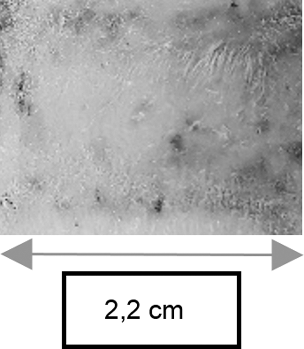
Skin of animal 5 after the 10th application of the laser.
Skin of animal 15 after the 10th application of the laser.
Skin of animal 25 after the 10th application of the laser.
Using ImageJ, the best results of each group were observed in the same animals (5, 15, and 25). Again, a better result was obtained using the laser with the G3 parameters, since after the 10th application of the laser, a lower number of black points were counted with this tool for animal 25 (Table 1 and Figs. 6 –8). According to the table of the numbers of black points that were counted by ImageJ (Table 1), sometimes after the laser application, the count increased; this outcome does not indicate the presence of more black pigmentation in that location but rather the breakage of the ink inside the macrophages that had not yet been eliminated.
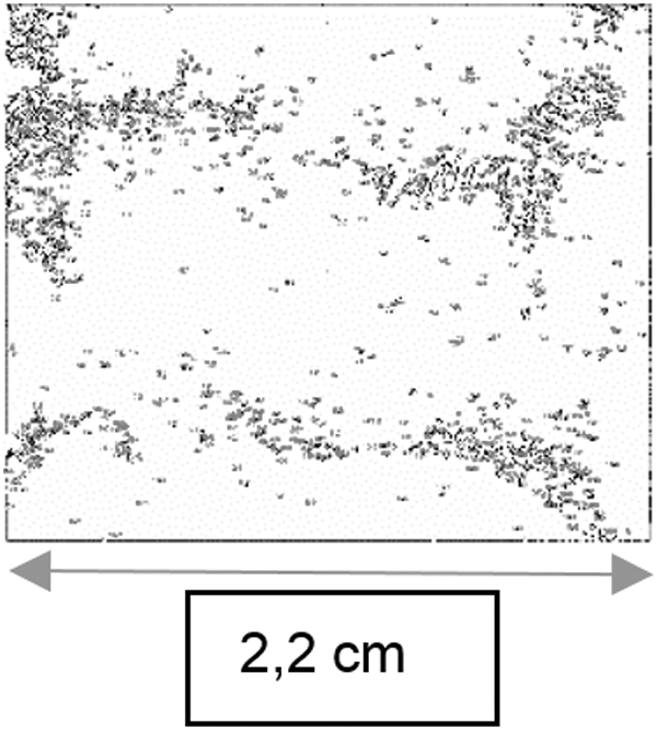
Study performed in ImageJ with animal 5 (G1) after 10 laser applications. Each point represents an analyzed black point of the prelaser tattoo pigment.
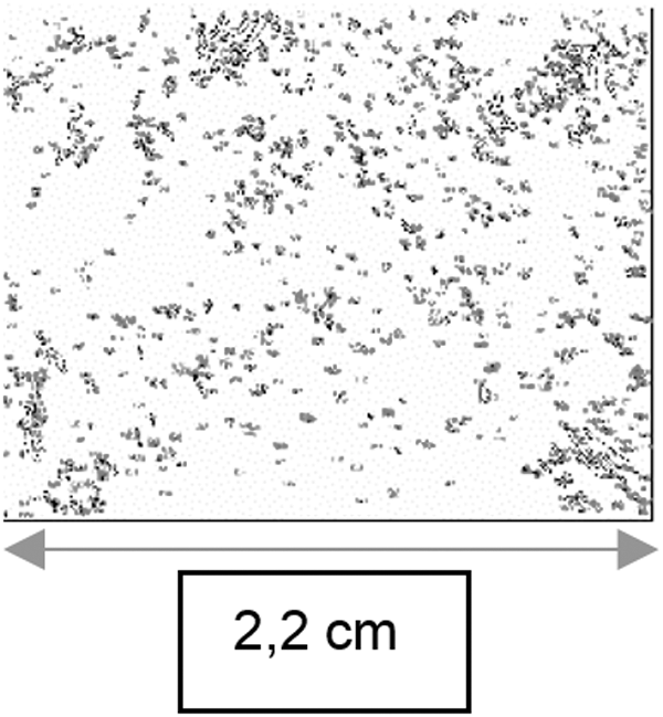
Study performed in ImageJ with animal 15 (G2) after 10 laser applications. Each point represents an analyzed black point of the prelaser tattoo pigment.
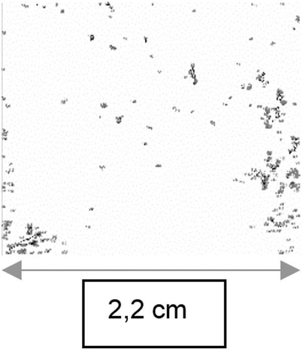
Study performed in ImageJ with animal 25 (G3) after 10 laser applications. Each point represents an analyzed black point of the prelaser tattoo pigment.
Finally, in the histological study, the best results of each group differed slightly from those that were previously observed. Animal 10 shows the best results in G1, animals 14 and 19 in G2, and animal 25 in G3. Despite this difference, a better result is obtained with the application of the laser with G3 parameters, since this yields the lowest number of black points (Table 2 and Figs. 9 –11).

Histological analysis of animal 10 (G1) at 40 × objective magnification.

Histological analysis of animal 19 (G2) at 40 × objective magnification.
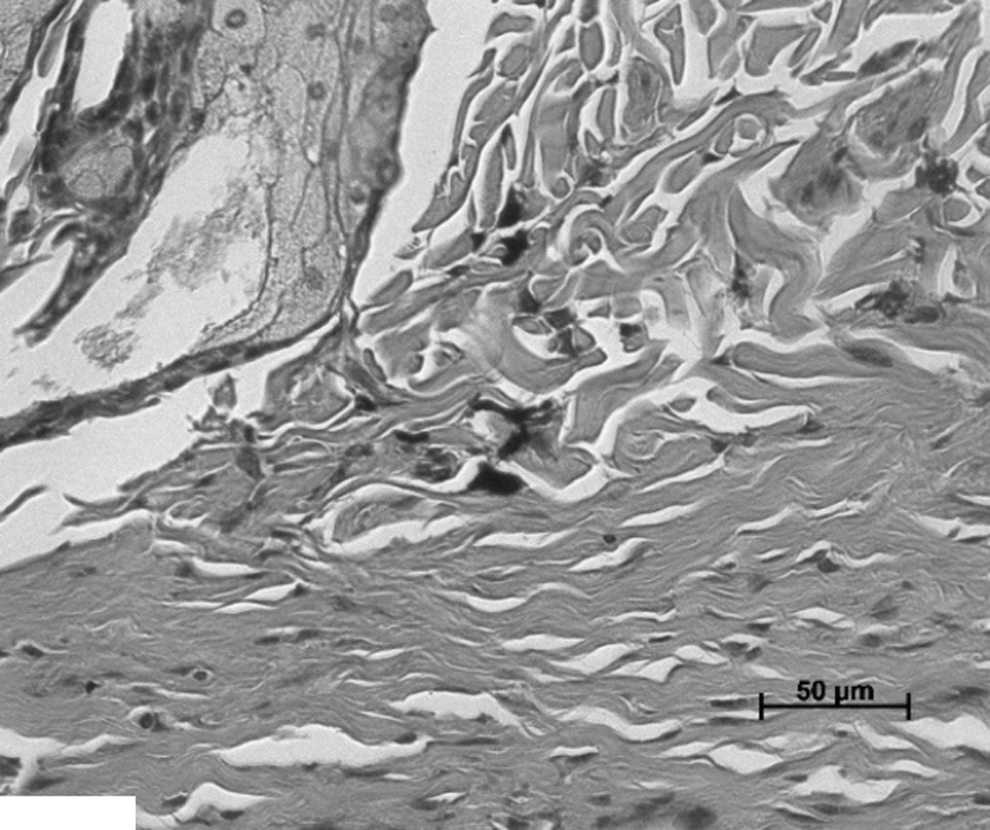
Histological analysis of animal 25 (G3) at 40 × objective magnification.
Counting was performed by applying a mesh of 9 vertical lines and 11 horizontal lines, with a total of 99 points of intersection, over the image that was obtained from the histological slide at 40 × magnification. The pigmentation was only considered when it was located exactly under any of the points of intersection of the mesh (Fig. 12). 7
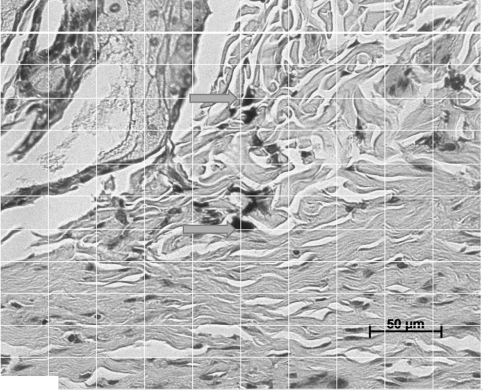
Histological image at 40 × objective magnification of animal 25 (G3), which exemplifies the method of counting black points indicated by the arrows at the exact intersections of the lines.
In a more detailed analysis, it is observed that the keratinocytes of the three groups are flattened in the epidermis but alive, thereby reinforcing the nonobservation of adverse events due to the use of laser. Comparing the obtained images, it is also observed that in G1 there is a substantial amount of ink inside the macrophages, in G2 there is ink both inside and outside the macrophages, and in G3 there is little ink and it is outside the macrophages. This outcome corroborates that the best results were obtained with the laser parameters that were applied in G3.
In the statistical analysis, a preapplied laser comparison is done to eliminate possible significant differences in the tattooing that could influence the design result. The results of the test (Table 3) yield a p-value of p = 0.7914, thereby proving that there is no significant difference between groups before the application of the laser; the results can also be analyzed through the box plot (Fig. 13).

Box plot of the statistical analysis of the numbers of black points that were counted prelaser via ImageJ, which demonstrates that there is no significant difference between the groups regarding the initial amount of black tattoo pigment in the dermis of the animals.
CI, confidence interval.
By statistical analysis of the numbers of black points after laser application, which are counted by the ImageJ tool (Table 4), p = 0.0010 indicates the significant difference between groups 1 and 3 and between groups 2 and 3. The box plot analysis (Fig. 14) shows similar results for both the medians and means of groups 1 and 2. Different, more satisfactory results were obtained for group 3, with lower numbers of black points.

Box plot of the statistical analysis of the black points that were counted postlaser via ImageJ, which demonstrates that there is significant difference between groups 1 and 3 and between groups 2 and 3.
CI, confidence interval.
Finally, the analysis of the postlaser results by counting black points by histological analysis (Tables 2 and 5) provided the value of p = 0.0012, which shows the significant difference between groups 1 and 3 and groups 2 and 3. Moreover, very similar values of p were obtained in the analyses, thereby corroborating the reliability of the result. The box plot (Fig. 15) shows group 3 with close and medium values, with low values of standard deviation and standard error, in contrast to the values presented by the other two groups.
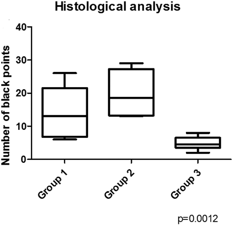
Box plot of the statistical analysis of the black points that were counted postlaser via histological analysis, which demonstrates that there is significant difference between groups 1 and 3 and between groups 2 and 3.
CI, confidence interval.
Discussion
Black color was chosen for this study because the population that wants to reverse the tattoo process consists mostly of individuals who obtained the tattoos a few years ago, when the techniques of tattooing were new and, for the most part, used black ink, as the developments in this area in the use of more advanced techniques and the use of colors have been made more recently. The type Tribal Black was chosen because it is usually used to fill tattoos. Starbrite Tribal Black has a larger particle aggregation diameter, a detectable production of reactive oxygen species and, among the studied inks, the highest concentration of benzopyrene, which is considered carcinogenic to humans by the International Agency for Research on Cancer (IARC). 8 Thus, it is one of the most difficult inks to remove.
A CO2 laser was used because it is one of the representative ablative lasers, treatment with it is of a reasonable cost compared with other lasers, it can be applied in the treatment of tattoos that have mainly water as the chromophore (it is not selective, but it is highly effective in the removal of black ink), and it acts mainly on the dermis (the place of deposition of the tattoo ink) due to its penetration. 9
The most commonly used animal in experimental studies of laser's skin effects is the pig, since it shows high similarity to human skin in terms of thickness and healing. However, it is essential to consider the large size of these animals and the difficulties that are involved in obtaining, handling, and maintaining them. In contrast, the Wistar rat is a small animal, which can be experimentally manipulated with greater ease, has low morbidity and mortality, and has already been used in experiments of similar methodology. 6
The number of sessions of laser use tends to vary because, in addition to the pigment, depth, and intensity, it depends on the response of the animal. However, the results tend to appear in the range of 6 to 10 sessions, based on which we chose the maximum number of laser applications. The 4-week intervals are essential for the observation of reactions in the animal's skin and its reconstitution for a new application to minimize issues. 10
Despite the limited CO2 laser dosimetry data for black ink tattoo removal, the literature parameters range from 9.9 W power, 250 mJ/pulse energy, and 300 μs (5 pulses/sec) time, 11 to a power of 6 W, energy of 1.2 J, and time of 0.2 sec. 12 In these studies, both sets of parameters yielded satisfactory removal results and few undesirable changes. Prior work with powers close to 20 W demonstrated hypertrophic scarring, redness, and hypopigmentation. 13 The only common parameter that was observed was the total energy of 1.2 J. It was then decided to vary the energy in a way to result in low-power applications, therefore minimizing collateral damages.
The ImageJ program was chosen for the macroscopic study of the images due to its use in similar studies. 14,15
The sample size calculation was based on a standard deviation of 25% with a difference to be detected of 50% at a significance level (α) of 5% with a test power (β) of 95% in a one-tailed hypothesis test. Thus, the sample size of each group should have been 5 animals; however, based on a prediction of the possible losses due to the long study time, it was decided to increase the sample size by 6 animals per group, for a total of 11 per group. At the end of the study, G1 had 6 animals, G2 had 8, and G3 had 6, which were sufficient for statistical analysis.
In the case of a preclinical surgical trial, some shortcomings of the project may be justifiable and do not interfere with the result, such as the quazi-randomization of the groups, since no randomization software was used to randomize the animals, but only the random division of the laser application; there was no secrecy of allocation or masking of the animals. Further, this is not a double-blind study.
In this study, the laser treatment aims at breaking the ink inside the macrophages, releasing the black pigment to be eliminated by the lymphatic pathway. Analyzing Fig. 16, one can observe the ink in the interstitial space, alongside the lymphatic vessel, suggesting that the black pigment will be absorbed by this vessel (as well as other cellular detritus) to be eliminated. As no specific histological marker was used to confirm it, we were not able to demonstrate that pathway; however, it allows new inquires to further researches.

Histological image of animal 30 (G3). The arrow represents the tattoo pigment in the interstitial space, alongside the lymphatic vessel.
Conclusions
Our results demonstrate that, among the considered parameters, the use of laser at 1 W power, a time of 0.15 sec, and an energy of 1.5 J yields the most satisfactory results in black tattoo removing. The comparisons proved that there was no interference in the result regarding the tattoo technique, and the p-values that were obtained in the comparisons of black points by visual and histological analysis are highly significant and close. The potential impact of these findings in clinical practice is that, from now on, it is possible to establish a uniform dosimetry to be used in CO2 laser treatments, leading to less expensive and more trustful procedures.
Footnotes
Acknowledgments
The authors thank Fundação de Amparo à Pesquisa do Estado de São Paulo—FAPESP—for the financial support of the project in the process, no. 2015/24459-8. They thank Laura Kawamura Demange, André Moreira Nicolau, Fernando Monicci Navas, Gunnar Willy Pereira Crepaldi, Stella de Aguiar Trigueirinho Ferreira, and Verônica Fernandes de Campos. They thank Full Professor of the Discipline of Histology and Structural Biology (Department of Morphology and Genetics of EPM/UNIFESP) Manuel de Jesus Simões and his staff for assistance in the preparation and analysis of histological slides. The authors also thank the professionals of the Centro de Desenvolvimento de Modelos Experimentais (CEDEME—UNIFESP) for all the assistance in caring for the animals. This research was carried out at Universidade Federal de São Paulo (EPM/UNIFESP).
Declaration of Responsibilities
The opinions, hypotheses, and conclusions or recommendations expressed in this material are the responsibility of the authors and do not necessarily reflect the views of FAPESP. The project is approved by the Ethics Committee on the Use of Animals (CEUA—UNIFESP) under number 2133301115.
Author Disclosure Statement
All the authors declare that they have no financial, commercial, political, academic, and personal conflicts of interest.