




Abstract
Objective:
The objective of the present study was to investigate the application of a carbon arc lamp on wound healing in a rat cutaneous full-thickness wound model.
Background data:
In clinical practice, wound healing has been promoted by irradiation with a carbon arc lamp. However, the corresponding mechanism has not been clearly defined.
Methods:
A cutaneous full-thickness wound on the back of rats was irradiated using a carbon arc lamp at a wavelength peak range of 620–740 nm with 54 J/cm2. Injured sham-irradiated control rats were used as the control. The rats were euthanized after 7, 14, and 21 days, while wound reepithelialization and healing quality were examined by histological analyses with comparison between groups. Cell proliferation was observed by 5-bromo-2′-deoxyuridine (BrdU) immunohistochemical staining.
Results:
Irradiation by the carbon arc lamp significantly accelerated wound healing. The wound-healing rate in the treated group at day 21 was 98.42% ± 0.56%, compared with 93.58% ± 1.26% in the control group (p < 0.05). Significant increases in the length of epithelial edges, collagen content, and microvessel density were observed in the wound sites in the treated group at days 7, 14, and 21 (p < 0.05). Moreover, the number of BrdU-labeled cells increased in the wound edge at days 7 and 14 due to irradiation (p < 0.05).
Conclusions:
The results demonstrated that the carbon arc lamp can promote wound healing together with improvement in its quality by stimulating cell proliferation.
Introduction
Phototherapy is a treatment using sunlight or artificial light (infrared, ultraviolet, visible light, laser) to prevent disease and promote recovery. 1 The carbon arc is an artificial light source, which consists of biomedical carbon. Electric heating of two carbon rods creates an arc of vaporized carbon between them, which is maintained by electrical heating, emitting light. The carbon arc is a broad spectrum light source, including visible light, infrared, ultraviolet, and magnetic pulses, and is thus more reflective of natural sunlight. In previous studies, the carbon arc has been demonstrated to be a mild, stable, and safe actinic radiation. The carbon arc lamp irradiation has been shown to be an effective and safe phototherapy in the treatment of dermatologic conditions, rickets, and surgical tuberculosis. 2 –4
The carbon arc lamp is a phototherapy instrument, which can produce radiant energy, thermal energy, and carbon particles in the treatment area of the human body. The carbon rod emits a broadband multimodal nanometer spectrum when burned. The emitted light energy and heat directly act on the human body and are rapidly absorbed by the skin and subcutaneous tissue, thereby generating thermal and photochemical effects. 2,3 Recently in clinical practice, we found that the carbon arc lamp has many effects, including anti-inflammatory, analgesic, killing of a variety of microorganisms, improving immune function, promoting epithelial cell growth, and promoting wound healing among others.
In phototherapy studies of wound healing, more attention has been paid to low-level laser in recent years. Low-level laser therapy has been reported to promote wound healing and improve wound-healing quality. 5 –8 Compared with the low level-laser with its narrow spectrum and monochromatic light source, the carbon arc is a broad-spectrum light source that may provide the therapeutic effect of integrated light. Therefore, we hypothesize that the carbon arc lamp will be an effective treatment instrument for wound healing. However, the function of the carbon arc lamp in accelerating wound healing has not been fully investigated, particularly at the cellular and molecular levels. Therefore, in the present study, our key objective was to investigate the effect of the carbon arc lamp on wound healing and the corresponding mechanisms through examining the gross morphology and pathology.
Materials and Methods
Animals
Thirty, male, 9- to 10-week-old Wistar rats (Southern Medical University Experimental Animal Center, Guangzhou, P.R. China) weighing 250–300 g were used. These experimental animals were maintained in separate cages without bedding. Food and water were provided ad libitum, and the animals were kept under a 12-h light/dark cycle. Before the experiment, the rats were fasted for 8 h while continuing to have free access to water. The Ethics Committee for Animal Research, Jinan University, Guangzhou, P.R. China approved the applied protocol. Based on the rules of the Animal Care Committee of Jinan University, all animals received appropriate humane care during the study.
Generation of cutaneous wounds and labeling of proliferative cells
On day 1, anesthesia was achieved by intraperitoneal injection of 3% pentobarbital sodium solution (2 mL/kg body weight), dorsal hair over the lower back was removed, and the skin was washed using an Er iodine disinfectant. A 2 × 2 cm template was used for consistent and full-thickness skin removal down to the panniculus carnosus. Before each irradiation, 20 mL of sterile saline was used to rinse wound surfaces.
Before surgery on day 1, the animals were randomly assigned to 2 groups, each with 15 animals. The first group was irradiated (treated group) and the second group sham-irradiated (control group). Proliferative cells of the rats were labeled by a label-retaining technique: Rats were injected intraperitoneally using 3 μg/g body weight 5-bromo-2′-deoxyuridine (BrdU; Product No. and Specification: B5002-250MG; Sigma Chemical Co.) at 12 and 2 h before euthanasia. 9
Wound-irradiation procedures
From day 1, wounds were irradiated or sham-irradiated once daily. On the day of euthanasia the animals were not irradiated. Irradiation was performed using an YW-0828A type carbon arc lamp with red medical carbon (GuangDong Ewan Biomedicine Technology Co. Ltd., Guangzhou, P.R. China). Using a thermopile power meter allowed the power output of the carbon arc lamp to be monitored before, during, and after the study. The parameters of carbon arc lamp are presented in Table 1.
The Parameters of Carbon Arc Lamp
Red medical carbon produces a broad light spectrum with a peak wavelength of 620–740 nm and the irradiance of the terminal is 45 mW/cm2. The light source was positioned 20 cm from the wound. The irradiation was applied for 1200 sec at an energy density of 54 J/cm2. Therefore, the daily cumulative dose of wounds was 169.56 J. The choice of this scheme is based on the conclusions of preliminary unpublished experiments.
Calculation of the wound area healing rate
The wound area healing rate was used to assess wound healing, being the most appropriate indicator in this process. The rat wounds in each group were photographed using standardized settings on days 7, 14, and 21. ImageJ analysis software (National Institutes of Health, Bethesda, MD) was applied to determine the size of experimental wounds. The wound area healing rate was calculated by the following formula:
In the present study, we define a healed wound as having a wound-healing area rate >95%.
Histologic assessment
The excised wound consisted of the original wound size and a surrounding circumference of 2 mm. Tissue specimens were fixed using 10% neutral-buffered formalin. The fixed wound tissue was dehydrated, embedded, and cut. The thickness of the cut sections was 5 μm. The sections of the central region of the wound tissue were subjected to hematoxylin and eosin (H&E) staining and photographed under a microscope. According to the criteria described previously, 10,11 five wounds (one wound was selected from each animal) were selected from each group at each time point to determine the length of the epithelial edges, the length of the epithelial gap and the area of the granulation tissue (Fig. 2B).
The sections of the central tissue of each group were Masson stained. Five sections (one section was selected from each animal) were selected at each time point, and in each section a grid of 200 points was evaluated in five random nonoverlapping regions from standardized photography. The positive areas of collagen and blood-vessel staining were calculated using ImageJ analysis software:
BrdU immunohistochemical staining
BrdU immunohistochemical staining was performed on sections of the wound tissue of each group. Following deparaffinization of 5 μm paraffin sections, they were placed in 3% hydrogen peroxide solution to quench endogenous peroxidase activity. The sections were blocked using 5% goat serum to prevent nonspecific binding and incubated with 1:100 diluted anti-BrdU antibody (Product No. and Specification: B2531-.2ML; Sigma Chemical Co.) at 4°C overnight. A streptavidin-biotin-peroxidase method was used for immunostaining (Histostain™—SP Kits, Product No. SPN-9002; Zymed Laboratories). Five sections (one section was selected from each animal) were selected at each time point, and in each section a grid of 200 points was evaluated in five random nonoverlapping regions from standardized photography to determine the mean value of positive cells in each section.
Statistical analysis
SPSS software was applied in analyzing all data in the experiment. The result of the analysis showed the mean ± standard deviation. To make the result of the experiment precise, an independent-samples t-test analysis of the repeated measurements was used to analyze group differences. A p < 0.05 (*) was considered statistically significant.
Results
Effect of a carbon arc lamp on reepithelialization of the wound in rats
We observed the wound area healing rate to investigate the effect of carbon arc lamp irradiation on epithelial regeneration in the wound of rats. The wound area healing rate in the treated group was significantly higher than that in the control group at days 7, 14, and 21 (p < 0.05) (Fig. 1). By day 21 postwounding, the wound area healing rate in the treated group was 98.42% ± 0.56% compared with 93.58% ± 1.26% in the control group. According to our definitions regarding the wound area measurement, the wound was healed in the treated group at day 21 (Fig. 2A). Histologically, the treated group wound had also completed epithelialization at day 21 (Fig. 2B).
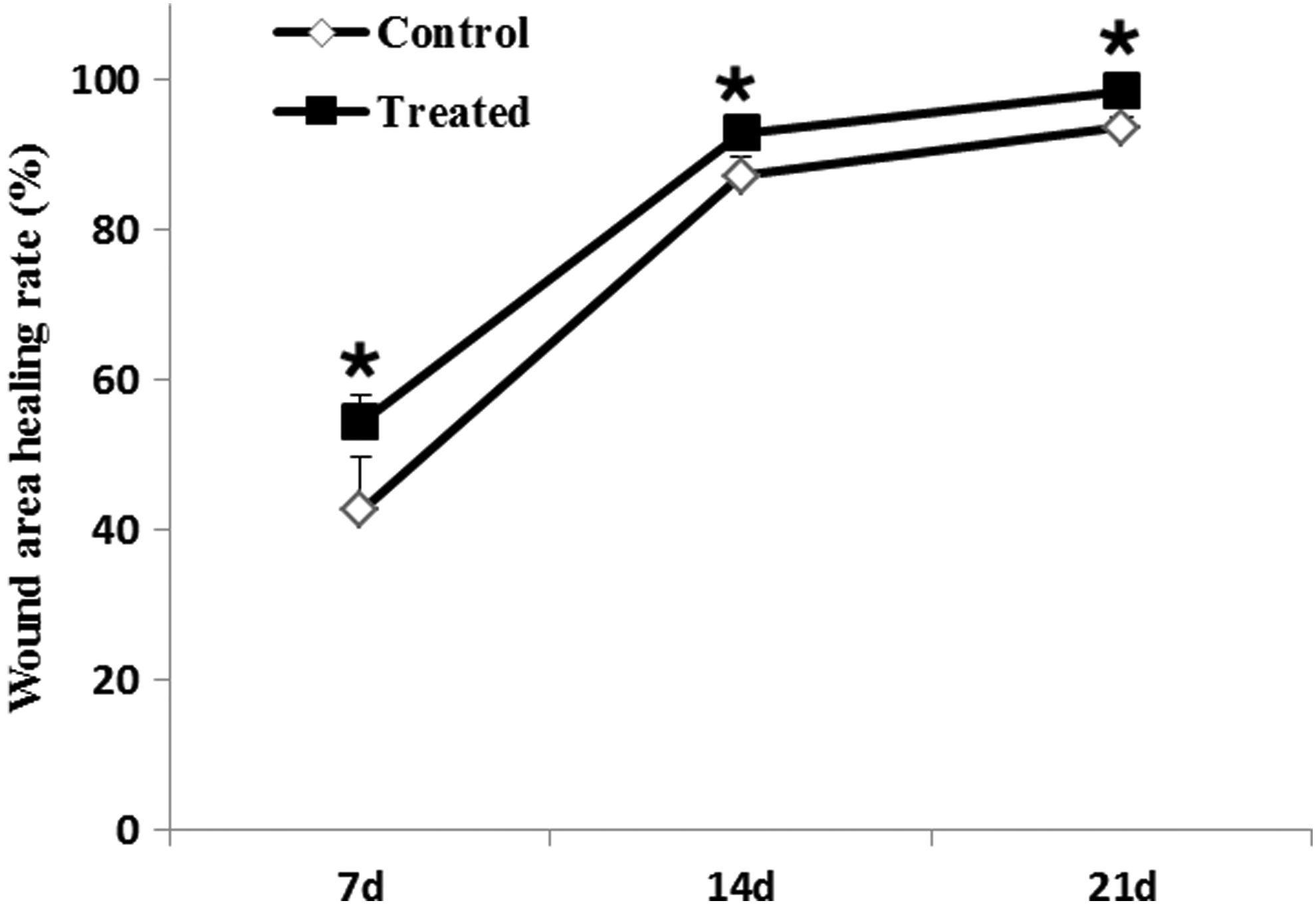
Wound area healing rate at days 7, 14, and 21 (%). Values are presented as the mean ± SD. Tested by independent-samples t-test (*p < 0.05). SD, standard deviation.
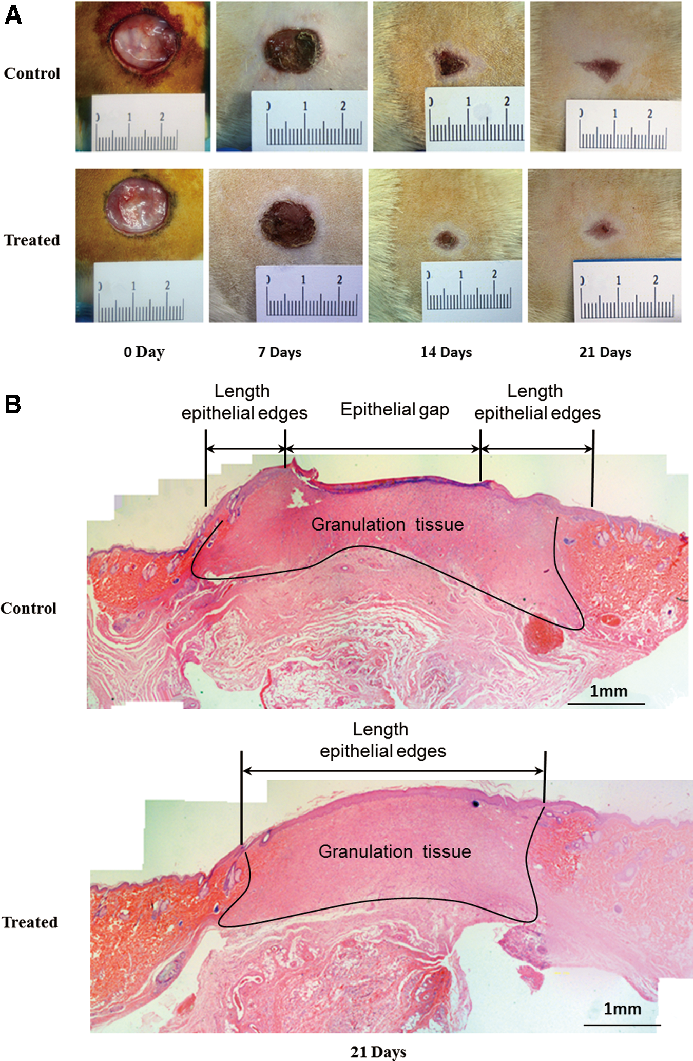
Morphological analysis of wound sites in rats at days 7, 14, and 21.
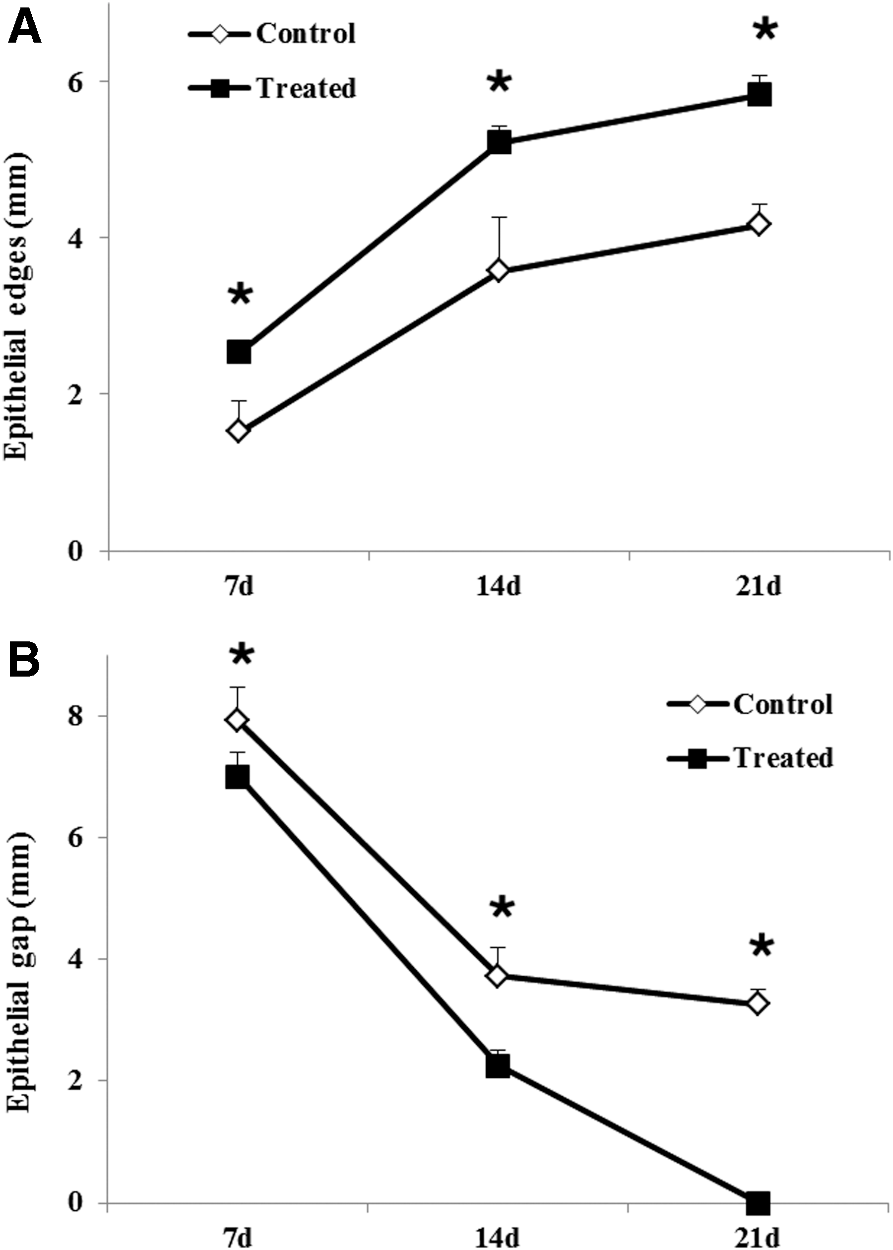
The length of epithelial edges is the most indicative index of reepithelialization. The irradiation of the carbon arc lamp promoted reepithelialization of rat wounds at days 7, 14, and 21 (Fig. 3A). Further, the length of the epithelial gap in the treated group was smaller than that in the control group at days 7, 14, and 21 (p < 0.05) (Fig. 3B). These results indicate that irradiation by the carbon arc lamp significantly accelerated epithelial regeneration of rat wounds during wound healing.
Effect of a carbon arc lamp on wound-healing quality in rats
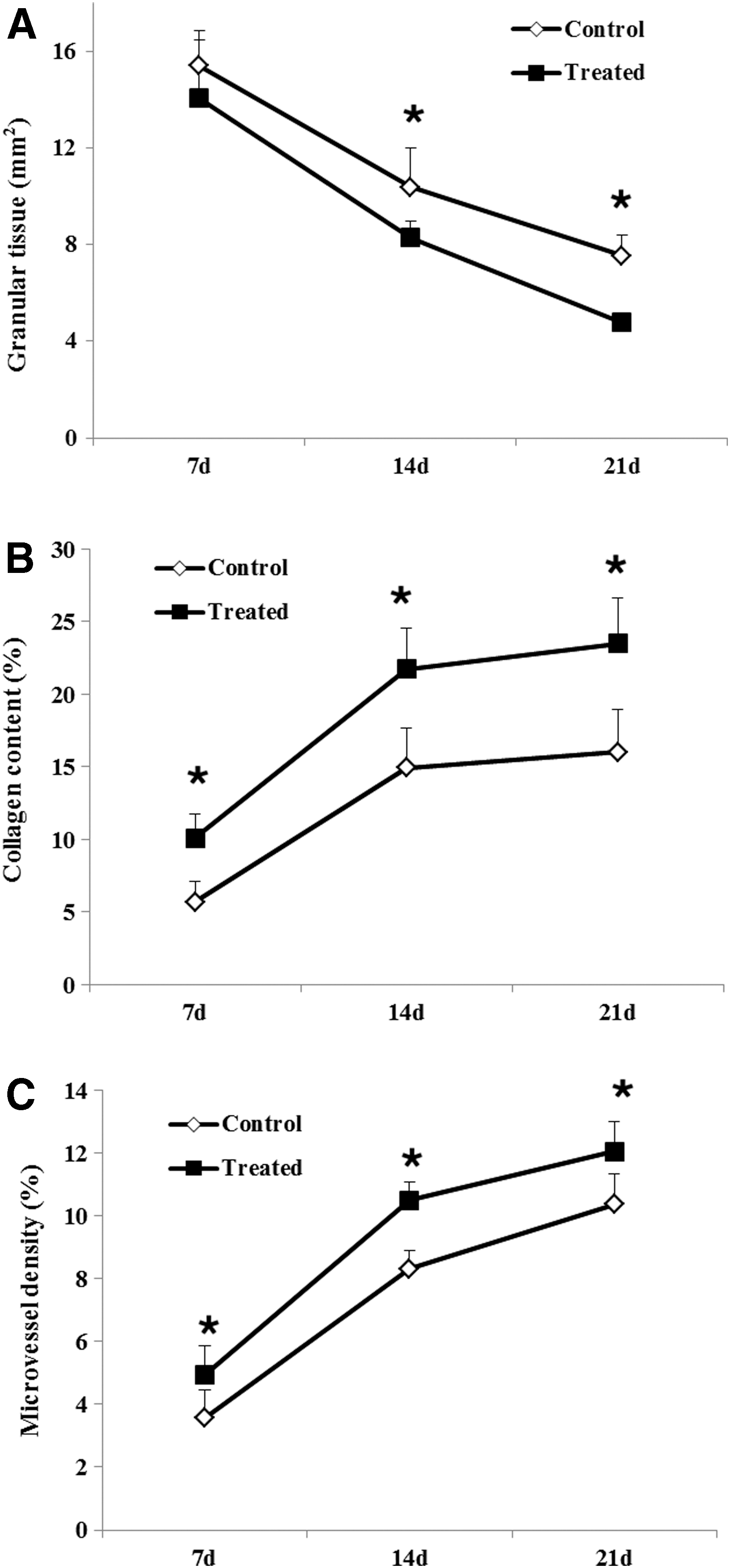
It was reported that epithelialization and granulation-tissue formation are active throughout the wound-healing process. 12 The proliferation of keratinocytes, fibroblasts, and endothelial cells contributes to the process. 12 The area of granulation tissue in the treated group was smaller than in the control group at days 14 and 21 (p < 0.05) (Fig. 4A). By contrast, the increase in collagen content and microvessel density in the treated group were greater than in the control group at days 7, 14, and 21 (p < 0.05) (Fig. 4B, C).
Granulation tissue in the control group was loose and clearly accompanied by edema (Fig. 5). By contrast, granulation tissue in the treated group was compact and highly cellular, mainly composed of fibroblasts and microvessels organized in a regular manner.

Masson-stained sections of wound specimens from rats in the control and treated groups at days 7, 14, and 21 (Masson staining, 20 × 10). Upper pictures showed the representative results from the control group. Note that granulation tissue was loose and clearly accompanied by evident edema. Lower pictures showed the representative results from the treated group. Granulation tissue was compact and highly cellular, mainly composed of fibroblasts and microvessels organized in a regular manner.
Effect of a carbon arc lamp on the proliferative cells in rats
The BrdU-labeled cells were assayed to determine whether the carbon arc lamp promoted wound healing by increasing cellular proliferation in the wound edge. BrdU-labeled cells were present in the epidermal basal layer, hair follicles, and glands (Fig. 6A) and were more abundant in the treated group than in the control group at days 7 and 14 (p < 0.05) (Fig. 6B). Therefore cellular proliferation in the wound edge of rats was greatly increased by irradiation from the carbon arc lamp.
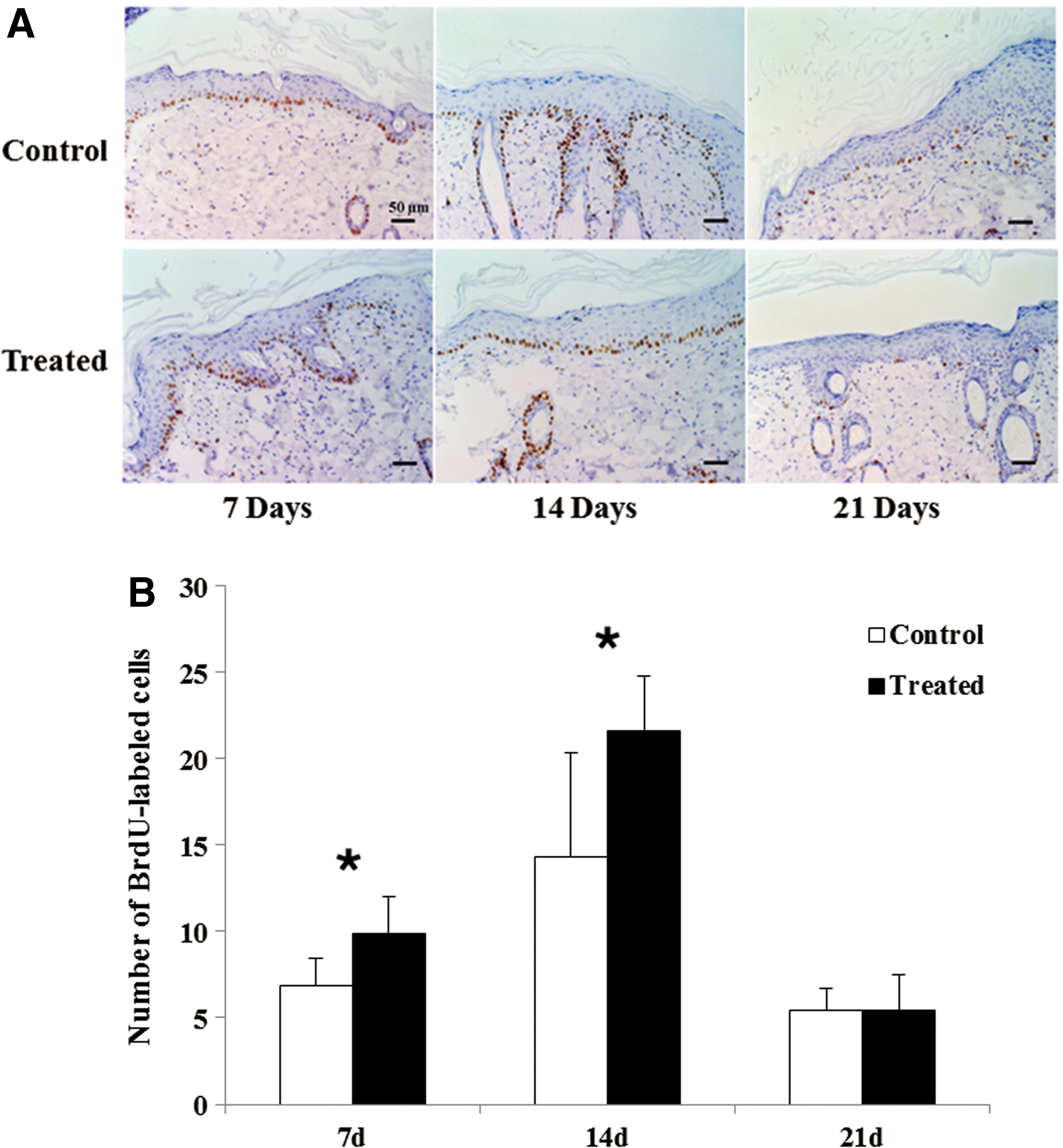
Effect of carbon arc lamp on cell proliferation in the wound in rats at days 7, 14, and 21.
In addition, the histological distribution of BrdU-labeled cells at the migrating keratinocyte leading edge was changed at day 5 (Fig. 7). BrdU-labeled cells were manifested as a detachment from the basal layer and were densely distributed around the spikes or granular layers of the epidermis. The number of BrdU-labeled cells in the margin was positively correlated with the wound-healing process. On day 21, the wound completely healed, and the BrdU-labeled cells were homing to the epidermal basal layer. Moreover, the number of BrdU-labeled cells was significantly reduced (Fig. 6).

Wound histology at day 5 (BrdU immunohistochemical staining, 20 × 10).
Discussion
The skin wound-healing process consists of three overlapping and characteristic stages, namely the inflammatory reaction (1–3 days), cell proliferation (4–21 days), and tissue remodeling (21 days to 1 year). 13 During wound healing, it has been observed that photostimulation influences the proliferation and migration of related cells, which promotes tissue repair and accelerates wound healing. 6,14
During the proliferative phase of wound healing, many cells are actively involved in the repair processes, in which epithelial cells, endothelial cells, and fibroblasts are important repair cells. 15 –17 They are involved in wound reepithelialization, granulation-tissue formation, and reconstruction of the skin barrier function. 16,18 In the present study, we observed that irradiation by the carbon arc lamp dramatically promoted keratinocyte proliferation and migration, which in turn accelerated wound reepithelialization. During the entire proliferative period in the present experiment, the collagen content and the microvessel density in the wound increased. Therefore, it is possible that irradiation by the carbon arc lamp activates extracellular matrix deposition and angiogenesis, contributing to an improved wound-healing quality.
Moreover, we demonstrated for the first time that irradiation by a carbon arc lamp enhanced the proliferation of BrdU-labeled cells in the wound edge in rats during wound healing. BrdU is a thymidine deoxynucleoside analog that penetrates into the nucleus of the cell during the S phase of the cell cycle, thus it has been proved to be a suitable marker for proliferating cells. BrdU-labeled cells are considered to be proliferative cells. 19 In the present study, we observed BrdU-labeled cells appearing in the epidermal basal layer, hair follicles, and glands, which were identified as epidermal stem cells located in the skin. 20 Therefore, it is possible to track epidermal stem cells during wound healing and to study their biological behavior, including proliferation and migration.
It has been reported that epidermal stem cells can be involved in wound repair, and the main function of these cells in wound edges may be to promote wound reepithelialization. 21 Indeed, we observed that reepithelialization and healing quality were accelerated and improved by the irradiation of a carbon arc lamp accompanied by an increase of BrdU-labeled cells. These results suggest that BrdU-labeled cells play an extremely important role in the proliferative phase of wound healing. With respect to this, we suggest that the carbon arc lamp regulates the wound-healing process by stimulating cell proliferation in the wound edge. This phenomenon may be closely related to the photobiostimulation of the carbon arc.
Taken together, our findings indicate that the application of a carbon arc lamp increases cell proliferation and enhances skin tissue repair. Therefore, irradiation by a carbon arc lamp is an effective method to aid wound healing. However, there remain many limitations in the study of the carbon arc lamp. For example, there is lack of related research about the influence of optical parameters, including how power density, spot area, and irradiation time, affect the therapeutic effect and the comparison with other light sources, such as a laser or LED, in terms of mechanism and efficacy. In response to these limitations, we intend to resolve this in the future in experimental research to provide an important theoretical basis for the safe and effective use of the carbon arc lamp for wound healing.
In phototherapy for wound healing, the carbon arc lamp has unique advantages compared with the current clinically applied phototherapy instruments. For example, the carbon arc lamp has the therapeutic effect of integrated light that includes visible light, infrared, ultraviolet, and magnetic pulses. The results of previous studies demonstrated that pathogens in acute wound infections can be killed by ultraviolet C (200–280 nm). 22 Therefore, it can be theoretically speculated that the carbon arc lamp will have a certain bactericidal effect, contributing to the healing of infected wounds. In addition, the area irradiated by the carbon arc lamp is 10-fold greater than by low-level laser therapeutic apparatus, so that it can be applied to the treatment of a larger area of the wound. In addition, the spectrum of the carbon arc is a natural light similar to sunlight, which is relatively mild and has no radiation effect on the human body. Patients can undergo wound irradiation treatment without any protective measures. Finally, it is cheap and readily available to patients. A family-type carbon arc lamp has been described, and this will lead to treatment that is not restricted by location.
Conclusions and Summary
This study is the first to have observed the effect of a carbon arc lamp on wound healing in rats, and completed the markers of proliferative cells in the wound-healing process. In particular, it is clear that the carbon arc lamp promotes wound healing and improves wound-healing quality by stimulating cell proliferation.
Footnotes
Acknowledgments
This work was supported by the National Nature and Science Foundation, P.R. China (No. 81372065), National Major Disease Control Science and Technology Action Plan Project (ZX-01-C2016021), Guangzhou Huadu District Science and Technology Program (HD14CXY001), the major project of Guangzhou Municipal Science and Technology Bureau (Nos. 201300000091 and 201508020253), and the foster research fund of The First Affiliated Hospital of Jinan University (No. 2017306). We thank GuangDong Ewan Biomedicine Technology Co. Ltd. for contributing the carbon photon therapy instrument.
Author Disclosure Statement
The authors have no conflict of interest to declare.