
Abstract
Objective:
To describe a clinical case of successful conservative management of Localized Juvenile Spongiotic Gingivitis Hyperplasia (LJSGH) using photodynamic therapy (PDT) and reviews the current literature on this pathology.
Background data:
LJSGH is a recently described rare disease with controversial treatment results. As of today, 13 publications report surgical treatment approaches. The use of CO2 laser and cryotherapy was reported only in one study. The use of PDT was not previously reported.
Patients and methods:
A 9-year-old male patient was referred to our institution with the chief complaint of asymptomatic “inflamed gingiva” starting 1 year before. Clinical examination revealed an erythematous line accompanying the gingival contour, with a certain degree of hyperplasia. The diagnosis of LJSGH was performed based on clinical features and later confirmed histopathologically. A novel approach using PDT was then proposed. The photosensitizer was methylene blue, and a semiconductor laser diode was used.
Results:
One week after starting PDT, gingival hyperplasia was partially reduced. Immediately after the end of treatment, a significant reduction of gingival hyperplasia was observed. PDT proved to be safe, quick and painless, with no esthetic harm.
Conclusions:
This case illustrates the benefit of a more conservative approach as opposed to surgical procedure, with good clinical response and decreased morbidity over a 2-year follow-up period.
Introduction
Localized Juvenile Spongiotic Gingivitis Hyperplasia (LJSGH) is a recently described inflammatory disease that mostly affects children and adolescents, predominantly on the anterior maxillary gingiva. Table 1 summarizes the previous reports of LJSGH. 1 –16 The main clinical feature of the disease consists of an erythematous line accompanying the marginal gingiva, and in some cases, gingival hyperplasia is present. Characteristically the disease is not associated with plaque-induced inflammation. 1,2,11 The absence of plaque is, therefore, an obvious sign of LJSGH, which means that the diagnosis of pubertal gingivitis should be ruled out. 1
Descriptive Analysis of the Previous Reports of Localized Juvenile Spongiotic Gingivitis Hyperplasia
Some cases occurred in both jaws (maxilla and mandible).
NR, not reported.
The histological examination shows intercellular edema in the spinous layer of the epithelium (spongiosis) and acanthosis. Other histological features similar to inflamed junctional/sulcular epithelium and periodontal pocket epithelium are generally found, such as elongation of rete pegs, atrophy of the epithelium, and a neutrophilic infiltrate. 1 Furthermore, the immunohistochemical analysis showed a pattern compatible with the profile described for the junctional epithelium. Possibly, the junctional epithelium exteriorized from the gingival sulcus would be more prone to irritation from a variety of sources, resulting in inflammation and hyperplasia, with the subsequent development of LJSGH. 2
Although the etiology of LJSGH remains unclear, it is clear that the disease has an inflammatory nature. The LJSGH was not correlated with HIV, HPV, estrogen, and progesterone levels nor with allergic factors, linear gingival erythema, or fungal infection. 1,3,9,10 Regarding treatment, although the surgical approach is still considered the gold standard, the technique is invasive with controversial results. 1,2,9,14 With this issues in mind, we adopted a more conservative and novel approach for the disease. Photodynamic therapy (PDT) has shown to be effective in treating similar inflammatory conditions, in addition to its selective toxicity, low invasiveness, and rare side effects. 17 This study aimed to report a clinical case of successful conservative management of LJSGH using PDT and reviews the current literature on this pathology. Previous cases were identified through a literature review using electronic databases such as PubMed, Scopus, Web of Science, Scielo, and gray literature (Google Scholar and dissertations databases). Databases were searched up to March 2018.
Case Report
A 9-year-old boy was referred to our institution with the chief complaint of asymptomatic “inflamed gingiva” starting 1 year before. There was no history of trauma or systemic infection. The medical history of the patients was not contributory. Extraoral examination revealed no abnormalities. Intraoral examination showed gingival hyperplasia and erythematous line along the marginal gingiva (Fig. 1A). All lesions had a granular surface texture without signs of bleeding or visible dental plaque. Thus, the clinical diagnosis of spongiotic gingivitis was established. Afterward, an incisional biopsy was performed. The histopathological examination demonstrated mucosal fragments covered by nonkeratinized stratified squamous epithelium, with papillary surface (Fig. 2A). The lesional epithelium was characterized by a prominent spongiosis in the spinous layer of the epithelium (Fig. 2B), irregular acanthosis, and neutrophilic exocytosis (Fig. 2C). The underlying connective tissue was richly vascularized with dense inflammatory infiltrations mostly by neutrophils, lymphocytes, and plasma cells. Neither dysplasia nor viral cytopathic effect, parasites, abscesses, or granulomas were observed. On the other hand, mixed inflammatory infiltrate was present. The histopathological findings confirmed the clinical hypothesis of LJSGH.
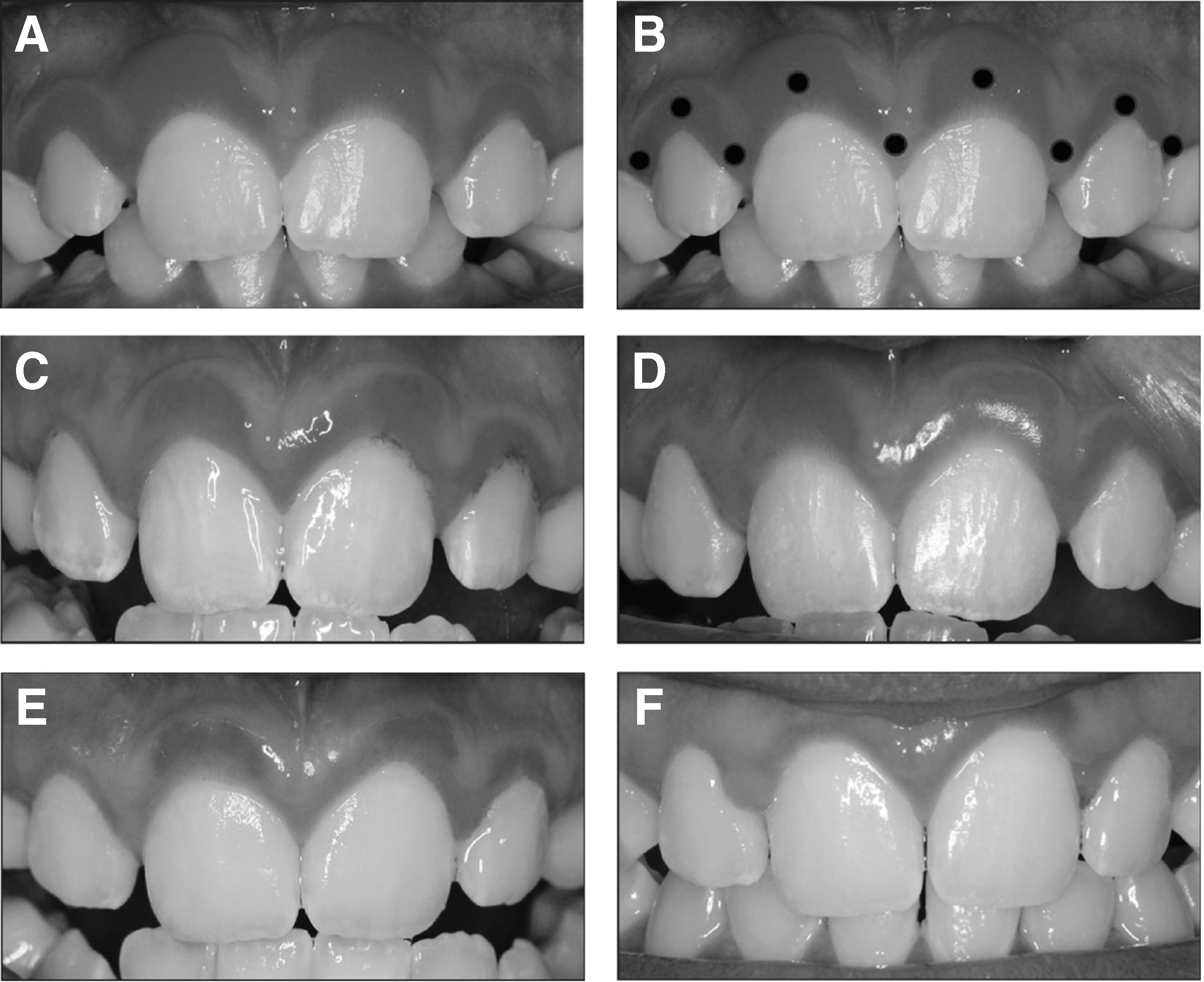
Frontal photographs demonstrating the improvement of LJSGH after PDT.
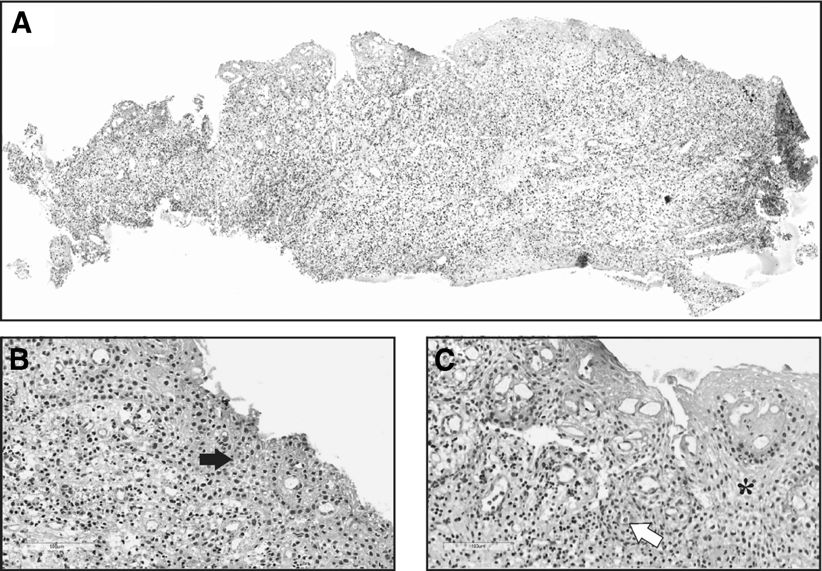
Microscopic examination.
Based on the final diagnosis, we proposed a more conservative treatment for the disease, as compared with the usual surgical procedure, the goal being to ensure better esthetic results. As a novel approach, we followed the same protocol for oral lichen planus (OLP) therapy used in our institution. This protocol consisted of topically placing methylene blue (0.1%) for 5 min throughout the gingival sulcus and the marginal gingiva surface, which was followed by low-level laser irradiation. The laser device used was DMC Therapy XT (São Carlos, São Paulo, Brazil) and the laser parameters are summarized in Table 2. There was a total of 9 laser applications points in the marginal sulcus of the affected incisors (Fig. 1B). In total, 12 sessions of PDT were performed (3 sessions per week on nonconsecutive days), during 1 month.
Laser Parameters for the Reported Case
CW, continuous wave.
The first signs of gingival hyperplasia reduction were detected 1 week after the start of PDT (Fig. 1C). Immediately after the end of treatment (12 weeks), a reduction of gingival hyperplasia was observed (Fig. 1D). The significant improvement of the erythematous line followed the criteria of thickness and color intensity. After 24 weeks, discrete areas of erythema persisted only in the area of the incisors, where the lesions were more evident. PDT was painless and did not cause any esthetic problems (Fig. 1E). After 2 years, a significant improvement of LJSGH was observed, and there was a slight hyperplasia in the interdental papilla between the maxillary incisors (Fig. 1F).
Discussion
Our case report first demonstrated PDT as a promising conservative approach to the treatment of LJSGH with a 2-year follow-up. Although it is a rare and recently described disease, the treatment described on most of the previous reports was based on invasive procedures, in which recurrence rates and inadequate esthetic results frequently occurred. On the other hand, PDT is safe, painless, noninvasive, inexpensive, and can be used without clinical contraindication. 18 –20
In our literature review, 221 previous reported cases of LJSGH were found with no clear gender preponderance (Table 1). 1 –16 This number may be an overestimation or an underestimation, as the disease is recently described and the diagnostic criteria has not been well established in some reports. Some authors have suggested that the lesion should be named as localized spongiotic gingival hyperplasia, as the disease may occur in both pediatric and adult population. 4 However, in this previous retrospective study, the higher frequency of LJSGH in patients over 21 years of age may be related to a later diagnosis, or to an overestimated frequency due to an unclear histopathologic diagnostic criteria. 4 Differently from this aforementioned study, most of the previously reported cases occurred in young patients, which justify the nomenclature of the disease. As a rare disease, the demographic factors related to LJSGH, such as gender, age, and true prevalence remain undetermined. The patient of the present case report was a 9-year-old boy, and the typical histopathological findings confirmed the clinical hypothesis of LJSGH.
There have so far been few studies demonstrating the clinical efficacy of the use of PDT in the treatment of periodontal disease. 21 –23 PDT influences periodontal healing and has an antimicrobial effect. 22 A previous study in an experimental rat periodontal disease model with ligature placement showed that PDT-treated animals presented decreased bone resorption, as well as reduced neutrophil migration and lower TNF-α expression. 23 Other study demonstrated that PDT is effective in increasing the expression of bFGF gene, an important factor in periodontal tissue regeneration and could indicate periodontal tissue regeneration. 24 In a recent clinical randomized controlled trial study, PDT significantly reduced inflammatory mediator levels in patients with severe chronic periodontitis under periodontal maintenance. 25 Therefore, PDT may have additional benefit in immunomodulatory response. The successful therapeutic effect of PDT in our case report was possible due to the anti-inflammatory and antiproliferative nature of the therapy. 21 Our PDT protocol for LJSGH was adapted from the protocols used to treat OLP. 17,26 –28 Similarly to LJSGH, OLP has an obscure etiology, with an inflammatory nature. 29
In 10 of the 16 previous studies found in the literature review, surgery was the main treatment modality. 1,2,5,8,9,11,13 –16 There were recurrence and esthetical issues in these previous case reports. 1,2,9,14 A previous study reported a sequential treatment with chlorhexidine mouth rise, topical corticosteroid, and systemic antifungal agent, without clinical improvement. 10 Cryotherapy and laser CO2 were also described as possible treatments of LJSGH. Despite the good clinical response, the authors reported moderate discomfort coming from the patient. 3,12,14,15 The spontaneous remission of the lesion has been mentioned in only two previous studies. 1,16 In the former study, the follow-up was not adequately documented. 1 In the second aforementioned study, the authors recognized that the spontaneous remission of LJSGH might be attributed to the elimination of a possible, yet unknown, causative factor. 16 The hypothesis of spontaneous remission is unlikely in our case, since the progressive improvement of LJSGH has been well documented, and has occurred throughout and after the PDT.
When compared with the invasive techniques most commonly used for treating LJSGH, PDT has several advantages. PDT's systemic effects are insignificant, its toxicity regarding normal tissue is minimal, and it presents reduced morbidity and excellent esthetic results. Furthermore, the technique is simple, cheap, and can usually be performed on an outpatient basis. 30
This report is limited in that it is a single-patient case observation. Since the proposed treatment was based on a combination of PDT with laser therapy, the effects of both treatment modalities might have been overlapped, or even the effect might be more related to the effect of laser therapy. Thus, further prospective studies to confirm our observations and to also optimize the treatment parameters should be performed.
Conclusions
In conclusion, our case report pointed out the benefits of the conservative approach of PDT in the treatment of LJSGH with a 2-year follow-up. Further clinical studies are required to ascertain its long-term validity in treating LJSGH.
Footnotes
Author Disclosure Statement
The authors declare that they have no conflicts of interest. No external funding, apart from the support of the authors' institution, was available for this study.