




Abstract
Objective:
The aim of the study was to compare the composite adaptation of three systems by using cross-polarization optical coherence tomography (CP-OCT).
Background data:
Most polymer-based restorations suffer from polymerization shrinkage that affects the interfacial seal. This shrinkage cannot be detected by conventional X-ray methods. Optical coherence tomography was proved to be a reliable non-invasive imaging tool to examine biological structures and biomaterials at micron scale.
Methods:
Twenty-four cylindrical class-V cavities were prepared on the buccal surfaces of the extracted human molars. After cavity preparation, samples were randomly divided into three groups (n = 8) according to the restoration system: one-step self-etch Clearfil Tri-S Bond Plus with Clearfil Majesty ES-2 composite (TS; Kuraray Noritake Dental), Single Bond Universal in self-etch mode with Filtek Z350 XT composite (SB; 3M ESPE), and one-step self-etch Plafique Bond with Plafique LX 5 composite (PB; Tokuyama Dental). The restoration placement was carried out according to the manufacturers' recommendations. Later, the specimens were immersed in a contrasting agent; then, image acquisitions were taken by CP-OCT to calculate the adaptation percentage by using an image analysis software.
Results:
Mann–Whitney U test showed no statistical significant difference in the adaptation percentage between TS (91.72 ± 11.6) and SB (93.43 ± 6.9) groups (p > 0.05). However, the adaptation percentage in PB (41.83 ± 28.5) was significantly lower than in the other tested groups (p < 0.05).
Conclusions:
Within the limitation of the study, TS and SB groups showed better adaptation than PB. Moreover, CP-OCT is a useful imaging tool that can display composite adaptation at micron scale.
Introduction
The etch-and-rinse adhesive has better performance in terms of bond durability to enamel over time than the self-etch adhesive system. However, the latter is getting more popular. 1 This popularity might be attributed to its simplified procedure and less sensitive technique, compared with the etch-and-rinse adhesive.
Regardless of the bonding system, dental adhesives are prone to micro-leakage that might be complicated by composite polymerization shrinkage. 2,3 This is known to cause postoperative sensitivity, marginal discoloration, fracture of the restorations, and secondary caries. 4
The volumetric composite shrinkage and micro-leakage are hard to be detected by clinical examination and conventional radiography. 5,6 Although micro-computed tomography and cone beam computed tomography are high-resolution imaging techniques, they are considered hazardous on biological tissues with radiation exposure. 7,8 Recently, optical coherence tomography (OCT) has been introduced as a non-invasive and non-hazardous high-resolution imaging system that can provide tomographic and volumetric images of biological structures and non-metallic biomaterials at micron scale. 9 This system was first used in Ophthalmology. 10 Later in 1998, this system was used to image hard and soft dental tissues. 9,11 After that, this imaging was widely used in dentistry for imaging enamel cracks, demineralization, biofilms, and restoration defects. 3,12 –16 In a study by Bakhsh et al., it was possible to image and quantify gap formation under composite restorations. 17 Moreover, a moderate positive correlation between OCT gap measurement and bond strength was reported showing that filling technique has a major influence on composite adaptation. 17
Cross-polarization OCT (CP-OCT) is a non-invasive imaging modality and has been mainly used in demineralization and remineralization studies. 16,18 –20 However, very few studies had investigated its usefulness in measuring the micro-gaps under the restorations.
Therefore, the aim of this study was to test the floor adaptation of three bonded resins restored in class-V cavities by using CP-OCT. The null hypothesis in this study is that there is no difference in composite adaptation among the tested restorations at the cavity floor.
Materials and Methods
Materials used
Three adhesive systems were used in this experiment: Clearfil Tri-S Bond Plus (Kuraray Noritake Dental), Single Bond Universal (3M ESPE), and Palfique Bond (Tokuyama Dental). Three resin composites were used: Clearfil Majesty ES-2 (Kuraray Noritake Dental), Filtek Z350 XT (3M ESPE), and Plafique LX 5 composites (Tokuyama Dental). The materials used in this study are listed in Table 1. The lot number and chemical composition of each material are according to the information provided by the manufacturers.
Composition of the Materials Used in the Study
3D-SR, methacyloyloxyalkyl acid phosphate; 10-MDP, 10-methacryloyloxydecyl dihydrogen phosphate; Bis-GMA, dimethacrylate; HEMA, 2-hydroxyethyl methacrylate; TEGDMA, triethyleneglycol dimethacrylate.
Sample preparation
This study was conducted in vitro on 24 extracted human non-carious molar teeth. The design of the study was implemented according to the guidelines of the Ethics Committee of King Abdulaziz University (KAU, Jeddah, Saudi Arabia) and in accordance with the principles of the Declaration of Helsinki.
Cylindrical class-V cavities were prepared on the buccal surfaces of each tooth with dimensions of 1.5 mm (depth) × 4 mm (diameter). After cutting the roots of the teeth, the prepared cavities were randomly divided into three groups (n = 8): TS group (Tri-S Bond Plus bonded to Clearfil Majesty ES-2 composite), SB group (Single Bond Universal adhesive in self-etch mode with Filtek Z350 XT composite), and PB group (one-step self-etch Palfique bond adhesive bonded to Palfique LX 5 composite). Each step of sample preparation was performed by the same operator to maintain the standardization of the methodology protocol. In addition, the filling technique for the composite restoration was carried out in a bulk increment (Bulk-filling technique—2 mm thickness), condensed gently against the cavity floor and walls, and cured according to the manufacturers' instructions, using quartz tungsten halogen light-cure with a ∼500 mW/cm2 light intensity (LITEX 680A; Dentamerica).
Contrasting medium preparation
All samples were stored in hydrated condition for 24 h at room temperature. Preparation of the contrasting solution was carried out by dissolution of 25 mg of silver nitrate in 25 mL of distilled water in a dark container. Then, ammonium hydroxide solution was added drop by drop to titrate the black solution until it became transparent. Before immersion in the prepared solution for 24 h, all specimens were coated with a nail varnish except the restoration area by 1 mm around the margins. Afterward, the specimens were washed with distilled water and placed in a photo-developing solution for 8 h under fluorescent light to convert the silver diamine ions into metallic grains. 21,22 A schematic illustration of the sample preparation and immersion is shown in Fig. 1.
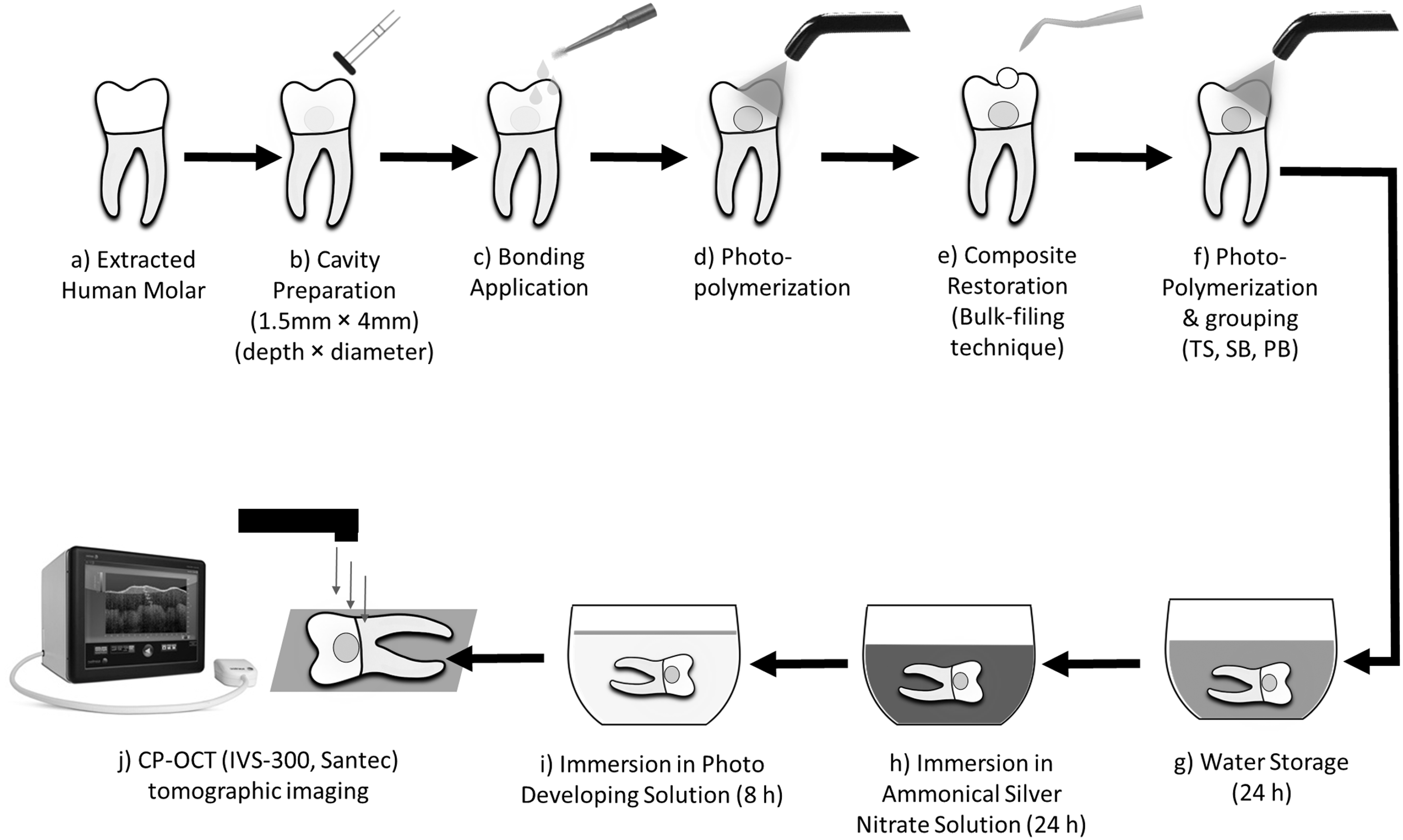
Schematic illustration showing specimen preparation, restoration, immersion in contrasting agent, and CP-OCT imaging. CP-OCT, cross-polarization optical coherence tomography.
CP-OCT system
The prepared specimens were imaged by using a CP-OCT system, which is a functional modification of swept-source OCT (SS-OCT) with a high scan rate (30 kHz) continuous wavelength centered near 1310 nm and a wavelength range of 100 nm. It uses a diode laser with axial and lateral resolutions of ∼12 and 30 μm, respectively. 16
All technical specifications are listed in Table 2. The size of the B-scan image was 500 × 924 pixels corresponding to 5 × 8.18 mm (x, z). Interferometric theories of CP-OCT image acquisition and processing are described elsewhere. 19
Technical Specifications of the Cross-Polarization Optical Coherence Tomography System
CP-OCT, cross-polarization optical coherence tomography.
Tomographic imaging and image analysis
Before tomographic imaging, all specimens were stored in a humid condition at room temperature for 24 h. The specimens were fixed on a micrometer stage, and the scanning probe and the laser beam were oriented perpendicularly to the buccal surfaces. Imaging of specimens is shown in Fig. 1.
Several sequential B-scans were obtained along the restoration at 250 μm interval distance. The presence of gap was observed as high signal intensities at the resin-dentin interface at the cavity floor in the form of bright white clusters of pixels. Quantification of the cavity floor length and the gap length on each B-scan was carried out by using a digital image software, as described in our previous reports (ImageJ v. 1.45q; National Institutes of Health). 2,17
The adaptation percentage parameter was defined as:
Statistical analysis
Statistical analysis was performed by using the Statistical Package for Social Science (SPSS for Windows, Version 23; SPSS), with the significance level defined as alpha = 0.05. Since the distribution of the data was not normal (Kolmogorov Smirnov; p < 0.05), non-parametric tests (Kruskal-Wallis test with Mann-Whitney U test) were carried out.
Results
After manipulation of the OCT data, some B-scans showed high signal intensities at the cavity floor in some areas in the form of a diffuse bright band of white pixels, which was interpreted as a loss of interfacial seal (micro-gap). Other areas showed no abrupt change in the signal intensities, which was an indication of no loss of interfacial seal (Fig. 2). The adaptation percentage results showed no statistically significant difference between TS (91.72 ± 11.6) and SB (93.43 ± 6.9) groups (p > 0.05), whereas there was a statistically significant difference between PB (41.83 ± 28.5) and the other tested groups (p < 0.05) (Fig. 3).
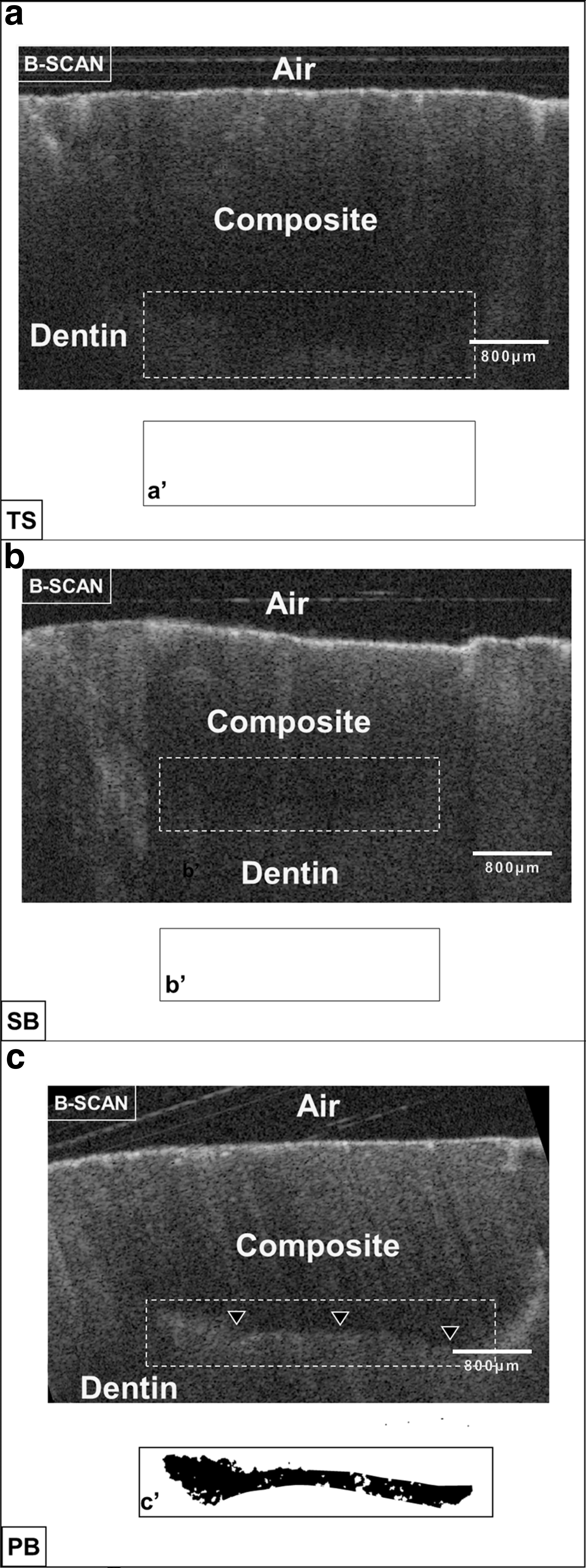
Representative B-scan images of the tested groups. (TS): In
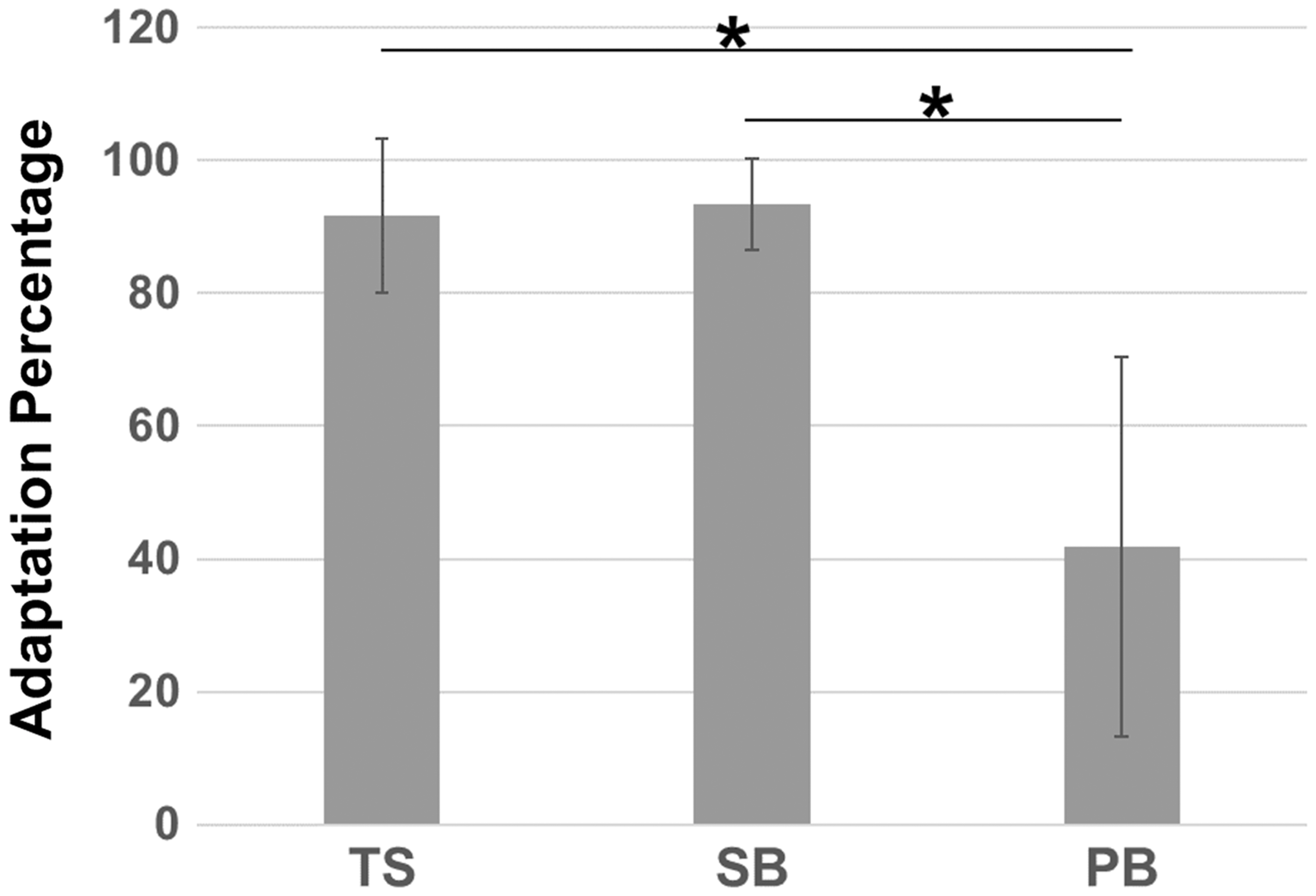
The adaptation percentage of the tested group. The asterisk means that there is statistical significance.
Discussion
In routine clinical and radiographic examination, detection of micro-gaps under composite restoration is a challenging procedure. With recent advancements in imaging modalities, the CP-OCT stands to show a great promise in ease of imaging with high resolution. It provides a comprehensive assessment of the biological structures using laser imaging without the need for traditional sectioning methods. 3,23
It has been reported in the literature that composite polymerization shrinkage is greatly influenced by the composite chemical composition, composite filler loading, and the cavity configuration (C-factor). 2,3,24,25 In this study, three different composites and their corresponding adhesives were examined from three different manufacturers to evaluate whether the differences in the composite composition and adhesive formula would influence the gap formation and composite adaptation when restoring class-V cavities.
C-factor is the number of bonded surfaces to the number of un-bonded surfaces in a dental restoration, as described in the literature. 26,27 Many reports showed that composite placement in the bulk-filling technique would induce the greatest polymerization shrinkage. 2 Therefore, the investigators in this study were assuming that this technique would test the performance of the composite in terms of cavity floor adaptation and gap formation under extreme conditions (High C-factor).
The results of the study showed no significant difference between the TS and SB groups. This could be related to the similarities between these two restorations. At the composite level, the filler contents of the composite in the PB group were around 71% by volume, whereas the filler contents in TS and SB composites were comparable (TS: composite filler contents 66% vol., SB: composite filler contents 63.3% vol.). Many studies demonstrated the effect of the filler contents on the volumetric shrinkage. 3,28 They found that increasing the filler contents will lead to lower volumetric shrinkage. However, it will increase the modulus of elasticity and contraction stresses as well. Since the cavities were filled with the composites in the bulk-filling technique, it was expected that high contraction stresses at the cavity floor would be generated. 2 As a consequence, the adaptation of the composite would be affected, especially in high C-factor cavities (as a class-V used in this study). This could explain the lower performance of the PB group in comparison to the TS and SB groups.
Other explanations would be related to the adhesive contents. Most self-etch adhesives contain solvents as acetone, alcohol, water, or combinations. 29 The role of these solvents is to remove the water contents from the organic dentin to enhance resin monomer infiltration into spaces previously occupied by water. 30 However, the rate of water removal from the dentin differs from one solvent to another. 31 In this study, the adhesive formula of TS and SB groups is alike, as they comprise 10-methacryloyloxydecyl dihydrogen phosphate (10-MDP) functional monomer and ethanol/water. Recent studies showed that ethanol has the ability to minimize the nanoleakage and improves the interaction of the adhesive to the hard tooth structure. 2 Moreover, it has a rapid water removal effect before curing the adhesive. 32 This would be attributed to the high vapor pressure of ethanol (5.95 kPa at 20°C), compared with the vapor pressure of isopropyl alcohol (4.1 kPa at 20°C) that was found in PB adhesive. This means that isopropyl alcohol requires more time to remove the water from the dentin, which would lead to incomplete water removal from the dentin before adhesive curing. This, in turn, could negatively influence the restoration adaptation in general. 2,21 Another finding of interest is in the adhesive monomers. The 10-MDP functional monomer in the adhesives of the TS and SB groups has a high binding affinity to calcium and hydroxyl apatite to form calcium-phosphate salts. 33 –35 Reports in the literature found that the formed chemical bond with 10-MDP tends to be more stable in water with low liability to bond degradation. 35,36
On the other hand, the adhesive in the PB group has a 3D-SR monomer, which has a three-dimensional structure with a functional group and a double bond. 37 The functional phosphate monomer group is capable of interacting with calcium in enamel and dentin, forming ionic bonds. 37 The interaction potential of the 3D-SR monomer with calcium is equivalent to or greater than that of mono-phosphate monomers, such as 10-MDP. However, the large molecular size of the 3D-SR monomer limits its penetration into dentin. 38 These findings can be interpreted to mean that the adhesive formula of self-etch adhesives and composite filler loadings have significant influences on the restoration adaptation. However, long-term adaptation durability studies are required.
The CP-OCT system was employed to detect the gaps between dentin-resin interfaces at the cavity floor. In this study, the use of a contrasting solution had improved gap detection. As the specimens were immersed in a contrasting medium, the silver ions diffused through the interfacial spaces to accumulate at the formed gaps under the restorations. In B-scan, these gaps were detected as diffuse reflections in the form of bright white clusters of pixels, which differs from the specular reflection. The appearance of the specular reflection (mirror-like reflection) is based on the flatness of the imaged surface. 3 This type of reflection is mainly seen with the SS-OCT system and not with the CP-OCT system. It is based on Fresnel phenomena that rely on the difference in the refractive indices of the imaged media. 3,39 On the other hand, the diffuse reflection is a result of the interaction of the incident light with a highly scattering medium (silver particles) that causes the reflected light to travel in different directions rather than a single direction as in the case of specular reflection. 39 It is worth mentioning that the specular reflection is not typically seen with the CP-OCT system due to the presence of a second polarizer that is positioned 90° to the first polarizer to remove the spectacularly reflected light from reaching the detector.
Within the limitation of the current research that includes a narrow study design within a short period and a limited number of the tested materials, the proposed null hypotheses were rejected. In future CP-OCT adaptation studies, thermal stress and cyclic loading should be considered.
Conclusions and Summary
It can be concluded that restoration adaptation is significantly affected by the adhesive formula and composite filler contents. The 10-MDP monomer might have a significant influence on the performance of one-step self-etch adhesives in terms of composite adaptation. CP-OCT is a great imaging tool that can display composite adaptation at micron scale.
Footnotes
Acknowledgments
This work was supported by the Saudi Dental Research (SDR) group and KAU. The authors, therefore, acknowledge and thank SDR and KAU for their logistic and technical supports. They would like to show their gratitude to Sarah Gashlan, Latifah Dughmash, Shada Ameen, Hatoon Bader, Ghufran Zaatarri, and Bayan Al-Jeffery for their technical assistance in this research. The authors declare no funding received for this research from any funding agency and no potential conflicts of interest and with respect to the authorship and/or publication of this article.
Author Disclosure Statement
No competing financial interests exist.