




Abstract
Background:
The search for alternative therapeutics against antibiotic-resistant bacteria is highly desirable. A promising approach is photodynamic antimicrobial chemotherapy.
Objective:
This work evaluated the photodynamic inactivation (PDI) efficacy of hypocrellin B (HB) on Gram-positive antibiotic-resistant bacteria.
Methods:
PDI efficacy of HB on Gram-positive standard and antibiotic-resistant Staphylococcus aureus, Enterococcus faecalis, and Streptococcus pneumonia and Gram-negative Escherichia coli and Klebsiella pneumoniae was assessed. HB photoactivity on biofilms formed by the Gram-positive bacteria and its cytotoxicity on mammalian CT26 cells were also investigated.
Results:
HB showed no obvious dark toxicity, but provided concentration-dependent inactivation of bacteria and mammalian cells. After irradiation with 72 J/cm2 light, 100 μM of HB achieved about 7 log10 reductions in bacterial survival of Gram-positive strains, but yielded only 2 log10 reductions in bacterial survival of Gram-negative strains. Gram-positive bacteria were as susceptible to PDI in biofilms as in planktonic suspensions, but the efficacy was attenuated.
Conclusions:
The results suggested that HB could serve as a potential antibacterial photosensitizer against Gram-positive antibiotic-resistant bacteria.
Introduction
The extensive use of antibiotics has led to the development of pervasive antimicrobial resistance. At present, infections caused by antibiotic-resistant bacteria are serious concerns for public health. 1 Even though existing antibiotics and vaccines are available, infectious diseases are still responsible for increased morbidity and mortality with ∼17 million global annual deaths. 2 Several classes of antibiotic-resistant bacteria are the main cause for these morbidity and mortality, including Gram-positive methicillin-resistant Staphylococcus aureus (MRSA), multidrug-resistant (MDR) mycobacteria, Streptococcus pneumoniae, and vancomycin-resistant Enterococcus faecalis (VRE), as well as Gram-negative extended-spectrum β-lactamase-producing Escherichia coli and Klebsiella pneumoniae, all of which are major threats to public health. 3
The increasing rate of antibiotic resistance necessitates the search for alternative therapeutics. A promising approach is photodynamic antimicrobial chemotherapy (PACT), which entails the utilization of innocuous light in combination with photosensitizer (PS), and the freely occurring oxygen in and around cells. 4 Following illumination with light of appropriate wavelength, the PS gets stimulated to an excited condition capable of undergoing molecular collisions with the freely available oxygen to generate reactive oxygen species (ROS). 5 Consequently, these ROS mediate the killing of neighboring microbial cells. 2
With respect to conventional therapies, the advantages of PACT include the following: (i) high selectivity since the hyperproliferative cells take PS in a selective manner and photodynamic action only occurs in the locations where light is administered; 6 (ii) PACT possesses a broad spectrum of antimicrobial action because ROS is toxic to almost all bacteria; 7 and (iii) bacterial resistance is regarded as an impossibility since ROS damages are entirely nonspecific. 5,8,9 For superiorities, PACT has great potential to be used in the superficial or localized infections caused by antibiotic-resistant bacteria.
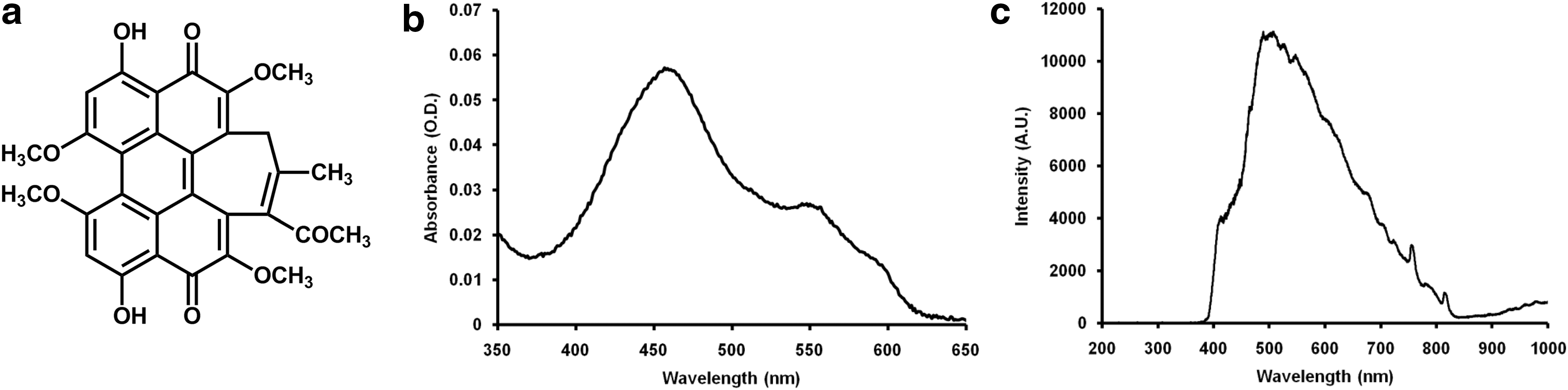
PS plays a crucial role in photodynamic inactivation (PDI) of antibiotic-resistant bacteria. 10 Traditional Chinese herbs are a rich resource of antibacterial drugs and potential PS. Hypocrellin B (Fig. 1a), isolated from natural fungus sacs of Hypocrella bambusae growing in the north-western region of Yunnan Province in China, is a good promising PS due to the high quantum yields of 1 O2, low dark toxicity, quick clearance rate, and availability in a pure monomeric form. 11 Recently, HB has been used as a PS to effectively kill several cancerous cells, 12 –14 but relatively little is known about its PDI efficacy on microorganism, especially antibiotic-resistant bacteria. Therefore, in this study, we investigated the PDI efficacy of HB against several important Gram-positive antibiotic-resistant bacteria, including MRSA, VRE, and MDR S. pneumoniae.
Materials and Methods
PS and light source
HB was prepared according to a previous report. 15 Stock solution (1 mM) was prepared by dissolving HB in 50 μL dimethyl sulfoxide and diluted into 1 mL sterilized phosphate-buffered saline (PBS, pH 7.4). The absorption spectrum of HB was recorded on an UV-Vis spectrometer (Agilent 8453). Light from a 50 W xenon lamp (CEL-HXF300; Ceaulight, China) was used for irradiation. Light with the wavelength of 400–780 nm was selected by an optical filter (CEL-UVIRCUT PD-145; Ceaulight, China) (Table 1). The emission spectrum of the Xenon lamp was recorded on a fiber optic spectrometer (S3000; Seemantech, China). A power meter (CEL-NP2000; Ceaulight, China) was used to adjust the density of 0.08 W/cm2 at the level of the samples. The distance between bacterial samples and optical filter was 10 cm. To prevent heating, a 1 cm ice-cold water filter was placed between samples and optical filter. Bacterial samples were irradiated for 900 sec, with the energy density of 72 J/cm2.
Equipment Parameters
Bacterial strains and culture conditions
Standard Gram-positive strains of S. aureus (ATCC 29213), E. faecalis (ATCC 29212), S. pneumoniae (ATCC 49619), and standard Gram-negative strains of E. coli (ATCC 25922) and K. pneumoniae (ATCC 700603) were stored in our laboratory. Clinical isolate of MRSA and VRE (ATCC 51299) and clinical isolate of MDR S. pneumoniae were collected from the First Affiliated Hospital of Xi'an Jiaotong University, Xi'an, China.
Standard and antibiotic-resistant S. aureus and E. faecalis, as well as E. coli (ATCC 25922) and K. pneumoniae (ATCC 700603), were grown on tryptone soy agar (TSA; Qingdao Rishui Biotech, China) at 37°C for 36 h. Colonies developed were transferred into 10 mL tryptone soy broth (TSB; Qingdao Rishui, China) and incubated to a log phase. The bacteria were transferred into a 15 mL tube, centrifuged at 4000 rpm for 15 min, washed twice with PBS, and diluted to a density of 1 × 10⁷ colony forming units (CFU)/mL. Standard and antibiotic-resistant S. pneumoniae were grown on blood agar plates (Beijing Kangqiao, China) at 37°C for 36 h. Colonies developed were transferred into 10 mL TSB supplemented with 5% sheep blood. The samples were incubated at 37°C to achieve a log-phase growth. Pellets were then collected by centrifugation, washed twice with PBS, and resuspended to a density of 1 × 10⁷ CFU/mL.
PDI of bacteria in planktonic cultures
Bacterial suspension (2 mL, 1 × 10⁷ CFU/mL) was centrifuged (4000 rpm, 15 min) and resuspended in 2 mL of HB with varying concentrations (0.1–100 μM). After incubation in the dark for 30 min, samples were placed in 35-mm polystyrene culture dishes (Corning) and irradiated with 400–780 nm light for 900 sec. After irradiation, pellets were centrifuged, resuspended with PBS, and serially 10-fold diluted. For S. aureus, E. faecalis, E. coli, and K. pneumoniae, 20 μL of each dilution was spread on TSA in triplicate. For S. pneumoniae, 20 μL of the dilution was spread in triplicate on blood agar plates. Colonies developed at 37°C for 48 h were counted. Bacterial survival was calculated as N PDI/N 0, where N PDI was the number of CFU/mL after PDI, and N 0 was the number of CFU/mL without treatment.
Biofilm formation of Gram-positive bacteria
Tissue culture plate method was used to screen for biofilm formation of Gram-positive bacteria. 16
PDI of Gram-positive bacterial biofilms
Gram-positive bacteria suspension (2 mL, 1 × 10⁶ CFU/mL) was inoculated into the wells of a 24-well microplate (Corning) containing sterilized glass coverslips and incubated at 37°C for 24 h. The medium was removed, and the biofilms on coverslips were gently washed twice with PBS. Then the coverslips were placed into the wells of a new 24-well microplate containing 2 mL HB (100 μM). The microplate was incubated for 30 min in the dark. After irradiation for 900 sec, the coverslips were placed into the wells of another 24-well microplate. The biofilms were resuspended in 2 mL PBS and dislodged by ultrasonication (Hangzhou Front Ultrasoni FRQ-1002 T, China) for 10 min, followed by rapid vortexing with a vortex mixer (Haimen Qilinbeier QL-901, China) for 1 min. Bacterial suspensions were serially 10-fold diluted with PBS, and 20 μL of each dilution was plated in triplicate on TSA. Colonies were counted after 24 h incubation. Bacterial survival was calculated as described above.
Cytotoxicity on mammalian cells
Colorectal carcinoma cell line CT26 was maintained in RPMI medium 1640, supplemented with 2 mM
Statistics
SPSS 22.0 (SPSS, Inc., Chicago, IL) was used to analyze the experimental data. The t test was conducted to validate the significance difference, and p values of <0.01 were considered as a significant difference.
Results
Figure 1b and c showed that the emission spectrum of the Xenon lamp covered the absorption spectrum of HB. The temperature of the samples before and after light irradiation was 36.3°C and 34.8°C, indicating that the light had no heating effect on the samples.
As shown in Fig. 2, HB exhibited no significant dark toxicity against the bacterial strains at the concentrations tested. However, HB in the presence of light produced a photodynamic killing effect, with the irradiated groups showing reduced bacterial survival with increasing concentrations of HB. In the presence of 72 J/cm2 light, 0.1, 1, 10 μM of HB yielded reductions in the bacterial survival of 1.26, 2.42, and 4.49 log10 for S. aureus (ATCC 29213), respectively, and 0.96, 2.02, and 3.92 log10 for MRSA, respectively. Under the same conditions, 1.20, 2.48, and 4.48 log10 reductions in the bacterial survival were observed for E. faecalis (ATCC 29212), and 1.34, 2.26, and 3.83 log10 reductions were observed for VRE (ATCC 51299), respectively. The log10 reductions of 0.41, 1.41, and 2.92 in bacterial survival were achieved for S. pneumoniae (ATCC 49619), and 0.78, 2.21, and 3.51 log10 reductions were achieved for MDR S. pneumoniae, respectively. When the concentration reached to 100 μM, about 7 log10 reductions in bacterial survival were achieved for all strains tested, suggesting that almost all bacteria were effectively inactivated.
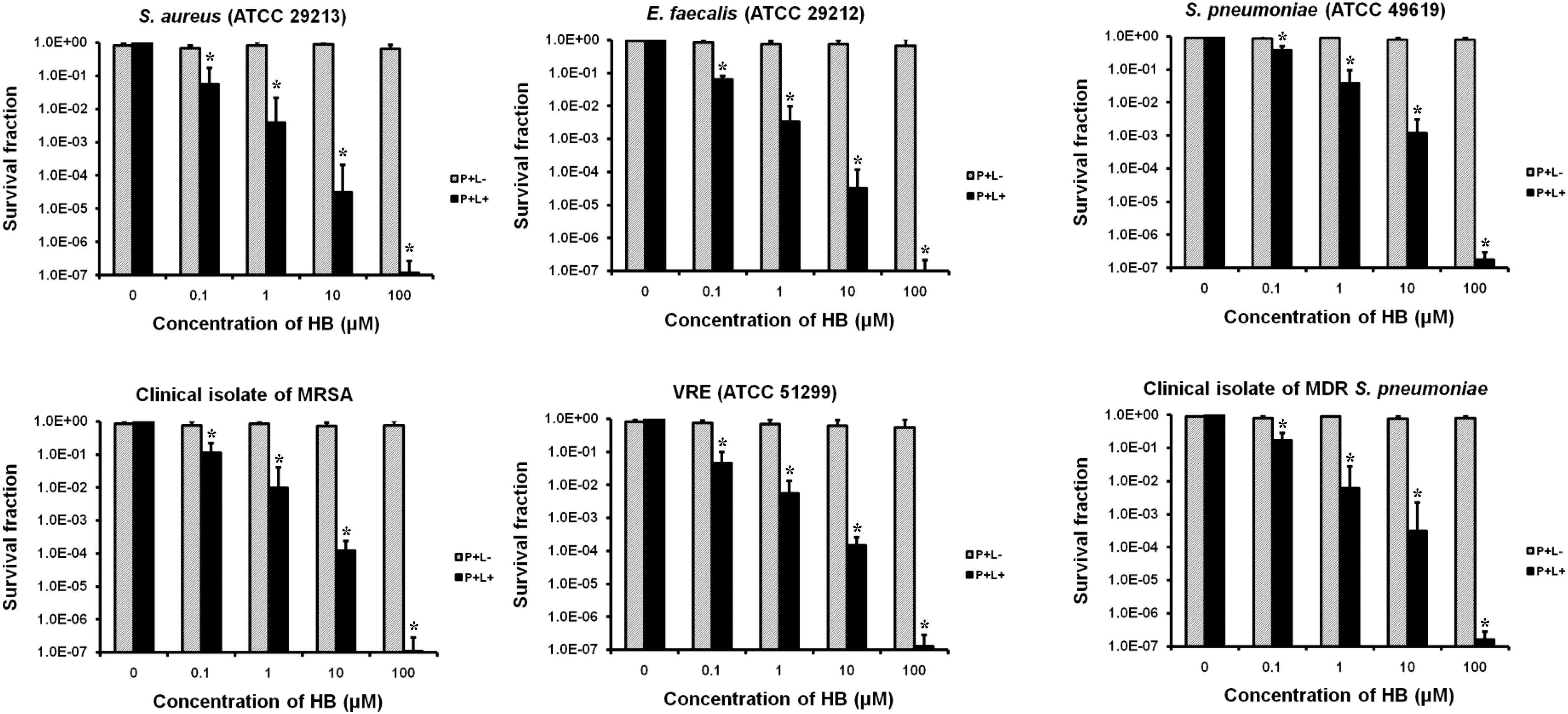
Bacterial survival fraction of Gram-positive antibiotic-resistant bacteria treated with different concentrations of HB. (P+L−): represent the survival fraction after incubation without illumination (dark toxicity); (P+L+): represent the survival fraction after 900 sec irradiation (72 J/cm2).
We also used two standard strains, E. coli (ATCC 25922) and K. pneumoniae (ATCC 700603), to assess the PDI efficacy of HB on Gram-negative bacteria. As shown in Fig. 3a and b, after 72 J/cm2 light irradiation, the PDI efficacy of HB against the two strains was much lower compared to that of Gram-positive strains. When HB reached to 100 μM, only around 2 log10 reductions were observed for E. coli and K. pneumoniae.

In tissue culture plate analysis, A492nm values for S. aureus (ATCC 29213), MRSA, E. faecalis (ATCC 29212), VRE (ATCC 51299), S. pneumoniae (ATCC 49619), and MDR S. pneumoniae were 2.372, 2.594, 0.544, 0.532, 2.674, and 2.834, respectively (Fig. 3c), which exceeded the A492nm limit of 0.240 that was indicative of biofilm producing strains. Biofilms formed by these strains were also treated with 100 μM of HB and irradiated with 72 J/cm2 light. As shown in Fig. 3d, 100 μM HB could yield 3.56, 3.71, 3.29, 3.35, 3.15, and 3.36 log10 reductions in survival fraction for biofilms of S. aureus (ATCC 29213), MRSA, E. faecalis (ATCC 29212), VRE (ATCC 51299), S. pneumoniae (ATCC 49619), and MDR S. pneumoniae, respectively.
Colorectal carcinoma cell line CT26 was used to test the cytotoxicity of HB on mammalian cells. As shown in Fig. 3e, HB exhibited no significant dark cytotoxicity for CT26 cells at the concentrations tested. After irradiation with 72 J/cm2 light, cell survival decreased with the increasing concentration of HB.
Discussion
Using traditional Chinese herbs as folk medicine for treating infectious diseases has a long history. Many active compounds isolated from traditional Chinese herbs have shown anti-infection and anti-inflammation effects. 17,18 HB has been previously used to treat rheumatoid arthritis, gastric diseases, and skin diseases related to fungal infections for several years. 19 Ma et al. evaluated the intrinsic antimicrobial activities of HB against MRSA, Pseudomonas aeruginosa, and Mycobacterium intracellulare and found that it showed weak antimicrobial activity. 20 However, upon light irradiation, HB showed effective photodynamic activities, and HB-mediated photodynamic therapy (PDT) was used as a potential clinical therapeutic method for treatment of several diseases. Jiang et al. reported that HB-mediated PDT could kill cancer cells by inducing severe damages on mitochondria. 13,14 Hu et al. found that HB-mediated PDT could induce significant keloid fibroblast apoptosis and decrease cell viability, indicating that HB has potential to be used for keloid treatment. 21
The photodynamic antimicrobial aspects of HB have been less investigated. The present study found that 100 μM of HB in the presence of 72 J/cm2 light irradiation could achieve about 7 log10 reductions in bacterial survival for S. aureus, E. faecalis, and S. pneumoniae suggesting that HB was an effective PS in PDI of Gram-positive bacteria, as well as standard and antibiotic-resistant strains were both sensitive to the HB-mediated PDI. However, at the same conditions, only 2 log10 reductions in bacterial survival were achieved for E. coli and K. pneumoniae, indicating that the HB-mediated PDI efficacy on Gram-negative strains was lower compared with Gram-positive strains. It is believed that neutral or anionic PS molecules are efficiently bound to and inactivate Gram-positive bacteria, whereas they are bound only to the outer membrane of Gram-negative bacteria, but do not effectively inactivate them after illumination. 1
The high susceptibility of Gram-positive bacteria to HB-mediated PDI could be explained by their physiology, as their cytoplasmic membrane is surrounded by a relatively porous layer of peptidoglycan and lipoteichoic acid that allows HB, a neutral PS molecule, to cross. But in Gram-negative bacteria, there is an outer membrane which forms physical and functional barrier to prevent the penetration by HB. 1 We proposed that when HB molecule is bound to and entered into Gram-positive bacteria, it could cause damages to the DNA and cytoplasmic membrane, allowing leakage of cellular contents or inactivation of membrane transport systems and enzymes (Fig. 4). Although detailed evidence is not enough, a previous study report indicated that HB-mediated PDI could remarkably damage the membrane integrity and lead to the loss of cytoplasm leakage. 22

Paragraph describing envelope structure of Gram-positive bacteria and possible mechanisms of HB-mediated photodynamic inactivation on the Gram-positive bacteria.
The PDI efficacy of HB against the Gram-positive bacteria was also investigated. Although these strains were also susceptible to HB-mediated PDI when grown as biofilms, the PDI efficacy was lower compared to planktonic cells. Bacterial biofilms are generally less susceptible than planktonic cells to antimicrobial agents like peptide and antibiotics, 23 as well as less susceptible to PDI. The affinity of PS to the cell envelope is considered as a determinant of the efficacy of photosensitization. 24 When bacterial cells are organized in biofilms, the affinity of PS to the matrix material will affect PS binding and, ultimately, affect the efficiency.
CT26 cell line was used to investigate the cytotoxicity of HB on mammalian cells. At the tested concentrations, HB exhibited no significant dark cytotoxicity against CT26 cells. However, after irradiation, cell survival decreased with the increasing concentration of HB. Although HB could not selectively eliminate bacteria over mammalian cells, a viable strategy is preparation of the drug-delivery systems to allow transportation of HB to the targeted bacteria, which could improve the selectivity of HB-mediated PACT. For instance, liposomal HB improved the aqueous solubility, tissue selectivity, as well as photodynamic efficacy.
A previous study in rat model of choroidal neovascularization showed that liposomal HB could result in sustained neovessel closure with minor damage under laser irradiation. 25 Besides, nanoparticles have also been fabricated by encapsulation of HB with targeting moiety for combating drug-resistant cancer cells. Chang et al. reported hyaluronic acid-ceramide nanoparticles encapsulated with HB and paclitaxel for targeted PDT through the binding of hyaluronic acid to CD44, an overexpressed protein in drug-resistance cancer cells. 26 HB encapsulated into nanovesicles conjugated with iRGD peptide as targeting moiety exhibited an improved accumulation in tumor and increased photodynamic antitumor efficacy. 27 Therefore, incorporation of HB into liposomes and nanoparticles which use anti-bacteria antibody or peptide as targeting moiety could enhance targeting delivery. These liposomes and nanoparticles might be prepared as injection for treatment of localized infections and as gels or spray for treatment of superficial infections caused by antibiotic-resistant bacteria.
The possible clinical applications of HB-mediated PACT might be probably focused on the treatment of superficial or localized infections, such as MRSA caused skin, wound, and burn infections, VRE caused oral cavity infections, MDR S. pneumoniae caused otitis media with effusion, and so on. Although many localized infections occur deep inside the tissue, it is possible to deliver both PS and light to these local regions through endoscopes and fiber optics. 28
In vivo assessments are also necessary to confirm that HB can meet the requirements for clinical practice. Huang et al. created burn wounds on BALB/c mice by applying preheated brass blocks. They found that polymer conjugated chlorin e6 (Ce 6)-mediated PDI was effective for treatment of superficial infection. 29 Dai et al., reported that polyethylenimine-Ce 6 conjugate-mediated PDI could induce 2.7 log10 inactivation of MRSA on a mice model of skin abrasion wound, which was made using needles by creating crossed scratch lines. 30 Zhou et al. subcutaneously injected VRE into the hind limb of BALB/c mice to create localized infection. They found that PDI could cause obvious infection regression and even complete eradication. 31 These pharmaceutical formulations and infectious models described in previous studies could also be used in our subsequent research.
Conclusions
In summary, this work assessed the PDI efficacy of HB against standard and antibiotic-resistant S. aureus, E. faecalis, and S. pneumoniae. Under light irradiation, HB inactivated these bacterial planktonic cells in a dose-dependent manner. In the presence of 72 J/cm2 light, 100 μM HB achieved about 7 log10 reductions in survival for these Gram-positive bacteria, but only yielded 2 log10 reductions in survival for E. coli and K. pneumoniae. Biofilms formed by these Gram-positive strains could also be effectively inactivated by HB-mediated PDI, but the efficacy was attenuated. The results obtained in this study showed that HB is a promising PS for the PACT treatment of superficial or localized infections caused by Gram-positive antibiotic-resistant bacteria.
Footnotes
Acknowledgments
This work was supported by the National Natural Science Foundation of China (81401710).
Author Disclosure Statement
No competing financial interests exist.
Funding Information
Funding was provided by the National Natural Science Foundation of China (grant no. 81401710).