
Abstract
Background:
Laser irradiation modifies the topography and composition of dentin surface aiming to increase the retention of bonded posts.
Objective:
To assess the effect of dentin irradiation with Er,Cr:YSGG or diode lasers on the bond strength of fiber posts using self-adhesive resin cement.
Materials and methods:
Sixty bovine root canals were root-canal treated, post spaces were prepared, and subsequently fiber posts were cemented. The samples were distributed according to the surface treatment: distilled water (control), Er,Cr:YSGG (1.5 W, 20 sec), or diode (1.5 W, 20 sec) lasers. Bond strengths were assessed by pull-out (n = 10) or push-out testing (n = 10). Pearson's correlation was calculated. Failure mode after testing and the depth of tags in the dentinal tubules were analyzed by confocal laser microscopy. Data were submitted to analysis of variance and Tukey's test. A p < 0.05 was considered significant.
Results:
In the push-out test, Er,Cr:YSGG laser had significantly different higher values (5.43 ± 0.10 MPa) compared to the control (4.79 ± 0.05 MPa). Diode laser values were not significantly different from the other groups (5.12 ± 0.27 MPa). In the pull-out test, there were no significant differences between Er,Cr:YSGG (6.86 ± 2.16 MPa) and diode (8.43 ± 1.77 MPa) lasers, and both had significant differences compared to the control (4.18 ± 1.29 MPa). No correlation was found. Adhesive failures were predominant in all groups, and no significant differences in tag penetration among the groups were found.
Conclusions:
The Er,Cr:YSGG laser increases the bond strength of resin cement and fiber post to dentin in both tests, compared to control group without laser treatment. Diode laser only enhanced bonding for pull-out test.
Introduction
Commonly, root-canal treated teeth have lost a large part of their coronal hard tissue due to decay, previous restorations, and fractures, thus often require specific restorative considerations. Coronal restoration following root canal filling is a crucial factor for treatment success. 1 Considering that teeth are submitted to masticatory shearing forces, previously root treated teeth might require an intracanal post to enhance retention for the extracoronal restoration. 2 An increase in request for metal-free post and cores has stimulated the development of restorative materials and systems, including transparent glass posts. 3,4 These aim to achieve more satisfactory esthetic outcomes, less treatment visits, and easier removal, in comparison to posts made of alternative materials. 5,6
The degradation of the interface has been associated with the failure of metal-free posts due to debonding at the adhesive resin–dentin. 7 –9 During endodontic treatment, irrigation with sodium hypochlorite (NaOCl) promotes deproteinization of dentin 10,11 and reduces the microhardness of intracanal dentin. 11,12 Therefore, NaOCl alters the structure and organic content of dentin and, consequently, the interaction with the adhesives commonly used to bond restorative materials to the natural tooth. 13
The use of high-power lasers for pretreatment of dentin before luting a fiber post to enhance adhesion has been proposed. 13 –18 Among new trends in the study of lasers in endodontology, diode lasers and Er,Cr:YSGG can adapt to the various complexities of root canal morphology. Diode lasers offer thin optical fibers that can fit into root canals, 15,19,20 thus promoting enhanced penetration to the less accessible areas of the tubular network. 20 –24 Diode lasers also can increase the dentin permeability by removing the smear layer and by potentially increasing cement penetration in dentinal tubules. 25 The wavelengths of 970 and 980 nm of diode lasers may increase bond strength in the root canal due to modification in dentin topography and composition. 24,25
Similarly, Er,Cr:YSGG laser has sapphire optical fibers designed for endodontic use, with thicknesses compatible with the limited dimensions of root canals. Er,Cr:YSGG laser treatment with the wavelength of 2.78 μm is capable to reduce hard dental tissues due to the high affinity for molecules of water, 26 is able to open dentinal tubules, 27 increase dentin permeability, 24,28 and can be used to enhance root canal disinfection. 29
Studies testing the bond strength of fiber posts bonded to dentin after pretreatment using diode 30,31 and Er,Cr:YSGG 21 lasers are scarce, and none compared the results of adhesion using pull-out and push-out tests in association. The pull-out test promotes a better stress distribution along the canal wall aiming to measure more accurately the bond strength between root dentin and fiber posts, 32 whereas the push-out test allows a good assessment of shear bond strength, as the load is applied parallel to the adhesion interface and results in shear stress. 32 Therefore, the use of both tests in the same study may allow a more comprehensive understanding of the effect of Er,Cr:YSGG and diode laser conditioning on bonding.
The aim of the present study was to evaluate the effect of dentin surface pretreatments with Er,Cr:YSGG or diode lasers on the bond strength of fiber posts through push-out and pull-out tests, compared to rinsing with water. The null hypothesis tested was that there are no differences in fiber post adhesion, based on push-out and pull-out bond strength testing, with or without dentin surface pretreatment with Er,Cr:YSGG or diode lasers.
Materials and Methods
Sample selection
Sixty bovine incisors were obtained from a slaughterhouse (Mondelli Food Industry S.A., Bauru, São Paulo, Brazil). This way, it was not necessary to obtain the approval of the ethics committee, since the teeth were destined to the discard. Bovine incisors freshly extracted and stored in a 0.1% thymol solution (pH = 7.0) at 4°C were cleaned with a scaler and water/pumice slurry in dental prophylactic cups, then rinsed in running water for 1 day and examined at × 20 magnification using a dental operating microscope, and subsequently radiographed to discard those with calcifications, longitudinal fractures, open foramen, and accentuated root curvature.
Root canal preparation, obturation, and post space preparation
These steps were carried out as previously described by Pelozo et al. 18
Dentin pretreatment
For laser application, the samples were placed in an adjustable acrylic device to maintain them in a standardized vertical position. The roots were randomly divided into three experimental groups as follows (n = 20) according to the pretreatment of root canal walls (Fig. 1).

Flowchart representation of the laboratory stages through.
In the control group without laser treatment, the root canals received a final rinse with 5 mL of distilled water using an Endo-Eze needle in syringe, until extravasation to entrance for 60 sec, followed by aspiration. The excess moisture was removed by sterile absorbent paper points before the bonding procedures.
In the Er,Cr:YSGG group, the 2.78 μm laser (Waterlase Millennium System; Biolase Technologies, San Clemente) was irradiated in the root canal with the following parameters: 1.5 W output power, 20 Hz frequency, 93.25 J/cm2, light in continuous mode, with 50/50% water/air flow rate. The focal area of the tip was 320 μm (MZ3, 19 mm, Biolase). The specimens were irradiated for 20 sec beginning with the tip at 10 mm depth and performing a helical motion along the same to the cervical third, ultimately reaching the apical part of the post space (Table 1).
Parameters Established for Laser Use
In diode group, a 970 nm laser (SiroLaser, Sirona, Bensheim, Germany) was irradiated in the root canal with the following parameters: 1.5 W output power, 20 Hz frequency, 238.85 J/cm2, and light in continuous mode. A 200 μm fiber optic tip was introduced up to the apical region. The laser was activated for 20 sec performing a helical motion, as previously described (Table 1).
Post cementation
The root canals were dried as above and filled with RelyX U200 self-adhesive resin cement (3M ESPE, St Paul), using a Centrix syringe (Kit aplicador precision; Maquira, Maringá, Paraná, Brazil). The posts (Exacto #3 Angelus, Londrina, Brazil) were marked at 10 mm of length, cleaned with 70% ethanol, and dried with compressed air, then inserted slowly into the previously prepared post space. Afterward, the excess of cement was removed, and a light-polymerizing unit (600 mW/cm2; Dabi Atlante, Ribeirão Preto, Brazil) was activated for 40 sec according to the manufacturer's instructions. The roots were stored in relative humidity of 37°C for 48 h. After this period, each group was randomly divided into two subgroups (n = 10), according to the test to be performed: pull-out or push-out (Fig. 1).
Push-out test
Each specimen was sectioned perpendicularly to its axis into 1 mm thick serial slices using a water-cooled low-speed saw (IsoMet 1000; Buehler, Lake Forest). Three slices were obtained for each post third. The first two slices from each post third, in the coronal-apex direction, were submitted to shear bond strength tests (push-out), and the third slice of each third was reserved for analysis by confocal laser microscopy.
The samples were subjected to the pull-out or push-out in the universal testing machine (Instron 2519-106; Instron Corporation, Norwood) a constant crosshead speed of 0.5 mm/min with 200 kgf load cell (Force transducer 2519-106; Instron Corporation), as previously studied. 28,30
A stainless-steel support was used to hold the specimens with the apical surface facing the punch tip and directly aligned toward the shaft. This aimed to ensure that loading forces were applied from the smaller to the larger part of the root slice (apical to coronal direction), avoiding any incident of interference caused by the motion, or contact of the shaft with the dentin that could affect the dislodging of the material and to align specimens in a precise and reproducible manner.
A stereomicroscope (with objective 1 × with final magnification of 10 × ) was used to select a compatible tip (Leica M165C; Leica Microsystems GmbH., Wetzlar, Germany) to determine the larger and smaller diameters of the intracanal material (apical and coronal side, respectively), together with a digital caliper of 0.001 mm accuracy (Mitutoyo Messgerate GmbH, Neuss, Germany), used to measure the slice height for each slice. A 6 mm long shaft with a tip diameter of 0.6 mm for the apical-third, 0.8 mm for the middle-third, and 1 mm for the coronal-third was used.
To calculate the push-out bond strength in megapascals the maximum failure load was recorded in Newtons or KiloNewtons based on the formula proposed by Nagas et al. 14 : Push-out bond strength (MPa) = N/A, where N, maximum load (N); A, area of post+cement (mm2).
The adhesion surface area was calculated as the lateral surface area of a cone using =
Confocal laser scanning microscopy
After the push-out test, the type of failure pattern that occurred in the slices was assessed using three-dimensional (3D) confocal laser scanning microscope (LEXT; Olympus Corporation, Tokyo, Japan) connected to a computer with specific software (LEXT, 3D Measuring Laser Microscope, software OLS 4000; Olympus Corporation). The 3D measuring laser microscope information was in high resolution and high accuracy. The images were in 3D with an objective of 5 × that was increased to 107 × . The observed failures were determined in percentages and classified as follows: (1) adhesive failure between post and cement; (2) adhesive failure between cement and dentin; (3) mixed failure between post/cement and cement/dentin, and (4) cohesive failure of post system. 34
The qualitative–quantitative analysis was performed on the third slice for each third by confocal laser microscopy. Each slice had the surface polished with sandpaper #600 and #1200 and finished with felt discs with alumina solutions at 0.3 and 0.05 μm (AROTEC Sj685/A Ind. And Trade, are Paulo, SP, Brazil). After polishing, the flat surfaces of the samples were etched with phosphoric acid for 10 sec, rinsed with water, and dried with absorbent papers before the microscopy analysis. Images were performed by means of 3D confocal laser microscope (OLS4000; Olympus LEXT) with a laser beam of 0.4 mm in resolution with 20 × magnification. The percentage of tag formation and linear cement penetration into dentinal tubules was measured in μm using the OLS4000 software, obtaining an average by section per sample. In addition, the present study assessed the uniformity of cement penetration in the dentinal tubules, aiming to assess flaws on the adhesive interface.
Pull-out test
The specimens had their external surfaces prepared with a diamond cylinder bur, embedded into the acyclic, and stored for 7 days at 37°C in 100% humidity. Pull-out tests were performed on the specimens using a universal test machine (Instron Model 4444, Canton, MA) with a speed of 2 mm per minute.
The maximum load at failure was recorded in Newtons and converted to megapascals by dividing the load by A, the bonded area. The bonded area was based on the formula: A = π (R + r)
Apart from the investigator carrying out the dentin pretreatments, all other investigators and assessors were blind to the groups being evaluated.
Data analysis
The data of the push-out and pull-out were normal and homogeneously distributed and were submitted to parametric analysis two-way analysis of variance (ANOVA) and one-way ANOVA, respectively. The correlation between the pull-out test and push-out test was analyzed through Pearson's correlation. Data of the depth of tags were analyzed by the Kruskal–Wallis test. For all the analyses, multiple comparisons were performed using the Tukey test. The probability level was 95%, and all analyses were performed in SPSS software version 19.
Results
Push-out bond strength test
Er,Cr:YSGG laser irradiation (5.43 ± 0.10 MPa) provided significantly different higher bond strength compared to control group without laser irradiation (4.79 ± 0.05 MPa) (p < 0.05). No significant differences were found when comparing bond strength values for the diode laser (5.12 ± 0.27 MPa) to the control group or Er,Cr:YSGG laser groups. Bond strength was not significantly different for the cervical, middle, and apical thirds when comparing the different treatments separately (p > 0.05) (Table 2).
Mean and Standard Deviation (in MPa) of Bond Strength After Surface Treatment in Root Canal Thirds (Cervical, Middle, and Apical) in Push-Out
Different letters indicate the significant statistic difference. Uppercase letter comparison of third in the same treatments—in row; lowercase letter comparison of treatments in the same third—in the column. Tukey test, p < 0.05.
Considering the interaction of treatments and thirds, it was observed that, in the cervical third, the Er,Cr:YSGG laser had significantly different higher bond strength (p < 0.05) compared to the control group and diode laser groups. No other bond strength values presented with significant differences (p > 0.05) (Table 2).
When assessing failure mode, there was a predominance of adhesive failures in Er,Cr:YSGG laser (60%), diode laser (50%), and control group (60%) (Figs. 2 and 3).
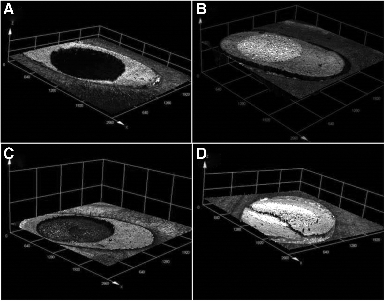
Confocal laser microscopy of the dentin surface illustrating the interface dentin/cement material to evaluate the type of failure pattern:
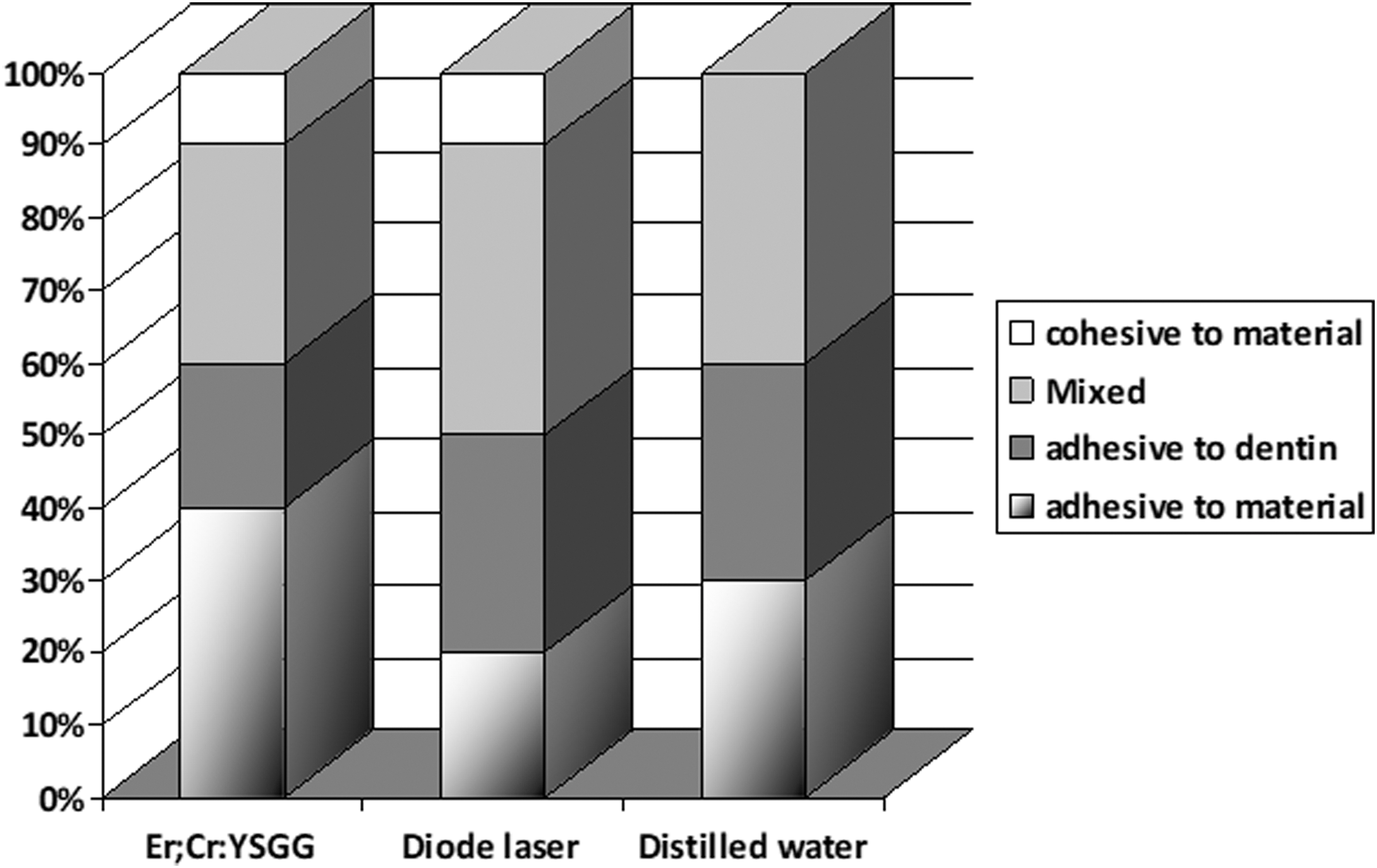
Percentage distribution (%) of the failure modes (adhesive, cohesive, and mixed) observed in fractographic analysis of cervical, middle, and apical slices, submitted to the push-out test.
In relation to depth of tags observed in cervical and middle thirds, Er,Cr:YSGG laser (1.81 ± 0.12) was significantly different lower than the control group (1.97 ± 0.03). Laser treatments presented with no significant differences among them (p > 0.05) for the apical third. No other bond strength values presented with significant differences f (p > 0.05) (Fig. 4 and Table 3).
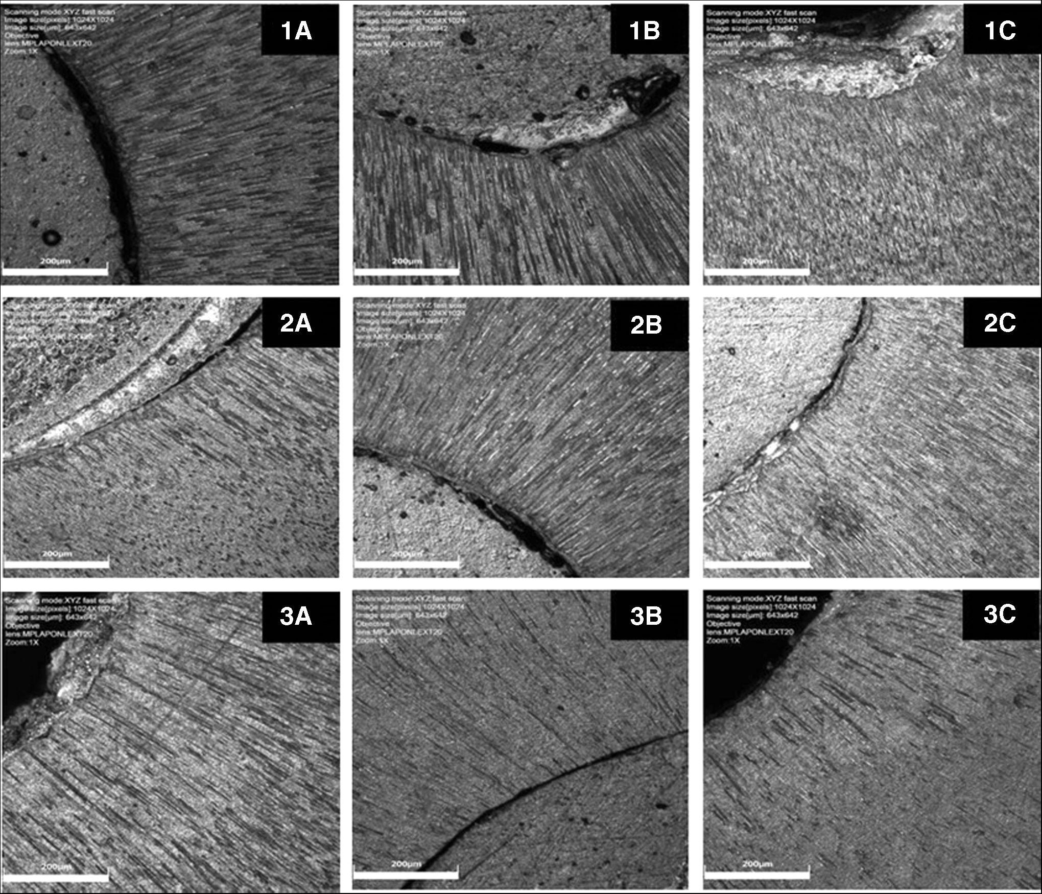
Images of confocal laser microscopy showing the depth of tags:
Penetration Depth of Tags (μm) Data of Cement in Dentin Irradiated in the Different Thirds
Different letters indicate a significant difference. Uppercase letter (column) comparison of treatments in the same third; lowercase letter (row) comparison of third in the same treatments. Tukey test, p < 0.05
Pull-out bond strength test
Pull-out strengths (mean and standard deviation) along the different post thirds are reported in Table 3. The diode (8.43 ± 1.77 MPa) and Er,Cr:YSGG laser (6.86 ± 2.16 MPa) values were not statistically different and presented with significantly different higher bond strength values compared to the control group (4.18 ± 1.29 MPa) (Table 4).
Mean and Standard Deviation (in MPa) of Bond Strength After Surface Treatment in Pull-Out
Different letters indicate significant difference among treatments. Tukey test, p < 0.05.
Pearson's correlation
There was a low degree of correlation between the pull-out test and push-out test through Pearson's correlation coefficient (r = 0.16).
Discussion
This study established that pretreatment with Er,Cr:YSGG improved bond strength for both tests, compared with the water rinse control without laser treatment. Pretreatment with diode laser exclusively enhanced bond strength values for pull-out testing. In accordance with the result of the study, the null hypothesis was rejected.
The increased bond strength after treatment with Er,Cr:YSGG laser was probably caused by the chemical changes of the dentin caused by irradiation. 28,35 This “thermomechanical process” provokes ablation and energizes water molecules to be propelled by the laser light and crashing with the tissue molecules. 27 The tissue is modified by the ablation and the surface temperature increases, inducing structural and chemical changes. 36 The adaptation of restorative materials in the dentin walls can be improved by the changes, since the surface becomes rougher and exposes the collagen matrix of the intertubular dentin. 28,35,37
Others studies confirmed the increase in bond strength associated with the effect of Er,Cr:YSGG laser on the dentin wall; 21,25,28,31 however, a different study concluded that Er,Cr:YSGG laser irradiation did not increase the bond strength of fiber posts when different intensities were used. 21 Disagreement in study results is likely related to methodological differences as the study in disagreement with the present results applied pulsed laser-powered irradiation, whereas our study used light in continuous mode.
Considering the diode laser, the mechanism of action is related to the high power penetration and high absorption picks in hemoglobin and melanin plus low interaction with water and hydroxyapatite. 22 The low absorption coefficient lets the energy cross into the intercellular water space, thus causing a profound hemostatic effect in the darkly pigmented tissues. 23 Diode laser (980 nm) modifies the smear layer without opening dentinal tubules of the canal walls. 21,24 Adhesive resin penetration may be obstructed due to the presence of a smear layer and closed dentin tubules, which can lead to decreased bond strength values. 21 Limited tag penetration following the use of both lasers was also noted in our assays, in particular for Er,Cr:YSGG, however their use was associated with enhanced bond strengths compared with the water rinse control.
In the push test it was possible to analyze the post thirds, and bond strengths were similar for the cervical, middle, and apical thirds overall. These results are in agreement with previous studies, which observed the same performance of high-powered lasers regardless of the root canal level. 28,38 However, when analyzing combined thirds and groups, bond strength at the cervical part was enhanced the most by the action of the Er,Cr:YSGG. This may be associated to the heterogeneity of the root wall dentin, which can influence the adhesive procedure. According to Camargo et al. 37 the more coronal dentin presents with a higher number and diameter of dentin tubules for bovine and human teeth, compared with more apical sections. It is proposed that dentin morphology can influence post adhesion because of greater diameter and density of tubules in the more cervical area, allowing for greater cement penetration and thus mechanical retention between dentin and cement, following smear layer and dentin modification due to laser action. 38 However, in the present study this association was not present.
The failure mode and its location give evidence regarding the quality of the bond between the dentin and the adhesive interface. In the present study there was a predominance of adhesive failures for all groups. This is consistent with previous studies. 14,30 Bonding to root canal surface is a recognized issue due to the endodontic morphology, handling characteristics of the adhesive systems, and overall adhesive procedure complexity. 39 In the root canal, the cavity configuration factor is critical, by increasing the stress polymerization of the resin cement. 2 The polymerization shrinkage forces inside a root canal might be greater than the adhesion of the cement to dentin, favoring the development gaps affecting the adhesive interface that may compromise the longevity of the restoration, even after the treatment of intraradicular dentin with high-powered lasers.
Confocal laser scanning microscopy demonstrated the differences between surface treatments. In the middle and cervical third, Er,Cr:YSGG laser treatment provided a lower tag penetration, significantly different from control group but not to diode laser. The diode laser was similar to the control group and Er,Cr:YSGG laser. Penetration of tags for the apical third was higher than the more coronal post space levels, which may be associated with fluid dynamics of the nonpolymerized resin cement following post insertion.
Although both high-powered lasers influenced the adhesion of the cement through the pull-out and push-out tests, there was no correlation between these tests, as seen in the obtained coefficient in Pearson's correlation test analysis. This could be justified by the fact that they measure adhesion strength differently. The pull-out test promotes a better stress distribution along the canal wall and is able to accurately measure the bond strength between fiber posts and root dentin, whereas the push-out test provides a good estimate of shear bond strength because the load is applied parallel to the adhesion interface and results in shear stress. 32
One possible limitation was the use of bovine teeth. Bovine specimens differ from human because of their higher and harder enamel prisms, 40 the greater dentinal tubule diameter average, 37 and the greater thickness of the peritubular dentin. 41 However, bovine teeth have been increasingly used in in vitro studies 18,42,43 due to legal issues, difficulty in accessing samples of the precise and consistent size, and difficulty in standardizing human teeth. Bovine teeth allow the use of an intratooth model with the advantage of obtaining samples from animals raised under the same husbandry conditions and of a specific age range, which is more likely to result in dentin of comparable characteristics. 44 On these concerns, a previous study assessed if the bond strength of root canal sealers using an intratooth bovine model produces comparable results to those in human dentin and confirmed this hypothesis. 45
Further in vitro studies will be required to assess the long-term bond strength values on adhesive interface of fiber post/laser-treated dentin, using thermal cycles and/or water storage.
Conclusions
The Er,Cr: YSGG laser increased the bond strength of resin cement and fiber post to dentin for both pull-out and push-out tests. Diode laser only enhanced bonding for pull-out bond testing.
Footnotes
Acknowledgments
The authors thank CAPES Foundation (Ministry of Education of Brazil, Brasılia/DF, Brazil) for granting a scholarship to Caroline Cristina Borges.
Author Disclosure Statement
The authors deny any conflict of interest. We affirm that we have no financial affiliation (e.g., employment, direct payment, stock holdings, retainers, consultantships, patent licensing arrangements, or honoraria) or involvement with any commercial organization with direct financial interest in the subject or materials discussed in this article, nor have any such arrangements existing in the past 3 years.
Authors' Contributions
C.C.B.: Execution of the study, confocal laser microscopy, and push-out and pull-out test writing of the article. R.G.P.D.: Responsible for the design and statistical analysis and supervised the confocal laser microscopy. F.C.C.R.: Help in the laboratorial phase and execution of the test. F.P.: Help in the laboratorial phase and execution of the test. G.R.-F.: Responsible for analysis, assistance in article writing, and corrections. M.D.S.-N.: Responsible for the design of the study and interpretation of the results. A.E.S.-G.: Responsible for the design, assistance in article writing, and corrections.
Funding Information
Coordenação de Aperfeiçoamento de Pessoal de Nivel Superior (CAPES) 88881.190153/2018-01.