
Abstract
Objective:
This research is conducted to clarify whether the action of low-power diode pumping solid state (DPSS) laser doses modify proteins of normal human blood serum in vitro.
Background data:
Low-power laser light is considered to act through biostimulation rather than through thermal effects. It was found that low-power laser light biostimulates various biological processes, such as increasing the blood flow within the microcirculation.
Methods:
Human blood serum samples were carefully collected and divided into five equal aliquots. One of them served as a control (nonirradiated serum) and the other four aliquots were irradiated by DPSS laser at a wavelength of 589 nm with different doses (50, 70, 90, and 110 J/cm2). The electrophoretic migration speeds of each specific protein were measured immediately after irradiation using protein electrophoresis. A paired Student's t-test was used between variables.
Results:
The protein concentrations were not significantly (p > 0.05) changed by the various doses of DPSS laser comparing with the nonirradiated counterpart. The electrophoretic migration speed of serum proteins was significantly decreased in almost all tested doses relative to the nonirradiated counterpart. Moreover, the irradiation of serum proteins (albumin, alpha1, alpha 2, beta, and globulin) with a laser dose of 70 J/cm2 was associated with a significant decrease (p < 0.003, 0.02, 0.002, 0.02 and 0.001, respectively) in protein migration speed compared with the protein migration speed of the control nonirradiated counterpart.
Conclusions:
Laser light at a wavelength of 589 nm induces processes that lead to decreases in serum protein migration speeds. Globulin protein was found to have the lowest migration speed among the other plasma proteins.
Introduction
Specific characteristics of low-power laser radiation have led to its use in many medical and research applications, such as monochromaticity, pulsed-mode emission or continuous-wave intensity, and collimation or directionality. Photobiomodulation therapy is used to attenuate body cellular activity through light irradiation.
1
Diode pumping solid state
Materials and Design
Blood samples and preparation
The research protocol and blood sample collections were approved by the local ethical committee at the Physiology Department, College of Medicine, Al-Mustansiryah University. All members were affirmed of the secrecy of all data acquired in the study. A total of six fresh human blood samples were used in this research. Fasting blood samples (9 mL) were collected by direct venepuncture and placed in a tube without anticoagulant. The human blood samples were centrifuged directly after collection at 1000 g for 10 min. After centrifugation, the supernatant fluid (serum) was carefully collected and divided into five equal aliquots. One aliquot served as a control (nonirradiated serum), and the other four aliquots were irradiated with different DPSS laser doses.
DPSS laser irradiation
Low-level DPSS laser was used under the following specifications: power output of 20 mW, wavelength of 589 nm, power density fixed at 60 mW/cm2, after warm-up (mrad), the pointing stability was <0.05, and the power stability was 0.322% for 4 h. These specifications are according to the manufacturer of this product (purchased from Changchun Dragon Lasers, model F Series, China). The peak power fluctuation of the DPSS laser was calibrated to be <0.3% for 1 h, and the output power stability of the laser (without any thermal isolation) was monitored using an optical power meter (Model 2936-C, Gentec-E, Mestro, Canada). The DPSS laser radiation with the beam spot area of 0.33 cm2 was provided to the serum sample tubes. The supernatant fluid (serum) samples, contained in 1 mL tubes, were irradiated with a DPSS laser light for 21, 29, 37.5, and 46 min. The delivered flounce was 50, 70, 90, and 110 J/cm2 for each irradiated group, respectively. The DPSS laser beam was focused at a single point in the center of the sample-containing test tube (usually centered from top to bottom). Irradiations were achieved in a dim room, and specified correct protective eyeglass was worn. Blood samples were given radiation at room temperature.
Protein electrophoresis protocol
The clinical use of electrophoresis in protein analysis is normally based on the simple electrophoretic separation of proteins according to their molecular weight and relative mobility. The electrophoresis system used consisted of an electrophoresis tank to hold the buffer for ready-made use of gels in the horizontal system (Mupid-2plus, Optima, Japan). The setup was provided with electrodes, support medium, and a power source supply of electricity at constant current and voltage. 13 Serum electrophoresis was performed using a commercially available kit (manufactured by Hellabio-Greece). The Hellabio Agarose Gels for protein electrophoresis were used to recognize five distinct bands (albumin, alpha1, alpha 2, beta, and globulin), and their concentrations are given as percentages of the total. The electrophoretic process for plasma proteins separation was run for 20 min. The resulted electrophoresis gel is shown in Fig. 1. The protein concentration of each fraction (band) was calculated in gm/dl by the computer program that fed into the Hellabio gel scanner, and then, the speed of migration was measured. The migration distance or speed (i.e., migration distance per 20 min) was calculated as follows: the scanned gel was fed to a “Paint” program that runs under Microsoft “Windows” operating system version 8.1. This program was able to measure the migration distance of each plasma protein band with high accuracy up to a pixel level. This measurement was achieved after careful calibration of the scanned gel image before measurement.
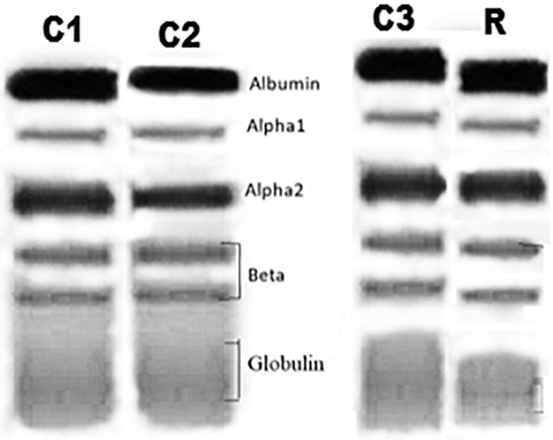
Photographs of the gel electrophoresis of serum proteins of control nonirradiated duplicate samples (C1 and C2). Gel electrophoresis of serum proteins of the nonirradiated (C3) and irradiated sample with 70 J/cm2 (R) is shown.
Statistical analysis
The Statistical Package for Social Sciences (SPSS) version 25.0 software, IBM Corp, Armonk, NY), was utilized to examine the data. All data are stated as the mean ± standard division. Data analysis was carried out using paired Student's t-test between the control and irradiated samples. The level of statistical significance was ≤5% (p ≤ 0.05).
Results
Protein content concentration in serum samples after exposure to low-power DPSS laser radiation at various doses
Plasma protein concentrations and migration speeds were assessed to check the influence of low-power DPSS laser irradiation on serum proteins. Table 1 indicates the plasma protein concentrations in serum blood samples exposed to the 589 nm DPSS laser for each dose (50, 70, 90, and 110 J/cm2), indicating that laser irradiation exposure did not significantly (p > 0.05) change the protein concentration in plasma when compared with the nonirradiated counterpart.
Protein Concentrations of Blood Serum Samples After Exposure to Low-Power Diode Pumping Solid State Laser at Different Doses
Values are expressed in g/L and are compared with the control (nonirradiated) plasma sample.
SD, standard division.
Variation of the migration speed of proteins at different laser doses in irradiated and nonirradiated serum blood samples
In vitro irradiation of human serum by a 589 nm DPSS laser significantly decreases the electrophoretic migration speed of albumin serum protein at all doses of 50, 70, 90, and 110 J/cm2. The albumin protein changes detected as a function of the irradiation dose are shown in Fig. 2A. The dose of 70 J/cm2 was observed to induce maximum change in albumin protein migration speed, and this dose was considered to be optimal relative to its nonirradiated counterpart.

Regarding alpha 1 serum protein, no significant changes in migration speed were found after laser irradiation contrasted with control (nonirradiated serum) samples at a laser radiation dose of 50 J/cm2, as shown in Fig. 2B. In contrast, irradiation with laser beam doses of 70, 90, and 110 J/cm2 was associated with a significant decrease in the alpha 1 migration speed (p < 0.05, 0.002, and 0.001, respectively) relative to the nonirradiated counterpart. The dose of 90 J/cm2 was found to induce maximum change in alpha 1 protein migration speed, and this dose was considered to be optimal relative to the nonirradiated counterpart. Figure 2C shows that laser doses of 50, 70, and 90 J/cm2 of 589 nm DPSS laser were associated with significant decreases in the migration speed of alpha 2 serum protein (p < 0.01, 0.002, and 0.001, respectively) relative to the nonirradiated counterpart. No significant differences in alpha 2 protein migration speed were found after laser irradiation relative to nonirradiated serum samples at a laser irradiation dose of 110 J/cm2. In addition, doses of 50, 70, 90, and 110 nm J/cm2 provided with the 589 nm DPSS laser showed a significant decrease in the beta protein migration speed (p < 0.03, 0.02, 0.01, and 0.01) compared with their control nonirradiated counterparts (Fig. 2D). The dose of 90 J/cm2 induced the maximum change in the beta protein migration speed, and this dose was considered to be optimal effective dose. Further, various radiation doses were found to induce significant changes in the globulin protein migration speed (p < 0.03, 0.01, 0.003, and 0.001, respectively) relative to its nonirradiated counterpart (Fig. 2E). The dose of 70 J/cm2 was observed to induce maximum change in the globulin protein migration speed, and this dose was considered optimal effective dose.
Variation of the protein migration speeds at a laser dose of 70 J/cm2 in irradiated and nonirradiated serum blood samples
The change in protein migration speeds as a function of the irradiation dose at 70 J/cm2 is summarized in Fig. 3. In vitro irradiation of serum human blood proteins (albumin, alpha1, alpha 2, beta, and globulin) with a laser dose of 70 J/cm2 was associated with a significant decrease (p < 0.003, 0.02, 0.002, 0.02, and 0.001, respectively) of protein migration speeds compared with their control nonirradiated counterparts. The globulin protein was found to have the lowest migration speed among the other plasma proteins.
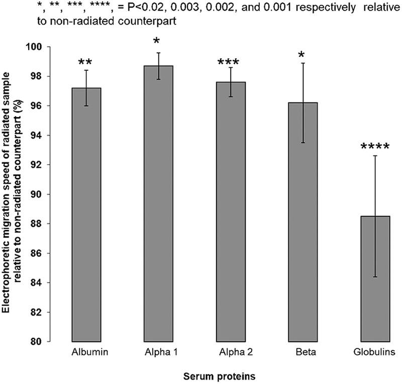
Effect of a radiation dose of 70 J/cm2 on the electrophoresis mobility of the serum proteins. N = 6 per group.
Discussion
In this study, electrophoresis techniques were used to evaluate the impact of low-power DPSS laser irradiation on the serum proteins in normal human blood. Electrophoresis is the migration of charged particles toward the opposite charged electrode in an electrical field. 14 The rate of migration of a given charged particle depends upon its net charge, size, and shape and upon the pH of the medium, the strength of electric field, the electrophoretic temperature, and the physical and chemical properties of the supporting medium. 15,16 Since proteins exist as charged particles, this method is widely used for the separation of proteins in a sample. 17 Four different doses were used to study the dose–response effect. The irradiated samples were compared with nonirradiated control groups. The data collected in this study propose that serum protein exposure to low-power DPSS laser (589 nm) at different doses does not change protein concentrations in serum samples (Table 1). The protein concentrations were not significantly changed by the various laser doses, indicating that the low-power DPSS laser actions at the doses of radiation used in this research did not change the concentration of serum proteins. Previous studies have reported that He–Ne laser exposure of human blood samples enhanced an alteration in plasma protein. 10,18,19 In vitro, influences of low-power laser irradiation on human blood have been described previously. The most significant differences in the cellular aggregation and viscosity of the plasma–cellular complex were described. 10 In another study, exposure to infrared diode laser beam at a wavelength of 810 nm increased plasma protein concentrations, which could depend on the doses of the laser used on blood samples, this indicates that laser light therapy procedures must consider doses, wavelengths, and frequencies of the laser light before starting treatment. 18 In addition, it was suggested that these wavelengths of laser interact differently. Therefore, each particular wavelength has its distinct biological effect on plasma proteins of human blood. In this study, the main reason for using the source radiating in the yellow (589 nm) spectral region is because irradiation with this wavelength of laser light brought about enhanced protein synthesis and accelerated cell division in various microorganisms. 20 In addition, other authors showed that a low-power DPSS laser at a wavelength of 589 nm is a special and distinctive quality of laser that has an impact on biological organisms through non-thermal means. 21,22 This study shows that the electrophoretic migration speed of all serum proteins was significantly decreased at almost all laser doses (50, 70, 90, and 110 J/cm2) relative to the nonirradiated counterparts (Fig. 1). However, the maximum significant decrease in the migration speed of serum proteins was observed mainly at the doses of 70 and 90 J/cm2 and, to lesser extent, at a laser dose of 110 J/cm2. The smallest decreases in the migration speeds of serum proteins were observed at a laser dose of 50 J/cm2. These results indicate that intermediate doses of laser energy produce more significant protein changes than higher or lower doses when the same wavelength is used to deliver low-power DPSS laser light. This finding is in accordance with the fundamental theory of the biphasic dose responses. 23 The biphasic dose curve is essential when finding the dose of the threshold (the energy level sufficient to obtain maximum biostimulation). Biostimulation is changed by bioinhibition when the administered dose is considerably higher than dose of the threshold. 24,25 To find out which serum protein migration speed is more affected than others by the DPSS laser used in this study, the optimum laser doses of 70 or 90 J/cm2 were plotted against the five serum proteins (albumin, alpha1, alpha 2, beta, and globulin). The result of this comparison is shown in Fig. 2. A similar trend to that shown in Fig. 2 was obtained if the laser dose of 90 J/cm2 was plotted against the five serum proteins mentioned before. The most marked change was in the migration speed of serum globulin protein, which was slower than the other serum proteins. This finding suggested that most of the photonic energy carried by laser radiation to serum proteins is absorbed by globulin protein. Thus, fewer photons are available to be absorbed by the other serum proteins.
The outcomes of this study could indicate that exposure to low-power laser induces changes in serum protein migration speeds. This change in the migration speeds of serum proteins is dose dependent. It is possible, therefore, to suggest that the DPSS laser may lead to a change in the electrical charge/or conformation of the serum blood proteins. Kujawa et al. 26 showed that near-infrared laser light radiation (810 nm) was able to induce long-term conformational changes of RBC membrane proteins and lipid structural states. The latter suggestion is supported by Genkin et al., who showed that the change in the electrical charge of serum proteins depends on the laser dose and exposure time. 27 Al Musawi et al. reported that nonirradiated RBCs that were resuspended in radiated serum resulted in a substantial decrease in the ESR. 7,22 The present results can explain the reduction in ESR. The low-power DPSS laser may change the electrical charge of the plasma proteins. This change in protein electrical charges may cause a decrease in the erythrocyte attractive bridging forces required for rouleaux formation, and consequently, it may decrease ESR. The implications of this research are the following: the tissue–laser interaction should be taken into consideration for the use of such laser radiation technique in clinical application, especially those involved in the fundamental functions of plasma proteins.
Conclusions
It was shown that irradiation with laser light at a wavelength of 589 nm induces processes that lead to decreases in serum protein migration speeds but without significantly changing the protein concentration of the samples. Our results clearly demonstrate that the globulin protein is the protein maximally affected by the laser compared with the other serum proteins analyzed.
Footnotes
Author Disclosure Statement
No competing financial interests exist.
Funding Information
No funding was received for this study.