
Abstract
Long-term alcohol consumption has been reported to increase oxidative stress in multiple organs and accelerate the aging process. A previous study in our laboratory has shown that Wu-Zi-Yan-Zong-Wan (WZ), a “Yang-invigorating” Chinese herbal formula, protected against ethanol-induced toxicity in HepG2 cells transfected to express human CYP2E1, presumably by enhancing mitochondrial antioxidant status and functional ability. The present study aims to investigate whether WZ extract treatment can afford protection against chronic ethanol-induced oxidative stress (a major risk factor of aging) and mortality in rats. The effect of the extract (1.8 g, 4.5 g, and 9 g raw materials/kg per day) on chronic ethanol hepatotoxicity was investigated in rats receiving steady intragastric infusion of ethanol-containing liquid diet. The results showed that long-term (42 days) herbal co-treatment protected against chronic ethanol-induced mortality and hepatotoxicity and in rats, as evidenced by decreased plasma transaminases activities. The extract also suppressed the pathological development of fatty liver, as assessed by histopathological examination and the ratio of liver weight to body weight. The hepatoprotection afforded by the extract was associated with decreases in the extents of reactive oxygen species production, lipid peroxidation, and oxidative modification of proteins, as well as the reversal of altered mitochondrial reduced glutathione level. The results suggest that the suppressive effect of WZ on chronic ethanol-induced oxidative stress and mortality may be attributed to the antioxidant action, particularly in mitochondria.
Introduction
Traditional herbal formulae have been clinically used for thousands of years in China. They are prescribed for the treatment of various patterns of body disorders according to the theory of Chinese medicine. The maintenance of a balance of Yin and Yang, a fundamental approach in the practice of Chinese medicine, is essential in achieving a healthy condition. The Yin–Yang relationship can be described in modern medicine term as the balance of antioxidant–oxidant. 20,21 It has been proposed that the pharmacological basis of “Yang-invigoration” in Chinese medicine involves the enhancement of mitochondrial adenosine triphophate (ATP) generation and the associated increase in mitochondrial antioxidant capacity. 22 In this regard, the long-term treatment with a “Yang-invigorating” Chinese formula has been shown to protect against oxidant injury in various tissues of rats. 23 Wu-Zi-Yan-Zong-Wan (WZ) is also a renowned “Yang-invigorating” Chinese herbal formula comprising Fructus Lycii (Lycium barbarum or L. chinense), Semen Cuscutae (Cuscuta chinensis Lam), Fructus Rubi (Rubus chingii), Semen Plantaginis (Plantago asiatica or P. depressa), and Fructus Schisandrae (Schisandra chinensis). In ancient China, the WZ formula was regarded as “the first and fundamental antiaging recipe.” Previous work in our laboratory has shown that an extract of WZ formula can protect against ethanol-induced toxicity in CYP2E1 cDNA-transfected HepG2 (E47) cells, presumably by enhancing the mitochondrial antioxidant status and functional ability. 24
In the present study, we investigated whether WZ extract could afford protection against chronic ethanol-induced hepatic oxidative damage and mortality in rats. The biochemical mechanism involving hepatoprotection against chronic ethanol toxicity was also investigated by examining the oxidative stress-induced biochemical changes in ethanol-intoxicated rat liver tissues.
Materials and Methods
Chemicals and reagents
Dihydroethidium (DHE) molecular probes were purchased from Sigma (St. Louis, MO). The Glutathione Assay Kit was manufactured by Cayman Chemical (Ann Arbor, MI), and the Quick StartTM Bradford protein assay kit was obtained from Bio-Rad Company (Hercules, CA). All other chemicals were of analytical grade. Solvents used for high-performance liquid chromatography (HPLC) were of HPLC grade.
Plant materials and preparation of WZ
Five component herbs were purchased from ZhiXin Chinese Pharmaceutical Co. Ltd., a Good Manufacturing Practice (GMP)-certified supplier of Chinese medicinal herbs: Fructus Lycii, the dried fruits of L. barbarum harvested from Ningxia Province, China; Semen Cuscutae, the seeds of C. chinensis Lam harvested from Hebei Province, China; Fructus Rubi, the dried fruits of R. Chingii Hu harvested from Zhejiang Province, China; Fructus Schisandrae, the dried fruits of S. chinensis (Turcz.) Baill. harvested from Heilongjiang Province, China; and Semen Plantaginis, the seeds of P. asiatica L. harvested from Jiangxi Province, China. Voucher specimens (WZ-01-05) were deposited at the School of Chinese Medicine for reference. The five herbs were ground into a coarse powder, and WZ was formulated by mixing various herbal powders in relative proportions according to the Pharmacopoeia of PRC (2005 edition). The herbal powder mixture was extracted by 50% ethanol (vol/vol, in H2O) in reflux for 60 min. The extraction procedure was repeated twice. The pooled extract was filtered to remove debris, and the ethanol was evaporated by rotary evaporator under reduced pressure. The concentrated extract was then dried by lyophilization to obtain the WZ extract. The extract was stored in the desiccator at 4°C until use.
Animal treatment
Male Sprague–Dawley (SD) rats, weighing 200–220 g, were obtained from the Laboratory Animal Services Centre, the Chinese University of Hong Kong. They were housed in cages under control conditions of temperature (21 ± 2°C) and relative humidity (55 ± 10%), and kept on a 12-h light/dark cycle. The experimental protocols for the present study have been approved by the Animal Experimentation Ethics Committee of the Chinese University of Hong Kong. Rats were fed a liquid diet without or with ethanol at doses gradually increasing from 8 to 16 g/kg per day for 6 weeks, using the enteral feeding protocol, as described previously by Tsukamoto and French. 25,26 The liquid diet was prepared by mixing corn oil (35% of total calories), protein (18% of total calories), carbohydrate (11% of total calories), minerals, and vitamins, plus ethanol (36% of total calories).
An alcohol-free isocaloric diet was prepared by replacing ethanol with maltose dextrin. The caloric content of the liquid diets was 1.0 kcal/mL. Sixty male SD rats were randomly assigned into three groups: Normal control group (n = 10), liquid diet without ethanol as the isocaloric liquid diet control (n = 10), and liquid diet with ethanol (n = 40). Gastric cannulation was conducted in all animals except for the normal control group. After 7 days of recovery, the liquid diet (with or without ethanol) was infused into respective animals. During the enteral feeding period, the rats fed with ethanol diet were divided into four groups: Vehicle (water) and WZ extract-treated groups at 1.8, 4.5, and 9 g raw material/kg per day, respectively, for 6 weeks. Body weight and mortality were recorded for each week. After 6 weeks, the rats were sacrificed and liver weight was recorded. Blood and liver samples were collected for biochemical and histopathological analyses.
Assessment of hepatic damage and serum contents of total cholesterol and triglycerides
Blood (0.5 mL) sample was drawn from the retro-orbital plexus at the beginning and the end of enteral feeding period. The blood samples were left at room temperature for 1 h to allow coagulation. The serum was collected by centrifugation (16,000 × g) at 4°C for 10 min. Serum contents of alanine aminotransferase (ALT), aspartate aminotransferase (AST), total cholesterol, and triglycerides were measured by Auto-Biochemical Analyzer (Roche Diagnostics GmbH, Mannheim, Germany) in The Hospital of People's Liberation Army Hong Kong Garrison.
Histopathological examination
Liver specimen was fixed in 10% neutral formalin. The tissue was embedded in paraffin and cut by a microtome at a thickness of 5 μm. The slice was stained by Hematoxylin & Eosin (H&E) staining and then examined using a light microscope.
Superoxide determination in situ
In situ production of superoxide was determined by the method of Minamiyama and colleagues. 27 Briefly, liver tissues were placed into 50 mM sodium phosphate containing 18% sucrose at 4°C overnight, and samples were frozen and sliced into 5-μm sections. The oxidation-dependent fluorescent dye DHE (40 μM; Molecular Probes, Eugene, OR) and icotinamide adenine dinucleotide phosphate (NADPH) (1 mM) were added to the incubation solution. Tissue sections were then incubated in a light-protected humid chamber at 37°C for 30 min. The sections were rinsed and viewed using a fluorescence microscope. Control tissue sections were incubated with 40 μM DHE without NADPH.
Lipid peroxidation assay
The hepatic content of malondialdehyde (MDA), an indirect index of lipid peroxidation, was measured by a HPLC method, which was modified from our previous publication. 28 Briefly, 250 μL of hepatic homogenate was mixed with 250 μL of phosphoric acid (1.22 M) and 500 μL of 0.5% (wt/vol) thiobarbituric acid. The mixture was heated in a boiling water bath for 30 min. After cooling, 400 μL of reaction mixture was mixed with 720 μL of methanol and 80 μL of 1 M sodium hydroxide, and the mixture was centrifuged at 16,000 × g for 10 min. The supernatant was analyzed by HPLC using an Alltech Alltima C18 column (particle size 5 μm, 4.6 mm × 250 mm). HPLC separation was achieved by an isocratic elution (1 mL/min) with a mobile phase consisting of equal volumes of phosphate buffer (25 mM, pH 6.5) and methanol. The eluate was monitored by a diode-array detector (DAD) detector set at a wavelength of 532 nm. The concentration of MDA was estimated using a calibration curve of standard solution prepared by acid hydrolysis of 1,1,3,3-tetramethoxypropane. The content of MDA was expressed as mg/g protein.
Preparations of mitochondria
Mitochondrial fractions were prepared by differential centrifugation, as previously described. 23 Briefly, hepatic tissue was homogenized in 5 volumes of isotonic buffer (0.25 M sucrose, 0.1 mM ethylene diaminetetraacetic acid [EDTA], 5 mM Tris/HCl, pH 7.4) using a Teflon-glass homogenizer. The homogenate was centrifuged at 600 × g for 10 min, and the pellet containing nuclear and cell debris was discarded. The supernatant was centrifuged at 8000 × g for 30 min to obtain the mitochondrial fraction. The mitochondrial pellet was resuspended in cold 2-(N-morpholino)ethanesulfonic acid (MES) buffer (50 mM MES including 1 mM EDTA (pH 6.0).
Measurement of glutathione in mitochondria and homogenate
The mitochondrial pellet was washed twice with MES buffer and resuspended in the same buffer. Metaphosphoric acid at a final concentration of 5% was used for deproteinization in the homogenate and mitochondrial fractions. After centrifuging at 15,000 × g, the supernatant was immediately measured for glutathione (GSH) content. GSH level was measured using the Glutathione Assay kit, and expressed as nmol/mg protein.
Protein oxidation detection
The content of protein carbonyls, an indicator of oxidative modification of proteins, was measured by the method of Dalle-Donne and colleagues using the 2,4-dinitrophenylhydrazine (DNPH) reaction. 29 Briefly, hepatic tissue was incubated with RIPA lysis buffer, containing 2-mercaptoethanol (final concentration 0.74 M) and proteinase inhibitor mixture, for 20 min at 4°C. The homogenate was centrifuged at 16,000 × g for 20 min at 4°C. An aliquot of the supernatant fraction (5 μL, containing 20 μg of protein) was transferred into Eppendorf tube and mixed with 5 μL of 12% sodium dodecyl sulfate (SDS) for deproteination. Ten microliters of DNPH solution (dissolved in 2 N HCl) was added into the reaction mixture. The mixture without DNPH was used as a blank control. The mixture was incubated for 15 min and then neutralized by 7.5 μL of 2 M Tris in 30% glycerol. The samples were loaded onto a 12% SDS-polyacrylamide gel for electrophoresis. The proteins were separated and transferred to polyvinylidene fluoride (PVDF) membranes. The membrane with transferred proteins was incubated with 1:150 primary antibody (rabbit anti-DNP) for 1 h, followed by incubation with 1:300 secondary antibody (goat anti-rabbit horseradish peroxidease [HRP]-conjugated) for 1 h. Then the DNPH–protein carbonyl-adduct was detected using a HRP-based chemiluminescence kit (ECL Plus™, Amersham Biosciences).
Protein assay
Protein concentration was determined using the Bio-Rad Quick StartTM Bradford protein assay kit. Diluted sample (5 μL) was mixed with 200 μL of assay reagent, and the mixture was left at room temperature for 10 min. The absorbance of the mixture was measured spectrophotometrically at 595 nm. Protein concentration was determined from a calibration curve using bovine serum albumin as standard.
Statistical analysis
Results were expressed as the mean ± standard deviation (SD). Multiple group comparisons were performed using one-way analysis of variance (ANOVA) followed by the Tukey test to detect intergroup differences by using SPSS version 13.0 (SPSS Inc., Chicago, IL). The difference was considered statistically significant when p < 0.05.
Results
Mortality and physical status in chronic ethanol-intoxicated animals
Ethanol intoxication for 6 weeks was found to be lethal in rats, as indicated by the 50% mortality rate observed at the end of treatment (Fig. 1). Co-treatment with WZ extract at doses of 4.5 and 9 g/kg ameliorated the ethanol-induced mortality from 5 rats to 4 and 2 rats, respectively. The growth was significantly retarded by chronic ethanol intoxication in rats, as indicated by the decrease in body weight (Fig. 2A). WZ extract co-treatment partially reversed the growth inhibitory effect of ethanol in a dependent manner. Figure 2B shows the ratio of liver weight to body weight (i.e., hepatic index), when compared with the nonethanol liquid diet group. Co-treatment with WZ extract at three doses (1.8, 4.5, and 9 g/kg) significantly (p < 0.01) decreased the hepatic index, when compared with the untreated ethanol group.

Effect of Wu-Zi-Yan-Zong-Wan (WZ) extract on survival rate of rats treated chronically with ethanol (EtOH).

Effect of Wu-Zi-Yan-Zong-Wan (WZ) extract co-treatment on the change of body weight (
Effect of WZ extract on chronic ethanol-induced hepatic damage
The extent of hepatic damage was assessed by measurements of serum ALT and AST activities. After 6-weeks of chronic ethanol intoxication, serum ALT and AST were significantly (p < 0.01) increased by 1-fold and 58%, respectively, when compared with the nonethanol group, indicative of hepatic damage. There were statistically significant changes in serum ALT and AST activities by treating the animals with WZ extract at 9 g/kg (Fig. 3A,B). However, there was no substantial difference among the treatments from a clinical point of view. Chronic ethanol treatment increased the contents of total cholesterol and triglycerides in serum (Fig. 3C,D). WZ extract at three doses (1.8, 4.5, and 9 g/kg) reduced the serum content of total cholesterol in ethanol-treated rats. Histopathological analysis of liver tissues indicated that ethanol intoxication caused macrovesicular and asystematic steatosis in the periportal and midzonal regions of the liver (Fig. 4C). In addition, cell swelling, fat accumulation, and degenerative and necrotic damage were observed in ethanol-intoxicated liver tissues (Fig. 4C). WZ extract co-treatment prevented the ethanol-induced macrovesicular steatosis, vacuole formation, and inflammation (Fig. 4D–F). The accompanying necrotic foci were much reduced when compared with the untreated ethanol-intoxicated group. Histological scores for comparing the pathological of the liver tissues are described in Table 1.

Effect of Wu-Zi-Yan-Zong-Wan (WZ) extract co-treatment on serum contents of alanine aminotransferase (ALT) (

Effect of Wu-Zi-Yan-Zong-Wan (WZ) extract co-treatment on histological changes in liver tissues of chronic ethanol-intoxicated rats. (Magnification, 400 × .) (
Histological examination was conducted by a pathologist who was blinded regarding the treatment groups. The liver pathology score was calculated as described previously. 15 Steatosis (the percentage of liver cells containing fat): <25% of the cells containing fat = 1, with 26–50% of the cells containing fat = 2, with 51–75% of the cells containing fat = 3, with >75% of the cell containing fat = 4. Lobular inflammation or necrosis: 0 foci = 0, <2 foci = 1, 2–4 foci = 2, >4 foci = 3. At least three different sections were examined per liver sample. The Mann–Whitney test was used for comparison of pathological scores.
Compared with nonethanol liquid diet group.
Compared with ethanol liquid diet group.
Effect of WZ extract on ROS production in liver tissue
In situ detection of ROS using the oxidation-dependent fluorescent dye DHE showed that the red fluorescence was the strongest around central veins. The fluorescence was the weakest in liver tissues from animals without ethanol intoxication (Fig. 5A,B). The fluorescence intensity of liver tissues from chronic ethanol-intoxicated animals was markedly increased (Fig. 5C). WZ extract co-treatment at a dose of 9.0 g/kg inhibited the ethanol-induced ROS production, as evidenced by a weaker fluorescence signal in liver tissues (Fig. 5F).
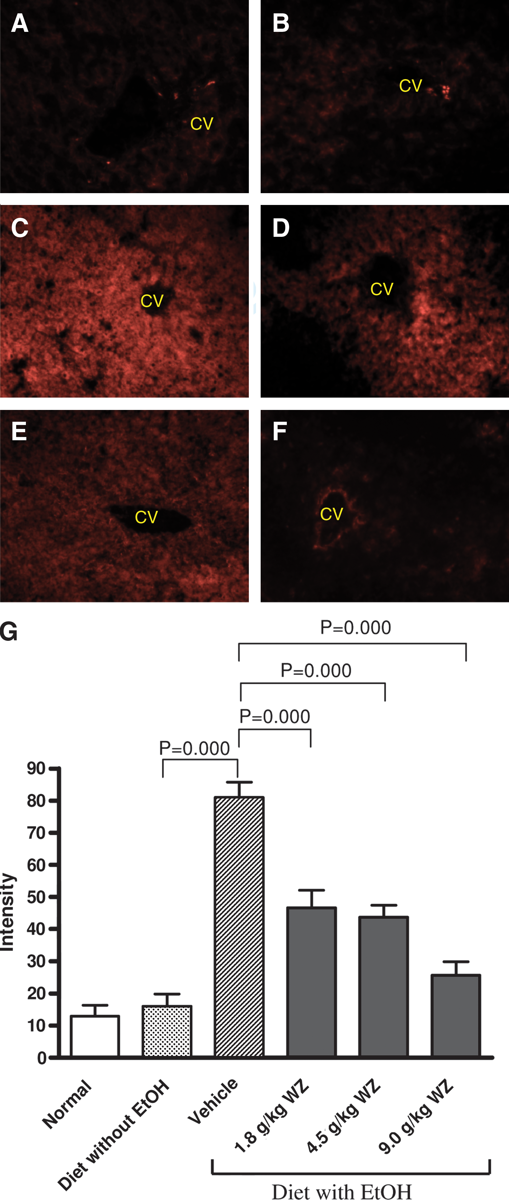
Effect of Wu-Zi-Yan-Zong-Wan (WZ) extract co-treatment on reactive oxygen species (ROS) production in liver tissues of chronic ethanol (EtOH)-intoxicated rats. (Magnification, 200 × .) (
Effect of WZ extract on hepatic MDA and GSH levels
Chronic ethanol intoxication enhanced lipid peroxidation in liver tissues, as evidenced by the increase (180%) in the extent of MDA production when compared with nonethanol-intoxicated group. WZ extract co-treatment caused a dose-dependent inhibition of lipid peroxidation in liver tissues of ethanol-intoxicated animals (Fig. 6A). Although no significant difference in hepatic tissue GSH level was found between the control and ethanol-intoxicated animals (Fig. 6B), the hepatic mitochondrial GSH level was significantly decreased by 49% in ethanol-intoxicated animals, when compared with the nonethanol control group. WZ extract co-treatment at 4.5 and 9.0 g/kg significantly (p < 0.01) reversed the ethanol-induced depletion in mitochondrial GSH, with the degree of protection being 62 and 67%, respectively (Fig. 6C).
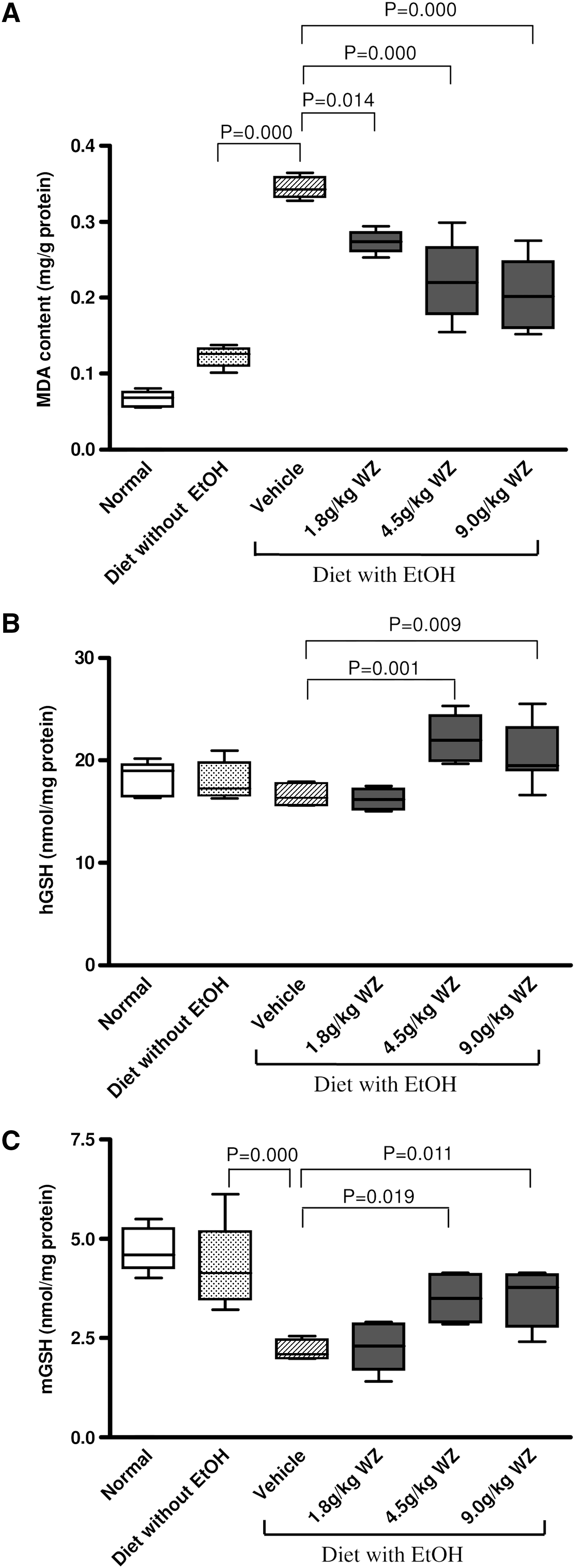
Effect of Wu-Zi-Yan-Zong-Wan (WZ) extract co-treatment on lipid peroxidation (
Effect of WZ extract on oxidative modification of proteins
Chronic ethanol intoxication intensified the expressive signals of DNPH–protein adducts, as an indication of increase in the extent of oxidative modification of proteins in liver tissues. WZ extract co-treatment at 4.5 and 9.0 g/kg markedly reduced the extent of oxidative modification of proteins in liver tissues (Fig. 7).
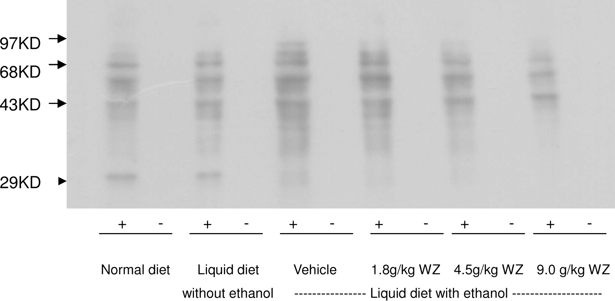
Effect of Wu-Zi-Yan-Zong-Wan (WZ) extract co-treatment on oxidative modification of proteins in liver tissues of chronic ethanol-intoxicated rats. Oxidative modification of proteins was assessed by noting the intensity of 2,4-dinitrophenylhydrazine (DNPH)–protein adducts. (−) Without DNPH; (+) with DNPH.
Discussion
Excessive alcohol consumption is a risk factor for chronic diseases and aging. 30 Prophylactic agents for alcohol toxicity have been searched actively, particularly from Chinese herbal medicine. Using a well-established rat model of chronic ethanol toxicity, 31 –34 we demonstrated the beneficial effect of WZ extract on chronic ethanol toxicity, as evidenced by decreases in mortality rate and the extent of liver oxidative damage. The hepatic damage was assessed by biochemical parameters and serum ALT and AST activities. In addition, the hepatoprotective effect of the WZ extract was also observed by histological examination of the liver and the development of fatty liver, as indicated by the ratio of liver weight to body weight in chronic ethanol-intoxicated rats. WZ extract co-treatment was able to reverse this pathological change, presumably by inhibiting the accumulation of lipids caused by chronic ethanol consumption in liver tissues. 35,36
It is well established that oxidative stress is involved in the pathogenesis of ethanol-induced liver damage. 33,34,37 –40 Consistent with this, increases in MDA and ROS production as well as a depletion of mitochondrial GSH were observed in liver tissues of chronic ethanol-intoxicated rats. Whereas cytochrome P450 2E1 is recognized as a critical contributor to ethanol-induced ROS generation, the mitochondrial respiratory chain and cytosolic enzymes, such as xanthine oxidase and aldehyde oxidases, have been implicated as sources of O2 − and hydrogen peroxide (H2O2) in parenchymal cells in ethanol-intoxicated liver. 41 GSH is critically involved in the defense against ROS, and chronic ethanol consumption has been shown to deplete mitochondrial GSH. 42,43
Our results suggest that mitochondrial GSH plays a critical role in chronic alcohol-induced hepatotoxicity. Chronic alcohol treatment did not produce a significant change of GSH in the hepatic cells, but caused a drastic (49%) decrease of GSH in the mitochondria (Fig. 6), which was probably due to the absence of enzymatic system for the synthesis and regeneration of GSH in the mitochondria and the higher rate of ROS production in this organelle. Ethanol increases the production of mitochondrial ROS, which in turn oxidizes mitochondrial GSH and causes oxidative damage. 44 It has been reported that both acute and chronic ethanol administration caused oxidative modifications of mitochondrial DNA in experimental animals. 43 The ability of WZ extract to protect against chronic ethanol hepatotoxicity may be related to the enhancement of mitochondrial antioxidant status and functional ability, as were the case for “Yang-invigorating” Chinese herbal formula. 22,23 In this connection, ingredient compounds present in the WZ extract, such as schisandrin B, verbascoside, astragalin, kaempferol, and hyperoside, have been shown to possess antioxidant activity. 45 –48 They may work synergistically and contribute to the hepatoprotective effect of the WZ extract
Oxidative modification of proteins by ROS occurs in pathological and aging processes. As a consequence of oxidative modification, carbonyl groups are introduced into the amino acid side chains of protein molecules by site-specific reactions. It has been reported that oxidative stress resulting from chronic ethanol consumption causes an increase in protein carbonyls in rat hepatic tissue. 39 Our findings have consistently shown a significant increase in oxidative protein modification in chronic ethanol-intoxicated rat livers, which was markedly suppressed by WZ extract co-treatment.
In conclusion, long-term WZ extract co-treatment was found to protect against chronic ethanol-induced mortality and hepatotoxicity in rats. The hepatoprotection afforded by WZ extract was associated with decreases in the extents of ROS production, lipid peroxidation, and protein oxidation, as well as a reversal of depleted mitochondrial GSH in chronic ethanol-intoxicated rats. The results suggest that the suppressive effect of WZ extract on chronic ethanol-induced mortality and oxidative stress (a major risk factor of aging) may be attributed to its antioxidant activity.