




Abstract
Background and Objective:
Skin, being a mirror of the body, is a major target for aging research. Aging is a complex process that involves the decline of function or dysfunction of many systems. The corticotropin-releasing hormone (CRH) system is involved in skin inflammation. In addition, CRH has a suggested role in age-associated conditions and in animal aging models. However, a consistent logic interaction between the different CRH system components and the aging process has, to our knowledge, never been examined before.
Methods:
The expression of CRH, CRH-binding protein (CRHBP), CRH receptor 1 (CRHR1), and CRH receptor 2 (CRHR2) in healthy skin samples of 42 patients of different ages (18–92 years) was evaluated by immunohistochemistry, and the age-related changes were assessed.
Results:
Compared with young skin, the aged skin displayed an upregulation of CRH in sebaceous glands and CRHR1 in hair follicles and the epidermis. Moreover, age-associated downregulation of CRHBP in the sebaceous and sweat glands was detected, whereas the CRHR2 showed no age-related changes.
Conclusion:
Our findings suggest that the age-associated changes in the expression of CRH system components reflect an exaggerated stress response reaction, putting the aged skin continuously in a stress-like situation.
Introduction
The reports concerning CRH expression in the brain are contradictory. Some studies have shown an activation of the CRH-secreting neurons by aging, and even a stronger activation in Alzheimer disease, an exaggerated form of aging-related neurodegeneration. 4 –6 Other studies, however, have reported a progressive decrease in CRH expression. 7,8 The hypophysial–pituitary–adrenal (HPA) axis affects the skin, 9 and a similar system to the systemic HPA axis exists in the skin. 10,11 The cutaneous HPA axis, and especially the members of the CRH system (CRH, CRH-binding protein [CRHBP], CRH receptor 1 [CRHR1], CRH receptor 2 [CRHR2]), 12 are expressed in the different skin compartments and function as a response mechanism to external stress factors. 13 They are also likely to play a role in the inflammatory processes in acne vulgaris, 12 which is a known stress-aggravated disease.
Indeed, CRH is also known to increase interleukin-6 (IL-6) and IL-8 release from cultured SZ95 sebocytes. 14 In the current study, the CRH system components were detected in the human skin and their expression showed age-associated alterations.
Materials and Methods
Patients
Waste healthy skin specimens from patients undergoing operations involving facial or scalp skin were obtained from 42 18- to 92-year-old patients (Table 1). The patients were recruited from the Departments of Dermatology, Venereology, Allergology, and Immunology, Dessau Medical Center, Dessau, Germany, and the Ain Shams Faculty of Medicine University Hospitals, Cairo, Egypt. Written informed consent was received from the patients to use the skin samples in experimental research work. This study was conducted according to the ethical standards of the Ethics Committee of the Charité Universitätsmedizin Berlin (# iPF-C 01GN0808) and the Faculty of Medicine Ain Shams University Research Ethics Committee (# FMASU 232/2009). Patients suffering from thyroid dysfunction, acne, atopic dermatitis, and seborrheic dermatitis or who were pregnant or breastfeeding and who were previously diagnosed with hyperandrogenism or polycystic ovary disease were excluded. Individuals receiving retinoids, antidiabetics (peroxisome proliferator-activated receptor agonists and insulin) or antihyperlipidemic agents (fibrates), hormone replacement therapy of all kinds, such as insulin, thyroxin, estrogens, progesterone, androgens; antiandrogens, such as finasteride and dutasteride; antihistaminic agents (within the last few days); opiates (as addictive or therapeutic drugs); or topical steroids, vitamin D3, retinoids, or capcaisin were also excluded.
Immunohistochemistry
The specimens were preserved with formalin and embedded in paraffin blocks. Before use, the blocks were cut into 3-μm-thick sections and mounted on positively charged slides (FLEX IHC Microscope Slides, Dako, Hamburg, Germany).
Sections from each biopsy were deparaffinized and then rehydrated. Antigen retrieval by steaming the specimens in pH 6.1 buffer (Target Retrieval Solution, Dako) or in pH 9.0 buffer (Target Retrieval Solution, Dako) at 99°C for 20 min was used when needed.
Staining of specimens with antibodies was performed in humidified chambers or Dako Autostainer. The optimal concentration of both the primary and secondary antibodies was predetermined by titration assay. The negative controls consisted of tissue incubated with antibody diluent instead of the primary antibody.
Antibodies
Antigen retrieval was with steaming in Tris-buffered saline buffer (pH 9.0). Polyclonal antibodies against CRH (c-20, CRH-BP (c-19), CRHR1 (c-20), and CRHR2 (N-20) (all goat antibodies from Santa Cruz; Heidelberg, Germany) were all at a concentration of 1:50. The antibodies were diluted with a background-reducing antibody diluent (Dako) and incubated at room temperature for 30 min.
Rabbit anti-goat biotinylated secondary antibody (Dako) was used at a concentration of 1:400 and incubated for 10 min. Sterptavidin alkaline phosphatase enzyme conjugate (Dako) was administered and incubated for 20 min. Finally the specimens were reacted with a fuchsin + substrate-chromogen system (Dako) or with liquid Permanent Red (Dako) twice for 5–10 min and then were counterstained with Mayer's Hematoxylin.
Evaluation
A semiquantitative method of immunohistochemical evaluation was used. Membrane or cytoplasmic staining of single cells was evaluated as positive. The intensity of the color was objectively evaluated according to a 4-level scale: 0, not stained; 1, weak staining; 2, moderate staining; and 4, intense staining. In addition, each of the skin structures—epidermis, dermis, hair follicles, sebaceous ducts, sweat glands, and sebaceous glands—was evaluated individually. The sebaceous glands were further divided into basal, differentiating, and mature cells.
All slides were examined randomly and evaluated using the same scale. The procedure of staining was repeated twice for each case and evaluated individually each time; the investigator was blinded.
Statistical analysis
The XLSTAT program was used. The statistical significance of the results of the immunohistological studies was calculated by the two-tailed Mann–Whitney test. Mean differences were considered to be significant at p<0.05.The correlation matrix (Spearman) was calculated to detect the statistically significant correlation between age and each of the positively stained structures.
Results
Corticotropin-releasing hormone
The entire epidermis, including the basal membrane, the dermis, and the blood vessels, was negative for CRH immunostaining, whereas the pilosebaceous unit (hair follicle, sebaceous gland, and sebaceous duct) and the sweat glands showed a weakly positive stain in all age groups. The sebaceous gland staining was differentiation-dependent, decreasing toward differentiated sebocytes, with a maximum intensity in basal undifferentiated cells. The terminal hair follicles showed a stronger staining intensity than the vellus ones.
The CRH expression in the sebaceous glands and ductus seboglandularis was significantly stronger in the eldest (≥70 years, n=13) compared to the youngest group (≤30 years, n=13) (p<0.0001) (Figs. 1 and 2). On the other hand, the sweat glands in the aged group exhibited a weaker CRH expression (p<0.0001) with a negative correlation between age and intensity of the stain (correlation matrix [Spearmann] −0.310, p=0.047] (Fig. 3).

Box plots representing the expression of corticotropin-releasing hormone (CRH) in the sweat glands, sebaceous glands, and sebaceous ducts of aged and young groups. , Minimum and maximum values;  , quartiles, and median; + mean.
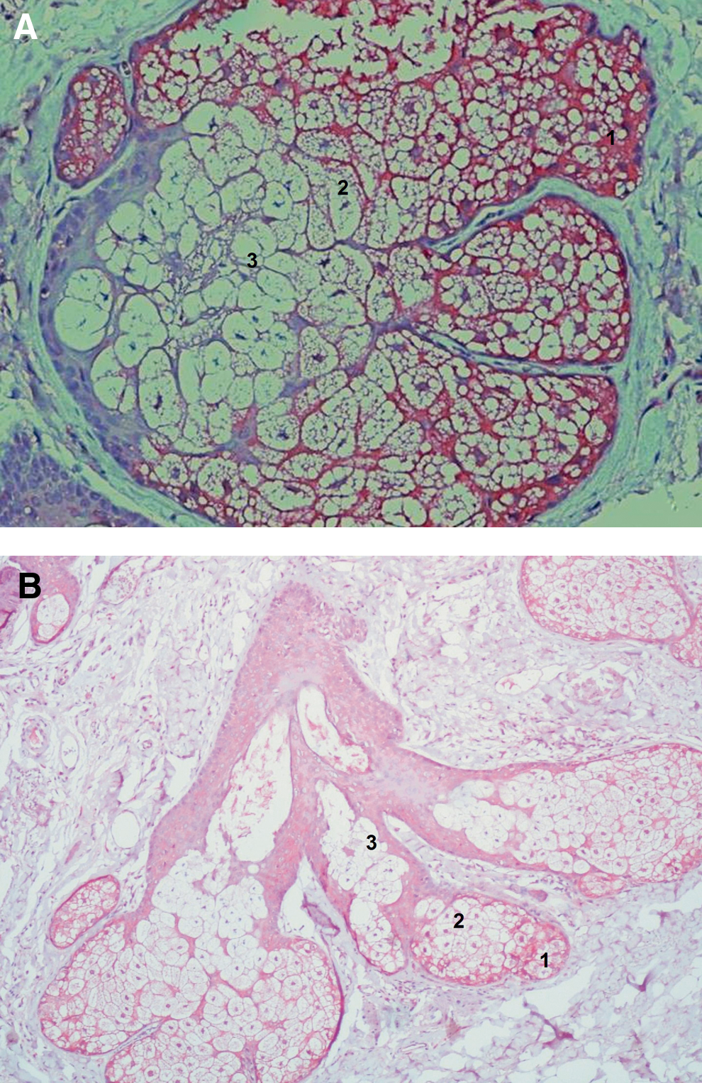
Localization of the immunohistochemical signals of CRH in the skin of the aged (>70 years) (

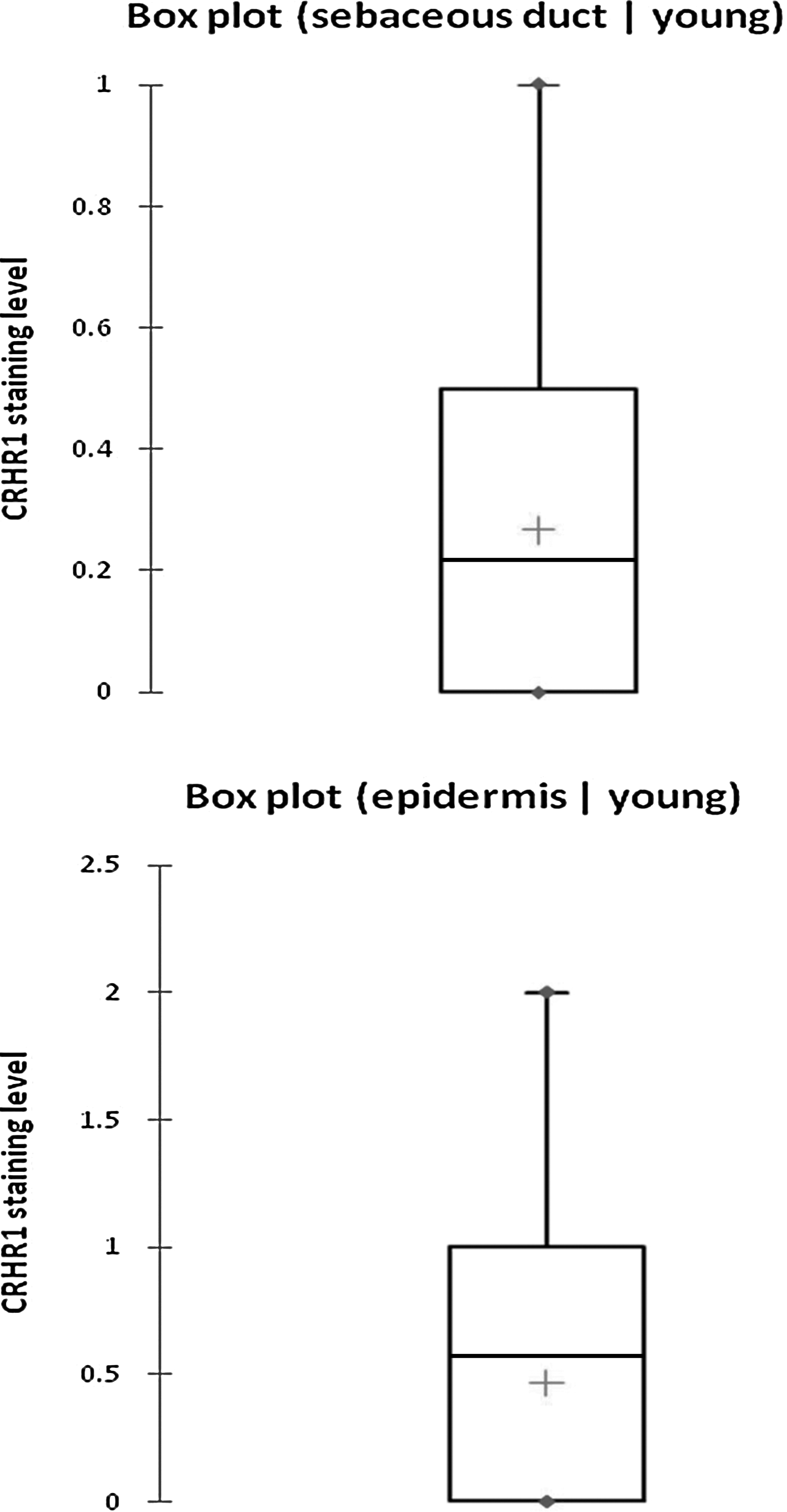
Box plots representing the expression of corticotropin-releasing hormone receptor 1 (CRHR1) in the epidermis and sebaceous ducts of aged and young groups. , Minimum and maximum values;  , quartiles, and median; + mean.
CRH receptor 1
Positive staining was observed in the epidermis, hair follicles, sebaceous ducts, sebaceous glands, and sweat glands. Cells in the dermis (fibroblasts and inflammatory cells) were also focally positive. The positive staining of the sebaceous glands ranged from weak to intense in different samples. CRHR1 expression in sebaceous glands was differentiation dependent, being strongest in the basal undifferentiated cells, with mature sebocytes almost presenting a negative staining result. Sebum was strongly stained (Fig. 4).
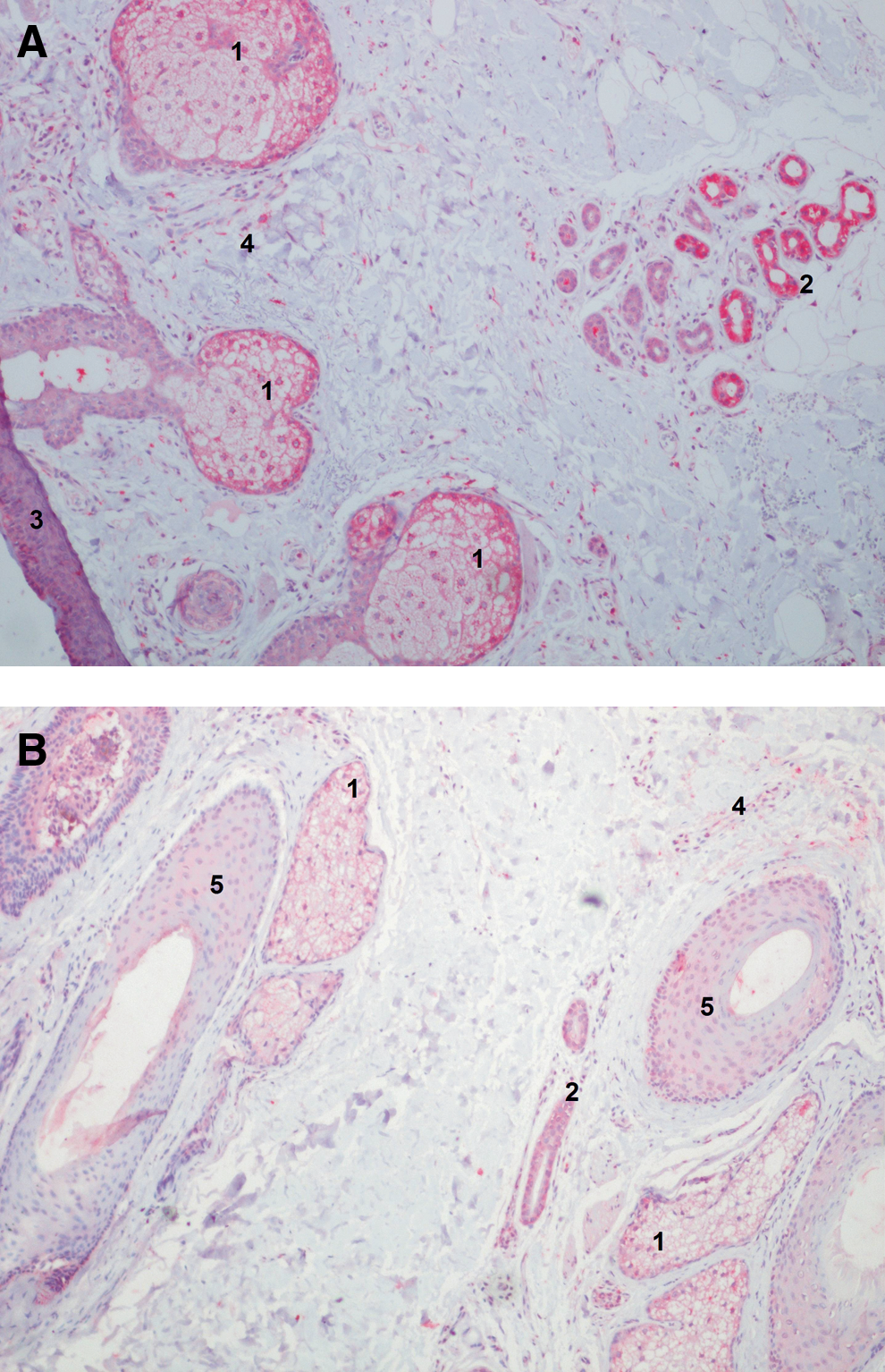
Localization of the immunohistochemical signals of corticotropin-releasing hormone receptor 1 (CRHR1) in the skin of the aged (>70 years) (
The sweat glands exhibited the strongest positive reaction for the CRHR1 among all skin structures. The secretory part of the glands showed stronger positive staining compared to the ductal portion. Like sebum, sweat also showed a strong positive reaction for the CRHR1 (Fig. 4).
The epidermis showed weakly positive CRHR1 staining in the stratum basale and stratum spinosum, gradually fading to negativity toward the stratum corneum. Basement membrane zone and stratum corneum showed no detectable expression of the CRHR1.
There was a correlation between age and intensity of the staining reaction in the hair follicles and the epidermis (correlation matrix [Spearmann] +0.308 and +0.435, p=0.043 and 0.003, respectively). A stronger staining was detected in the eldest (≥70 years, n=13) compared with the youngest (≤30 years, n=13) age groups regarding CRHR1 expression in epidermis (p=0.016) and ductus seboglandularis (p=0.061) (Figs. 3 and 4).
CRH receptor 2
Sebaceous glands, sebaceous ducts, and the secretory portions of the sweat glands expressed CRHR2. In addition, 66% of the smooth muscles stained positively for CRHR2. Epidermis, dermis, blood vessels, and hair follicles were negative for CRHR2. The CRHR2 expression in the sebocytes was constitutive and showed a homogeneous stain throughout differentiation phases (Fig. 5). There was no correlation between age and intensity of the staining reaction comparing the eldest (≥70 years, n=13) and youngest (≤30 years, n=13) age groups.
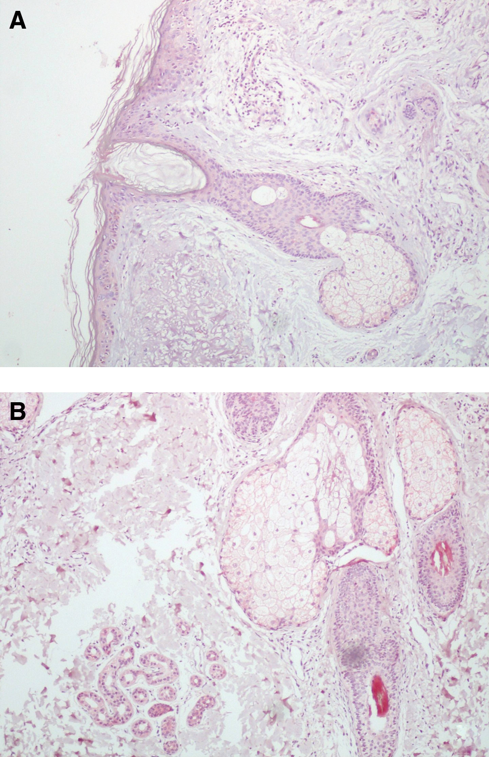
Localization of the immunohistochemical signals of corticotropin-releasing hormone receptor 2 (CRHR2) in aged (>70 years) (
CRH binding protein
CRHBP was strongly expressed in sebaceous glands in a differentiation-dependent manner. Basal undifferentiated cells showed the strongest expression, whereas mature sebocytes were almost negative. Sweat glands showed much weaker staining compared with the sebaceous glands. CRHBP expression was undetectable in the epidermis and hair follicles, whereas it was weakly positive in the dermal smooth muscle bundles.
There was a significant correlation between the age and dermal exhibition of CRHBP (correlation matrix [Spearmann]=0.612, p<0.0001) (Figs. 6 and 7). In contrast, CRHBP staining of sweat glands and sebaceous glands was weaker in the aged (≥70 years, n=13) compared to the young group (≤30 years, n=13) (Figs. 6 and 7).

Box plots representing the expression of corticotropin-releasing binding protein (CRHBP) in the sebaceous glands and dermis of aged and young groups. , Minimum and maximum values;  , quartiles, and median; + mean.
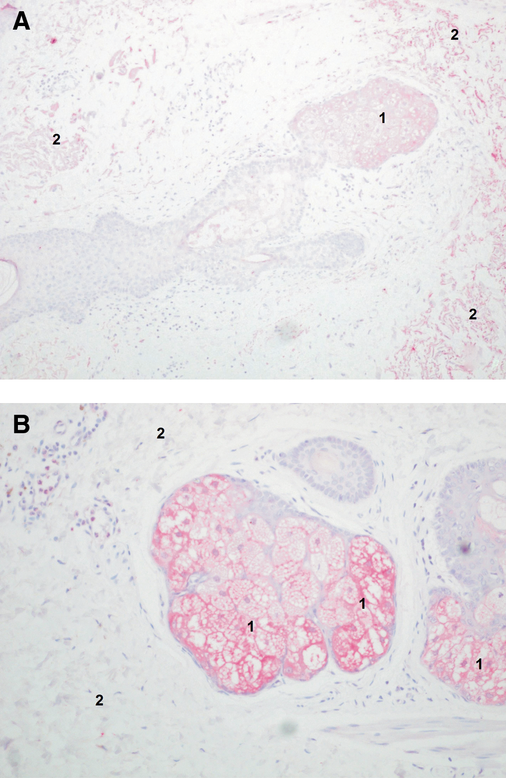
Localization of the immunohistochemical signals of corticotropin-releasing hormone binding protein (CRHBP) in aged (>70 years) (
Discussion
This study defined the changes of the CRH system occurring in the aging skin at the protein level (Table 3). The protein expression of molecules of the CRH system has previously been shown by different groups, including ours, to be compatible with mRNA changes. 9,10,13,15 Also expression of the CRH system on the gene level was extensively studied. 9,10,13
The CRH system components were selected according to their proven role in influencing the synthesis and quality of skin lipids, 15 which are greatly variable for the maintenance of skin quality throughout life, 16 their effect on proliferation and differentiation of skin cells, as well as the influence on chronic inflammation 9,13,14,17,21 and age-related diseases. All antigens tested were expressed mainly in the sebaceous gland area, hence making it the "maestro" that regulates the aging process in the skin.
The sebaceous gland is the major endocrine gland of the skin, and its cells, the sebocytes, are known to produce CRH. 15 The age-associated increased expression of CRH in the sebaceous glands, which was found in this study, as well as the elevated age-dependent expression of CRHR1 in epidermis and hair follicles (Table 2) are likely to correspond to the exaggerated systemic HPA stress response detected in aging. 3 Moreover, the increased expression of CRHR1, which is considered the major coordinator of stress response, in the specimens of aged individuals suggests a chronic stress-induced pathothysiology of aging. Because CRHR1 expression was unchanged in a sebocyte model of hormonal aging, 18 its increased expression is probably not driven hormonally.
x, Expressed: −, not expressed.
CRH, Corticoptropin-releasing hormone; CRHR1, CRH receptor 1; CRHR2, CRH receptor 2; CRHBP, CRH binding protein.
CRH, Corticotropin-releasing hormone; CRHBP, CRH binding protein; CRHR1, CRH receptor 1; CRHR2, CRH receptor 2.
In neuronal culture however, the CRH protects neurons against the amyloid toxicity. 19 By analogy, the detected CRH age-dependent downregulation in the highly innervated sweat glands may reflect the changes occurring in the aging neurons of the brain leading to Alzheimer disease, because both the neurons and the sweat glands are of ectodermal origin. 20 CRHBP, which has a control and buffering action for CRH, exhibited an age-associated decline in sweat and sebaceous glands.
The CRHR1 is the main CRH receptor in the skin, and it binds CRH with a much greater affinity compared to the CRHR2 13 ; in addition, CRH does not influence the expression of both receptors. 15 In our study, CRHR2 is totally absent in the epidermis, in agreement with Slominski et al. 11 The CRHR2 surprisingly showed no age-associated change, although it is responsible for hair growth. 10
In summary, CRH as well as the effector receptor CRHR1 are upregulated in aging skin, supporting the CRH system hyperactivation theory of aging. From previous literature and from our data, we can conclude that in aged skin a defective upregulated stress response signaling is detected, with CRH and CRHR1 being the upstream players. This defective stress response puts the skin in a continuous stress-like condition. This inefficient local attenuation of the CRH system activation in the aged skin may induce a local proinflammatory response. 21 A relationship between the CRH and elevated IL-6 and IL-8 expression was already detected in the acne vulgaris 12 and sebocyte cultures, 14 but this does not seem to be the proinflammatory pathway in aging (manuscript under review).
Footnotes
Author Disclosure Statement
No competing financial interests exist.