




Abstract
Introduction:
Previous studies have indicated that resveratrol, a natural phytoestrogen, can act as an anti-aging therapy to resist age-related changes of several body tissues. However, the anti-aging effects of resveratrol on bone have been poorly investigated in this natural aging population. Accordingly, this study was design to evaluate the effects of resveratrol on bone mass and biomechanical properties in old rat femora.
Methods:
Twenty 22-month-old male Wistar rats were divided into two randomly assigned groups (n=10). The first group was treated for 10 weeks with resveratrol (10 mg/kg per day) and the second group was left untreated (control). Rat femora were collected. Bone mass and bone microestructure were investigated by microcomputed tomography and histomorphometry. Biomechanical properties were determined by a three-point bending test. Plasma levels of CTX (carboxy-terminal telopeptide of type I collagen) and osteocalcin were also determined. Statistical analyses were performed by a Student two-tailed unpaired t-test. In all experiments, a value of p<0.05 was considered significant.
Results:
Microcomputed tomography analyses demonstrated that resveratrol-treated rats had significant higher bone volume, bone trabecular number, and cortical thickness and lower spacing between trabeculae in comparison to the control group. Histomorphometric analyses confirmed the increase of bone volume in resveratrol-treated rats compared to controls. Resveratrol-treated rats had significant higher bone flexural modulus, stiffness, and ultimate load compared to control group. Treatment was not associated with changes in plasma CTX or osteocalcin.
Conclusion:
These findings demonstrate that resveratrol increases bone microstructure and bone mechanical properties in old male rats, suggesting that resveratrol might be used as anti-aging therapy to resist age-induced bone loss.
Introduction
B
Resveratrol is a natural phytoestrogen produced by certain plants, including berries, grapes, and nuts. 4 Resveratrol expresses potent anti-oxidant and anti-inflammatory properties, and hence, it has been suggested to be a therapeutic agent for several age-induced diseases. 4 –9
Several previous in vitro and in vivo studies have pointed out the positive effects of resveratrol on bone. 10 In vitro studies have indicated that resveratrol is able to stimulate osteoblast proliferation and differentiation directly, 11 –14 inhibit osteoclast formation, and promote osteoclast apoptosis. 12,15 In vivo studies have revealed that resveratrol was able to promote bone mineral density and inhibit bone loss in ovariectomized rats 16 –18 and in young rats under tail suspension 19 or old rats under hind limb suspension conditions 20 (unloading animal models).
However, to our knowledge, the potential role of resveratrol as an anti-aging therapy for bone loss in old populations growing naturally has not been investigated. Accordingly, we have conducted this in vivo study to investigate the effects of dietary supplementation of resveratrol on architecture, bone mass, and biomechanical properties of the bones of old rats.
Materials and Methods
Animals
This study was carried out in accordance with the guidelines for Ethical Care of Experimental Animals of the European Union, approved by the Ethical Committee for Animal Studies of the Complutense University (Madrid, Spain). Twenty 22-month-old male Wistar rats, equivalent to 60 years of age in humans, 21 were used in this study. All rats were housed in polycarbonate cages, subjected to a 12-hr light–dark cycle at the constant temperature of 23°C, and fed a standard laboratory rat diet (A.O4 Panlab, Barcelona, Spain) and water ad libitum. Animals were randomly divided into two groups of 10 rats each. The first group of rats was treated with resveratrol at dosages of 10 mg/kg per day. The second group of rats was left untreated as a control group (only water and vehicle-treated). After 10 weeks of treatment, which is equivalent to 6 years in human age, 21 animals were sacrificed. Rat femora were collected and fixed in buffered formaldehyde for further analyses. Experimental analyses were conducted on femurs because the femur has been shown to be a good model to assess age-related bone changes. 22 Body weight was also measured after the treatment in both groups.
Chemical
Resveratrol was obtained from Actafarma Laboratories, Madrid, Spain. The purity of the product was 99% as checked by high-performance liquid chromatography.
Resveratrol administration
Resveratrol was supplied to the animals at a dose of 10 mg/kg per day. It was dissolved in absolute ethanol and added to the drinking water in a final ethanol concentration of 0.066%. The drinking water had a final resveratrol concentration of 15 mg/100 mL. Water bottles were covered with aluminium foil for protection from light, and the drinking fluid was changed every day. Untreated animals received 0.1% alcohol in tap water. The total amount of water used every day was used to determine the total amount of resveratrol taken by the animals. After 70 days of treatment, animals were killed by decapitation, and blood and femora were collected.
Plasma determinations
Trunk blood was collected and centrifuged to obtain plasma. The marker of bone resorption CTX (carboxy-terminal telopeptide of type I collagen) and the marker of bone formation osteocalcin were measured by a commercially available rat-specific enzyme-linked immunosorbent assay (ELISA) kit (Biomedical Technologies, Stoughton, MA).
CTX is a specific and sensitive marker of bone resorption that is produced by cathepsin K during normal action of osteoclasts and is involved in systemic bone resorption. 23 Osteocalcin is the major non-collagen protein found in bone. It contains three γ-carboxyglutamic acid residues that confer a very strong ability to bind to hydroxyapatite and calcium. Osteocalcin is synthesized by osteoblasts during the process of bone formation; it is mostly incorporated into bone matrix, but some escapes into the blood. Thus, osteocalcin measurement reflects new protein synthesis and provides a valuable tool for assessing bone formation. 23
Microcomputed tomography scan analysis
Microcomputed tomography (micro-CT) scan was used in our study to assess bone phenotypes of old rat femora. Micro-CT scan was chosen because of its ability to measure cortical and trabecular tissues separately. 24 Proximal right femurs were scanned at 40× magnification with a SkyScan 1072 (Bruker-Microct, Kontich, Belgium) and analyzed using bone analysis software (Skyscan, v. 2.2f, Kontich, Belgium). 25 The X-ray source was adjusted at 100 kV and 98 μA. The degree of rotation between each picture was 0.7°. The segmentation of the image was made by a global threshold and a voxel size of 21.90×21.90×21.90 μm. The following three-dimensional morphological parameters were evaluated for the 20 femurs: Bone volume-to-tissue volume ratio (BV/TV), trabecular thickness (Tb.Th), trabecular number (Tb.N), cortical thickness (Ct.Th), spacing between trabeculae (Tr.Sp), bone marrow area, and bone surface density. 26
Bone histology and histomorphometry
Left femurs were used for histology analyses. Femurs were fixed in 4% paraformaldehyde at 4°C for 24 hr and then dehydrated in graded ethanol series before being embedded in methyl methacrylate resin, as previously described. 27 Fuchsin Methylene Blue stain was added to 7-μm un-decalcified sections of rat femurs. BV/TV was measured.
Mechanical test
Biomechanical properties of rats femurs collected from our experiments were analyzed by a three-point bending test, the most popular method for the characterization of long bone biomechanical properties. 28 A three-point breaking test was performed using an Instron 5569 tester (Instron Corp., Canton, MA) on the midshaft of the right femora obtained from both control and treated rats. 25,29 The span of two support points was 20 mm, and the deformation rate was 0.1 mm/min. The extrinsic parameters were young modulus, stiffness, ultimate force, and work to failure and were calculated from the resulting load-displacement curves.
Statistical analyses
Descriptive outcomes (mean±standard deviation [SD]) of each test for the studied groups were calculated. Statistical analysis was performed by SPSS v. 21.0 program. A Shapiro–Wilks statistical test was applied to assess the normal distribution of data. The Student two-tailed unpaired t-test was applied to assess significant differences between the studied groups. In all experiments, a value of p<0.05 was considered significant and is indicated by a single asterisk.
Results
Resveratrol improves microstructure and biomechanical properties of old rat femora
Bone phenotype and mechanical properties of rat femora treated with resveratrol and control were examined using three-dimensional micro-CT scan, histomorphometry, and the three-point-bending test. In Fig. 1, micro-CT analyses revealed that resveratrol-treated rats had significant higher bone volume (BV/TV=31.9±3.7%), bone trabecular number (Tb.N=10.2±1.6/mm), cortical thickness (Ct.Th=0.57±0.13 mm), and lower spacing between trabeculae (Tr.Sp=0.075±0.01 mm) than the control group (BV/TV=25.0±3.7%; Tb.N=8.3±0.5/mm; Ct.Th=0.4±0.14 mm; Tr.Sp=0.1±0.02 mm). However, no significant changes in trabecular thickness, bone marrow area, and bone surface density between the groups can be seen. In agreement with these results, histomorphometric analyses confirmed the increase of bone volume in old rats following resveratrol treatment (BV/TV=31.5±1.5%) compared to control (BV/TV=27.3±1.4%; Fig. 2). Three-point bending analyses of rats demonstrated higher bone stiffness (485±153 N/mm), flexural modulus (3363±1198 N/mm2), and ultimate load (183±37 N) in resveratrol-treated rats than in controls (stiffness=307±75 N/mm; flexural modulus=1332±976 N/mm2; ultimate force=148±36 N; Fig. 3). However, femurs of resveratrol-treated rats did not exhibit a significant change in work to failure (276±109 Nmm) compared to controls (244±126 Nmm).
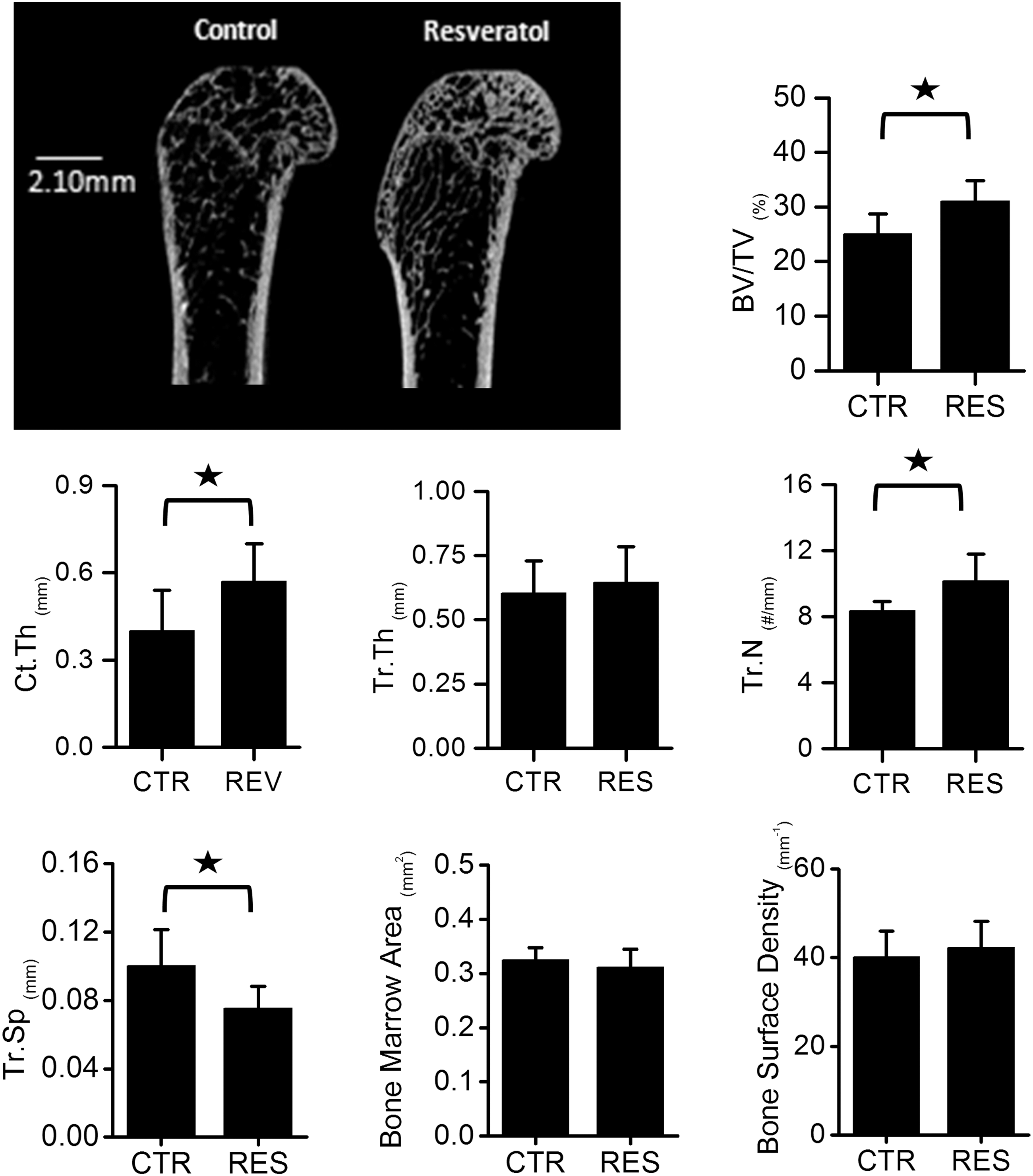
Microcomputed tomography (micro-CT) analyses of bone mass. Micro-CT sagittal sections of the femurs retrieved from old rats treated with resveratrol (RES) or left without treatment (control [CTR]). Resveratrol-treated rats had higher bone volume percentage (BV/TV), cortical thickness (Ct.Th), trabecular number (Tb.N), and lower spacing between the trabeculae (Tr.Sp) than the control group. No significant differences in trabecular thickness, bone marrow area, and bone surface density were observed between the groups. Values are mean±standard deviation (SD). (*) p<0.05.
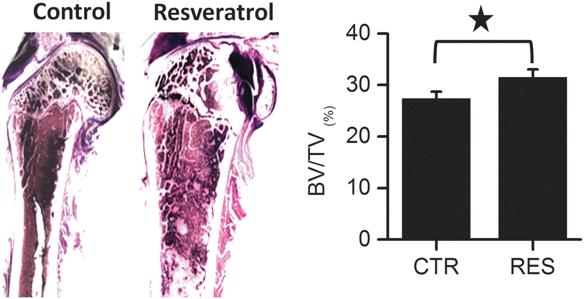
Histological and histomorphometric analyses of bone mass. Histological sagittal sections of the femurs retrieved from aged rats treated with resveratrol (RES) or left without treatment (control [CTR]). Resveratrol-treated rats had significantly higher bone volume percentage (BV/TV) than the control group. Values are mean±standard deviation (SD). (*) p<0.05. Color images available online at
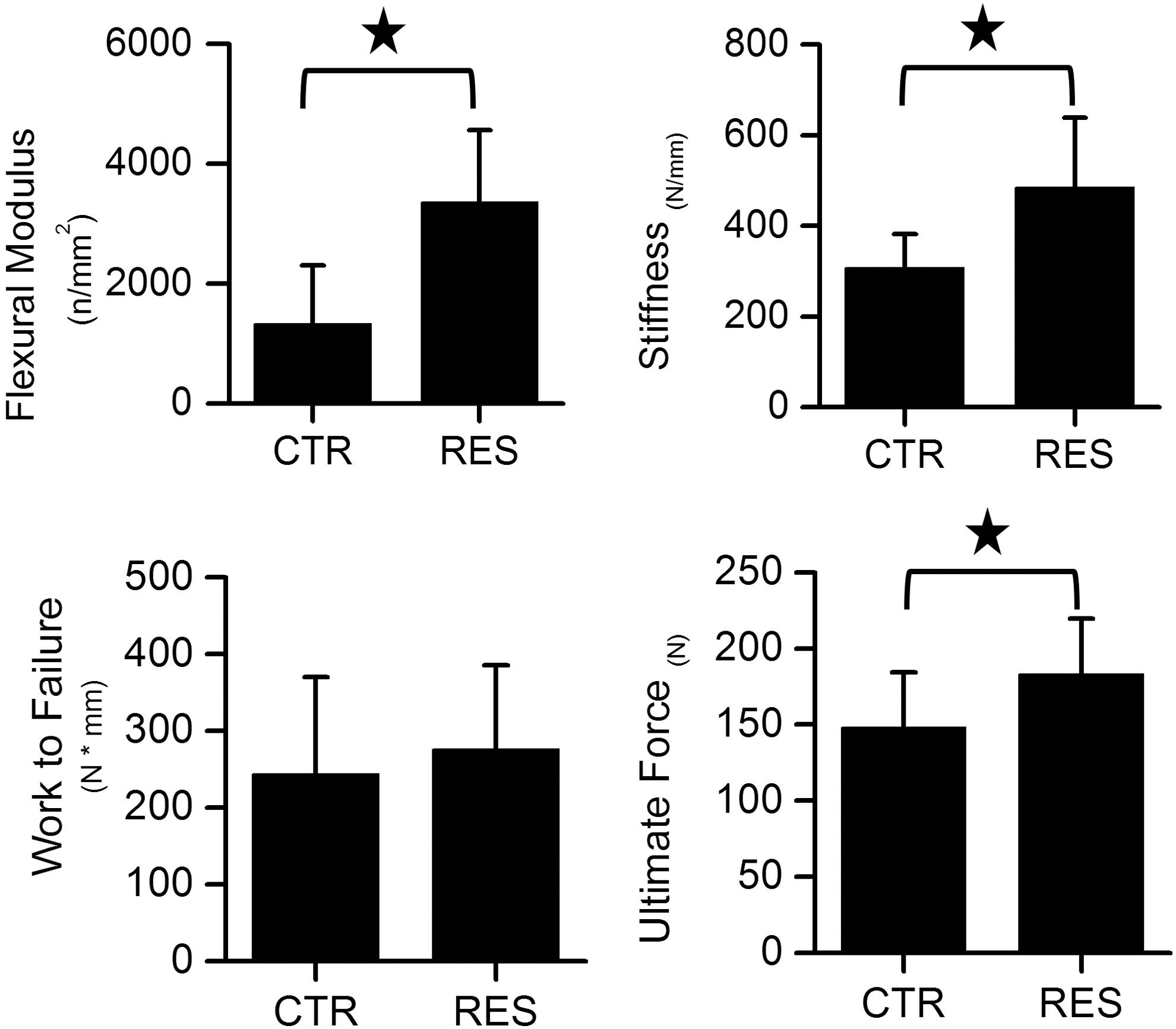
Bone mechanical properties. Mechanical analyses conducted by three-point bending test on femurs collected from aged male rats treated with resveratrol (RES) or left without treatment (control [CTR]). Resveratrol-treated rats had significantly higher bone stiffness, flexural modulus, and ultimate force than control. Values are mean±standard deviation (SD). (*) p<0.05.
Body weight
Resveratrol administration for 2.5 months was not able to modify the body weight significantly (524.7±3.5 grams in resveratrol group versus 528.7±63.6 grams in controls).
Plasma bone markers
Administration of resveratrol for 10 weeks was not able to modify neither plasma CTX values nor osteocalcin levels significantly (Fig. 4). (CTX, control 689±32 μg/mL and resveratrol 789±42 μg/mL; osteocalcin, control 8.9±3.6 ng/mL and resveratrol 4.82±2.8 ng/mL).
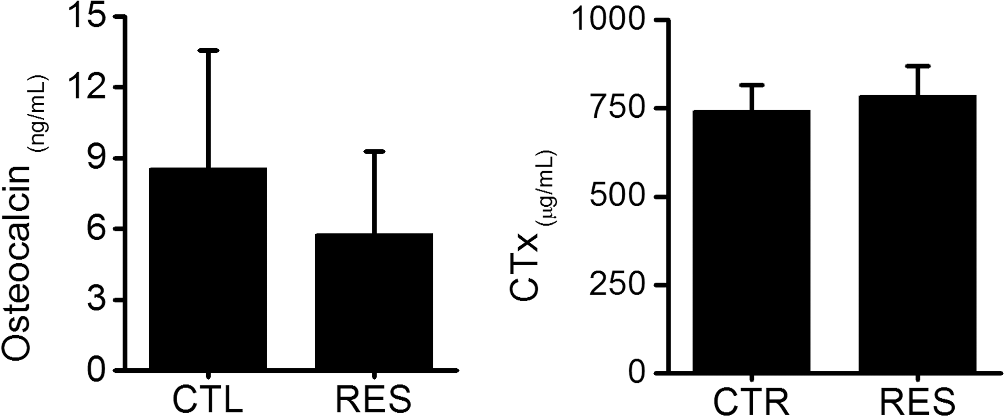
Effect of resveratrol (RES) on plasma osteocalcin and CTX (carboxy-terminal telopeptide of type I collagen). No significant differences in osteocalcin or CTX were observed between the groups. Values are mean±standard deviation (SD). (*) p<0.05. CTL and CTR, control.
Discussion
Aging is a progressive decline of body tissues hemostasis, leading to deterioration of these tissues with adverse health outcomes. Age-related changes are associated with impared DNA repair after damage by oxidative stress. 30 Accordingly, several studies have been carried out by our group 31 –36 and by others 7,9,16,17,18,19,37 to find new therapeutic approaches to slow down age-related changes by providing the body with molecules that express anti-oxidant and anti-inflammatory properties. 37 One of these molecules is resveratrol. Resveratrol is a polyphenolic phytoestrogen that is capable of substituting for estrogen decline, 38 and its consumption seems to be safe with no reported toxicity in humans or animals. 39,40 Accordingly, resveratrol was extensively studied in the literature to evaluate its anti-aging properties for various body tissues. 4,5,6,7,9,16,17,18,19,20,41
Resveratrol supplementation has been shown to exert anti-inflammatory effects in various mammalian models of aging. 42,43 Studies from other groups have previously established that resveratrol can exert significant cardiovascular protective effects in various models of myocardial injury, 44,45 hypertension, 44,46 and type 2 diabetes. 47
Aging is associated with chronic, low-grade increases in circulating levels of inflammatory markers. Specifically tumor necrosis factor-α (TNF-α) and interleukin-6 (IL-6) have been described to be not only indicators of inflammation but also causes of morbidity and mortality in the elderly. 48 Pro-inflammatory cytokines released from several tissues such as IL-1β, IL-6, and TNF-α and pro-inflammatory transcription factor nuclear factor kappa B (NF-κB) have been implicated in the pathogenesis and progression of tissue alteration and failure. A rise in inflammatory status was observed in previous studies in our old Wistar rats, 31 which showed an increase in protein expression of IL-1β and TNF-α and a decrease in IL-10. These cytokines exert cytotoxic and inhibitory effects on several tissues being responsible for its dysfunction and damage. Pro-inflammatory cytokines might induce the formation of reactive oxygen species (ROS), which could trigger an inflammatory response through the activation of transcription factor NF-κB that then translocates into the nucleus, where it activates a variety of inflammatory genes such as inducible nitric oxide synthase (iNOS), COX-2, cytokines (IL-6, IL-1β, TNF-α, etc.) and chemokines (monocyte chemoattractant protein-1 [MCP-1], IL-8, etc.). 49,50 The pro-inflammatory cytokines IL-1βand TNF-α could activate in turn NF-κB, and their expression is possibly induced in response to NF-κB activation, thus forming an amplifying feed-forward loop and a vicious cycle, eventually leading to tissue dysfunction and cell death. 50 Oxidative stress markers like nitric oxide (NO) and heme oxygenase-1 (HO-1) were also increased with age in old Wistar rats. 31
Several cytokine receptors are also linked to NF-κB signaling and enhancement of their inflammatory responses. Previous studies of our group found NF-κB p52 protein expression to be elevated in pancreas 35 and heart 33 of senescence-accelerated prone (SAMP8) mice. Furthermore, other groups have also reported an increase in nuclear NF-κB p52 binding activity in the liver of old rats. 51
Resveratrol seems to impinge on the most basic and evolutionarily conserved genetic pathways responsible for the control of metabolism and life span of eukaryotic organisms, conferring resistance against oxidative stress, injury, and cell death. Therefore, resveratrol exhibits diverse mechanisms of action and targets a great number of intracellular molecules. 52 In our case, we were interested in the effect of this substance on bone.
Bone, as with any body tissues, is susceptible to deterioration with advancing age. 2,53 –55 Age-related bone changes are associated with loss in bone mass and increase in susceptibility to fractures. 53 –55 Accordingly, our study has focused on resveratrol to evaluate its potential benefecial effects on slowing down age-related bone loss.
In this study, the authors have investigated the effects of resveratrol on femurs of 22-month-old male Wistar rats. This model was chosen because bone mineral density (BMD) and bone mineral content (BMC) were previously determined by our group in femora of male Wistar rats from different ages. 56 A maximum was detected for both BMC and BMD at 12 months of age with a significant reduction at 18 and 24 months, in agreement with Iida and Fukuda, 57 although the current bone loss animal model was set with male rats of 24–27 months of age. 58
Resveratrol supplement enhances bone microstructure in old rats
In our study, elderly rats growing naturally under normal conditions were treated with resveratrol and compared to a similar group of untreated rats. No significant variation in body weight was observed after resveratrol administration, in agreement with Durbin et al. 20 Results from micro-CT scan and histomorphometric analyses showed that resveratrol-treated rats expressed better bone quality compared to controls. These results are in agreement with previous studies conducted in ovariectomized rats (a model of menopause), 16,17 old-mice, 43 and young rats under tail suspension 19 or old rats under hind-limb suspension conditions 20 (models of mechanical unloading). However, we are the first to report the protective effect of reseveratrol in resisting age-related bone loss in old rats growing naturally under normal conditions.
Resveratrol-treated rats demonstrated higher biomechanical properties than those that were untreated. Our results are in agreement with previous studies in which it has been shown that resveratrol treatment is able to improve bone mechanical properties in old mice. 43 Other previous studies could not confirm the effect of resveratrol on biomechanical properties on femurs. 20 This might be related to the fact that resveratrol benefecial effects were assessed in old rats for a short period (3 weeks) 20 in comparsion to a relatively longer period (10 weeks) of treatment conducted in our study.
The results of the current study showed significantly higher bone volume, bone trabecular number, cortical thickness, and lower spacing between trabeculae than the control group. However, although no significant changes in trabecular thickness were seen, there was a tendency for it to be higher in resveratrol-treated rats. However, a larger sample size is needed to confirm that. Moreover, all of the mechanical properties were increased, except for work to failure, which is a mechanical property that relies on the elasticity of the collagen and mineral crystals. The elasticity of collagen might not be affected following resveratrol treatment in our study, which could explain why there was no significant difference in work to failure in resveratrol-treated rats compared to control. However, bone quality was increased with resveratrol.
In the current study, resveratrol was supplied to the animals at a dose of 10 mg/kg per day. The dose of resveratrol provided is similar to Durbin's study 20 (12 mg/kg per day), and much lower than 400 mg/kg per day used by Habold et al. 19 This dose has been considered as moderate and safe. 20 Moreover, Su et al. 59 investigated various doses of resveratrol and found a significant increase in BMD with 10 mg/kg per day during 10 weeks in ovariectomized female rats.
Mechanism by which resveratrol improves the health of bones in old population
The mechanism by which resveratrol promoted bone mass is still not clearly understood. However, previous studies indicate that resveratrol can affect both osteoblasts (bone-forming cells) and osteoclasts (bone-resorbing cells). 10 In vitro studies have demonstrated that resveratrol can directly promote osteoblast proliferation and differentiation. 11 Indeed, resveratrol was able to enhance the expression of bone morphogenic protein (BMP)-2, 59 runt-related transcription factor 2 (Runx2), 13,14 and Sirtuin-1, 60 thereby affecting the Wnt signaling pathway, a key promoter for osteoblast differentiation. Moreover, resveratrol was able to enhance osteocalcin and osteopontin synthesis, 59 markers for osteoblastogenesis. In addition, it has been shown that resveratrol is capable of reducing receptor activator for NF-κB ligand (RANKL) expression by osteoblasts, inhibiting osteoclast proliferation. 12,13,15
In this study, neither plasma osteocalcin nor CTX was modified by the treatment. However, bone microstructure and biomechanical properties were enhanced. These facts indicate better bone quality after 70 days of resveratrol supplement, although bone remodeling seems to be not affected, perhaps due to senescence cellular. These data were partially in agreement with Durbin et al., 20 who did not find changes in plasma CTX after resveratrol treatment for 21 days, although osteocalcin levels were increased.
However, these current plasma data could show limitations of the use of biologic markers. These plasma markers could not reflect resveratrol actions at the tissue level. In agreement with Garnero, 61 this could be because these bone markers do not allow the investigation of bone quality. New bone markers like sclerostin and fibroblast growth factor-23 (FGF-23), markers of osteocyte activity, would be needed to study changes on bone quality in old bones, an important determinant of skeletal fragility.
Future research
This study is the first evidence indicating that resveratrol supplementation is able to improve bone microstructure and biomechanical properties in old animals growing naturally under normal conditions. However, the exact effects of resveratrol on bone cell proliferation and differentiation in vivo are still unknown. The possible interference with inflammation, oxidative stress, or apoptosis is one of the possibilities, since resveratrol is able to reduce plasma TNF-α in old male Wistar rats, raising levels even lower than young rats (our unpublished data). Accordingly, future studies should be conducted to determine the exact mechanism by which resveratrol favors bone mass. Also, future studies should be conducted to evaluate the optimum dose and treatment time of resveratrol to maximize the beneficial effects of resveratrol to resist age-related bone loss. In addition, clinical trials should be conducted in a large scale to investigate resveratrol as a potential therapy for age-related bone loss.
Conclusion
Within the limitations of this experimental study, resveratrol was able to improve the microstructure and biomechanical properties of old male rat femora, suggesting its potential use as an anti-aging therpy to slow down age-related bone loss.
Footnotes
Acknowledgments
The authors wish to extend their gratitude to Mrs. Rocio Campon for her expert technical assistance.
Author Disclosure Statement
No competing financial interests exist.