




Abstract
A traditional herbal prescription Kyung-Ok-Ko (KOK), composed of Rehmannia glutinosa Liboschitz var. purpurae, Lycium chinense, Aquilaria agallocha, Poria cocos, Panax ginseng, and honey, has been widely used in Oriental medicine as an invigorant for age-related diseases, such as amnesia and stroke. However, the beneficial value of KOK on uterine dysfunction related to hyperandrogenism is largely unknown. We investigated the effect of KOK (2.0 g/kg/day, per os) on endometrial abnormalities in a dehydroepiandrosterone (DHEA, subcutaneous)-induced polycystic ovary syndrome (PCOS) rat model. Preadministration of KOK significantly (p<0.05) decreased the elevated body weight, uterus weight, and endometrial thickness by PCOS induction, corresponding to reduced apoptosis and the infiltration of immune cells (CD4+ T cells, CD8+ T cells, and macrophages) in the endometrium. These results were associated with reduced mRNA expression of interleukin (IL)-1β, IL-6, IL-8, and matrix metalloproteinase-3 and increased mRNA expression of IGF-β1, transforming growth factor (TGF)-β, TGF-β1, and vascular endothelial growth factor in the uterus after DHEA injection. These multiple effects of KOK may synergistically prevent the development of endometrial abnormalities in DHEA-induced hyperandrogenism via anti-inflammatory action, indicating that KOK has preventive and therapeutic potential for suppressing PCOS.
Introduction
P
Current strategies to reduce endometrial cancer risk in PCOS are aimed at ameliorating risk factors, such as obesity, anovulation, and endometrial hyperplasia. 4 Clinically, symptoms and features of PCOS can be cured pharmacologically with medications, including clomiphene citrate, raloxifene, tamoxifen, metformin, aromatase inhibitors, oral contraceptive pills, and glucocorticoids. They can also be cured surgically by laparoscopic ovarian drilling as a second-line treatment after medical therapy failure. These treatments, however, are not always completely effective. 9 –12 Clomiphene citrate has been widely used to induce ovulation in women with PCOS. 13,14 However, it can cause side effects, such as P4 resistance in the endometrium, ovarian hyperstimulation, multiple pregnancy, ectopic pregnancy, and congenital malformations. 15 In contrast, oral contraceptive pills are commonly used to treat chronic PCOS to induce regular menses and protect the endometrium, 9 –12 although their long-term safety in PCOS needs to be evaluated. 1 Because PCOS can be reduced by improving insulin resistance, metformin (insulinomimetic or insulin-sensitizing agent) can be prescribed to reduce the risk of endometrial hyperplasia in PCOS. However, metformin can produce side effects, such as nausea, diarrhea, bloating, flatulence, vomiting, and lactic acidosis. 16 In addition, even with treatment with these medications, endometrial dysfunction in patients with PCOS still can lead to higher risks of implantation failure, spontaneous miscarriage, and endometrial hyperplasia or endometrial cancer. 1 –4,17,18 Nevertheless, endometrial dysfunction stemming from PCOS has not received the attention that it deserves. A safe and effective way to prevent endometrial hyperplasia in women with PCOS is important because of the disease burden caused by endometrial cancer and its economic costs. Complementary and alternative medicine (acupuncture, herbal medicine, dietary supplements, etc.) can prevent or alleviate PCOS or minimize the adverse effects of these chemicals for women with PCOS. 19
Kyung-Ok-Ko (KOK; Qiong-yu-gao in Chinese; Kei-gyoku-kou in Japanese) is a traditional Oriental prescription consisting of a decoction of the following six ingredients: Rehmannia glutinosa Liboschitz var. purpurae Makino (Scrophulariaceae), Lycium chinense Miller (Solanaceae), Aquilaria agallocha Roxburgh (Thymelaeaceae), Poria cocos Wolf (Polyporaceae), Panax ginseng C.A. Meyer (Araliaceae), and honey. 20 In eastern Asia, such as Korea, China, and Japan, KOK is prescribed as a tonic medicine to give energy to the body or as a traditional medicine to cure various age-related symptoms, including lack of vigor and immunity, emaciation or weakness, stroke, amnesia, and dementia. 21 According to recent studies, KOK has antioxidative, 22 antityrosinase, 23 anticancer, 24 and cognitive-enhancing effects. 25 Recently, we have reported that KOK may effectively prevent and improve dehydroepiandrosterone (DHEA)-induced PCOS in rats via anti-inflammatory action. 20 That is, KOK can significantly decrease the elevated body weight, size and number of follicular cysts, elevated level of estradiol in serum, elevated percentage of CD8+ T lymphocytes/cells in the retroperitoneal lymph nodes, elevated mRNA expression of CD11b and CD3, and activated inflammatory mediators in the ovaries after DHEA injection. 20 These results suggest the possibility that KOK can display beneficial effects against PCOS-related endometrial dysfunction. Therefore, we investigated whether KOK had a positive effect on endometrial hyperplasia in a DHEA-induced PCOS rat model. Here, we demonstrated that preadministration of KOK reduced the elevated uterus weight, endometrial thickness, endometrial infiltration of immune cells, and expression of inflammatory mediators by DHEA-induced hyperandrogenism. Taken together, KOK may be applied as beneficial material to prevent or treat endometrial dysfunction, including endometrial hyperplasia.
Materials and Methods
Animals and ethical statements
Sprague Dawley dams (Narabiotec Co., Ltd., Seoul, Korea), each with eight to nine female pups, were housed under 12-hour light–12-hour dark cycle (light on 07:00–19:00) at room temperature (23°C±2°C) and humidity of 55%±10%. They were provided food and water ad libitum. These animals were allowed to habituate to the housing facilities for 1 week before the experiments were commenced. All experimental procedures were reviewed by the Institutional Animal Care and Use Committee (IACUC) or the Ethics Committee of Animal Experiments of the Kyung Hee University, Republic of Korea. The committees specifically approved this study [KHUASP (SE)-12–030]. Proper randomization of laboratory animals and handling of data were performed in a blinded manner in accordance with the recent recommendations from an NIH workshop on preclinical models of neurological diseases. 26
Animal sacrifice
All efforts were made to prevent undue stress or pain to rats. Rats with signs of imminent death were euthanized to avoid suffering. Each rat was deeply anesthetized with an intraperitoneal overdose of sodium pentobarbital (50 mg/kg, body weight). Their ovaries, uteri, and blood were sampled.
Hyperandrogenism induction, KOK preparation, and KOK treatment
PCOS induction and KOK treatment were carried out as previously described. 20 Briefly, prepuberal rats (23 days old, female) were divided into the following four groups (n=6 per group): (1) sham [sesame oil, subcutaneous (s.c.)+saline, per os (p.o.)], (2) DHEA [DHEA, 60 mg/kg body weight/day, s.c.+saline, p.o.], (3) DHEA+KOK [DHEA, 60 mg/kg body weight/day, s.c.; KOK, 2.0 g/kg body weight/day, p.o.], and (4) KOK alone [sesame oil, s.c. +2.0 g/kg body weight/day of KOK, p.o.]. Hyperandrogenism in PCOS rat model was induced by injecting DHEA as previously described. 20,27 Briefly, rats in the DHEA group received a daily injection of DHEA dissolved in 0.2 mL sesame oil beginning at 23 days of age. KOK (lot no. SQ12, 1200 g) was obtained from the Kwang Dong Pharmaceutical Company (Pyongtaek, Korea). Its preparation was described previously. 20,28 KOK was administered orally daily for 20 consecutive days 2 hours before the first injection of DHEA. Rats in the sham or KOK group were administrated saline or KOK without a DHEA injection. The dose of KOK was determined based on a preliminary experiment for this study, traditional prescription, 21 well-known formula, 29 and the best effective dose in our previous report. 28 Body weight was measured daily during the experimental period before treatment with DHEA, KOK, or saline. The stage of estrus cyclicity was determined by microscopic analysis of the predominant cell type in vaginal smears obtained daily beginning on day 10 (33 days after birth) in KOK preadministrated rats and day 25 (48 days after birth) in rats postadministrated with KOK after DHEA injection as described in our previous report. 20
Histological and immunohistological examination
Histological examination was carried out as previously described.
20
Briefly, 20 days after the DHEA injection, the left uteri (n=6 per group) were immediately removed and fixed in 4% paraformaldehyde solution. The middle portion (about 0.5 cm) from each uterus was cut, dehydrated, and cleared in ethanol and xylene, embedded in paraffin, and cut into 5-μm-thick sections in a transverse plane. These sections were stained with hematoxylin–eosin (H&E) dye. The stained sections were scanned. The longest outer diameter of each uterus was measured using NIH ImageJ program (
TUNEL staining
Apoptotic cell death was evaluated by TUNEL staining according to predescribed protocols 20 and manufacturer's instructions (ApopTag Peroxidase In Situ Apoptosis Detection Kit, S7100; Millipore, Billerica, MA).
Real-time polymerase chain reaction (PCR)
To investigate the induction level and uterine function recovery in PCOS, mRNA levels of proinflammatory cytokines were assessed at 20 days after the DHEA injection and KOK administration. Rats (n=6 per group) were anesthetized. Their right uteri were immediately removed and deep-frozen. Real-time polymerase chain reaction (PCR) was performed using SYBR Green PCR Master Mix (Applied Biosystems, Franklin Lakes, NJ) as previously described. 20,30 The mRNA levels of each target gene [interleukin (IL)-1β, IL-6, IL-8, matrix metalloproteinase (MMP)-3, insulin-like growth factor (IGF)-1β, transforming growth factor (TGF)-β, TGF-β1, and vascular endothelial growth factor (VEGF)] were normalized to that of glyceraldehyde 3-phosphate dehydrogenase mRNA. Fold induction was calculated using the 2−ΔΔCT method as previously described. 31 Oligonucleotide primers used in real-time PCR are summarized in Table 1.
GAPDH, glyceraldehyde 3-phosphate dehydrogenase; IGF, insulin-like growth factor; IL, interleukin; TGF, transforming growth factor; MMP, matrix metalloproteinase; VEGF, vascular endothelial growth factor.
Statistical analysis
Results are expressed as mean ± standard error of the mean. Statistical significance in differences between values was determined using analysis of variance with a Fisher's post hoc test. Statistical difference was considered when p<0.05 unless otherwise indicated.
Results
KOK inhibits the development of DHEA-induced PCOS in rats
Based on a previous study, 20 2.0 g/kg body weight/day of KOK was the most effective dose in DHEA-induced PCOS rat model. Therefore, we used this dose in this study. To confirm whether PCOS was induced by DHEA, we investigated whether DHEA treatment increased body weight, one of the major clinical features. 2,3,17 We induced cyst formation in the ovaries of rats with PCOS (Fig. 1). The body weight was significantly (p<0.05) increased at 20 days after DHEA injection compared to the age-matched sham group. However, the increased body weight was decreased by preadministration of KOK (Fig. 1A), corresponding to our previous report. 20 In addition, the ovaries from the rats in the DHEA group exhibited a swollen appearance with multiple cystic follicles (Fig. 1C), consistent with fully developed PCOS. 32 –34 Interestingly, the size and number of follicular cysts in the ovaries were decreased after the administration of KOK (2.0 g/kg/day) (Fig. 1E), which was consistent with our previous study. 20 Preadministration of KOK (2.0 g/kg/day) alone did not significantly (p>0.05) affect body weight or cystic formation in the ovaries compared with the sham group (Fig. 1). Since regular estrus cycle is a main index of normal uterine function in humans, we confirmed the stage of estrus cycle by vaginal smear. All rats (n=12) in the normal and KOK groups exhibited regular estrus cycle at 10–20 days of age after starting the experiment. The estrus cycle of all rats in the DHEA group (n=6) was stopped, while four rats in the DHEA+KOK group exhibited regular estrus cycle. Additionally, at the day of euthanasia, six, one, two, and three rats displayed diestrus, proestrus, estrus, and metestrus stages, respectively, in the normal and KOK groups. All rats in the DHEA group displayed diestrus or metestrus stage. In the DHEA+KOK group, two, three, and one rats displayed diestrus, proestrus, and metestrus stages, respectively. These results indicated that hyperandrogenism and PCOS were successfully induced by DHEA and that preadministration with KOK inhibited the development of PCOS by DHEA.
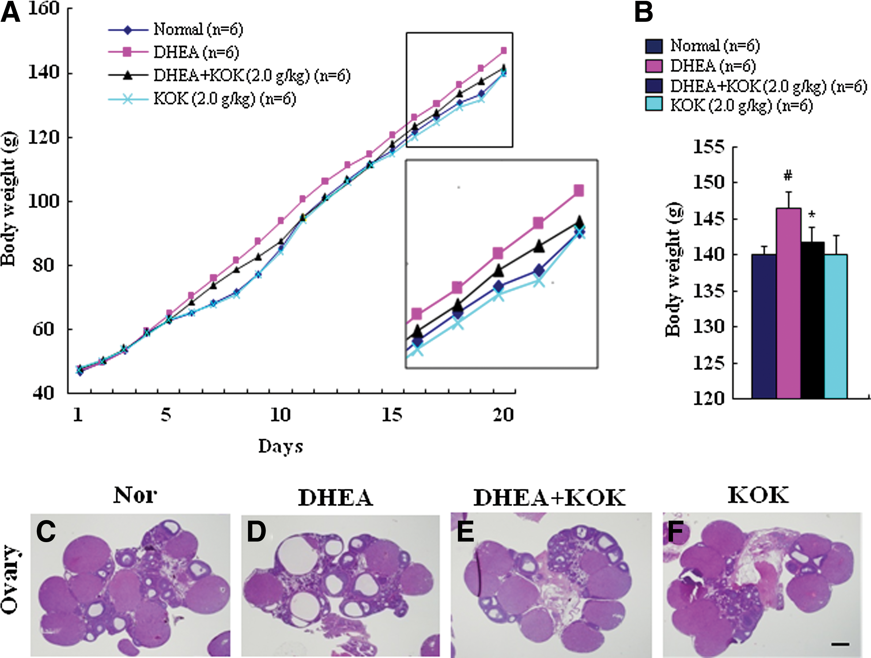
Effect of KOK on body weight gain and ovarian morphology in rats with PCOS.
KOK reduces abnormality of uterine weight and uterine morphology
Because the mean thickness of the endometrium was significantly increased in the PCOS group compared to that of the control group, 35 we examined uterine weights in each group. The weights in the DHEA group (68.8±11.4 mg/cm) were higher than those in the sham group (40.4±1.8 mg/cm), whereas the uterine weight in the DHEA+KOK group (44.6±9.4 mg/cm) was significantly (p<0.05) decreased compared to that in the DHEA group (Fig. 2A). Administration of KOK alone did not increase or decrease uterine weight compared to the sham group. Because endometrial malformation mirrors the pivotal clinical features of women with PCOS, 3 we investigated whether DHEA changed the appearance and the pathohistological structures of uteri and whether KOK inhibited these changes. Representative images from the middle portion of the uteri from each group are shown in Figure 2C–F. The sizes of the uteri in the DHEA group were larger than that in the sham or KOK group. The sizes of the uteri in the DHEA+KOK group were smaller than that in the sham group. To determine the exact change in size, we measured the longest outer diameters of the uteri using H&E-stained images. The outer diameters of the uteri in the sham group (1.04±0.01 mm) were similar to those in the KOK group (1.03±0.00 mm), whereas the diameters were significantly (p<0.05) increased in the DHEA group (1.82±0.02 mm). However, the elevated diameter was significantly (p<0.05) reduced after the administration of KOK (1.21±0.00 mm) (Fig. 2B). Microscopic examination of the uteri from the sham and KOK groups revealed normal endometrium (En), myometrium (My), and epimetrium (Ep) with normal cystic glands (Fig. 2G, K, O). Glands were lined with simple cuboidal epithelia (Fig. 2O). However, the endometria from DHEA-treated rats revealed glands with extended lumens (Fig. 2H, L, P). Glands were lined with pseudostratified or stratified epithelia with atypical cells and nuclei (Fig. 2P). These abnormal structures were alleviated, showing cystic glands (Fig. 2Q) as those observed in the sham and KOK groups (Fig. 2R). These results indicated that preadministration with KOK inhibited endometrial malformation by DHEA.
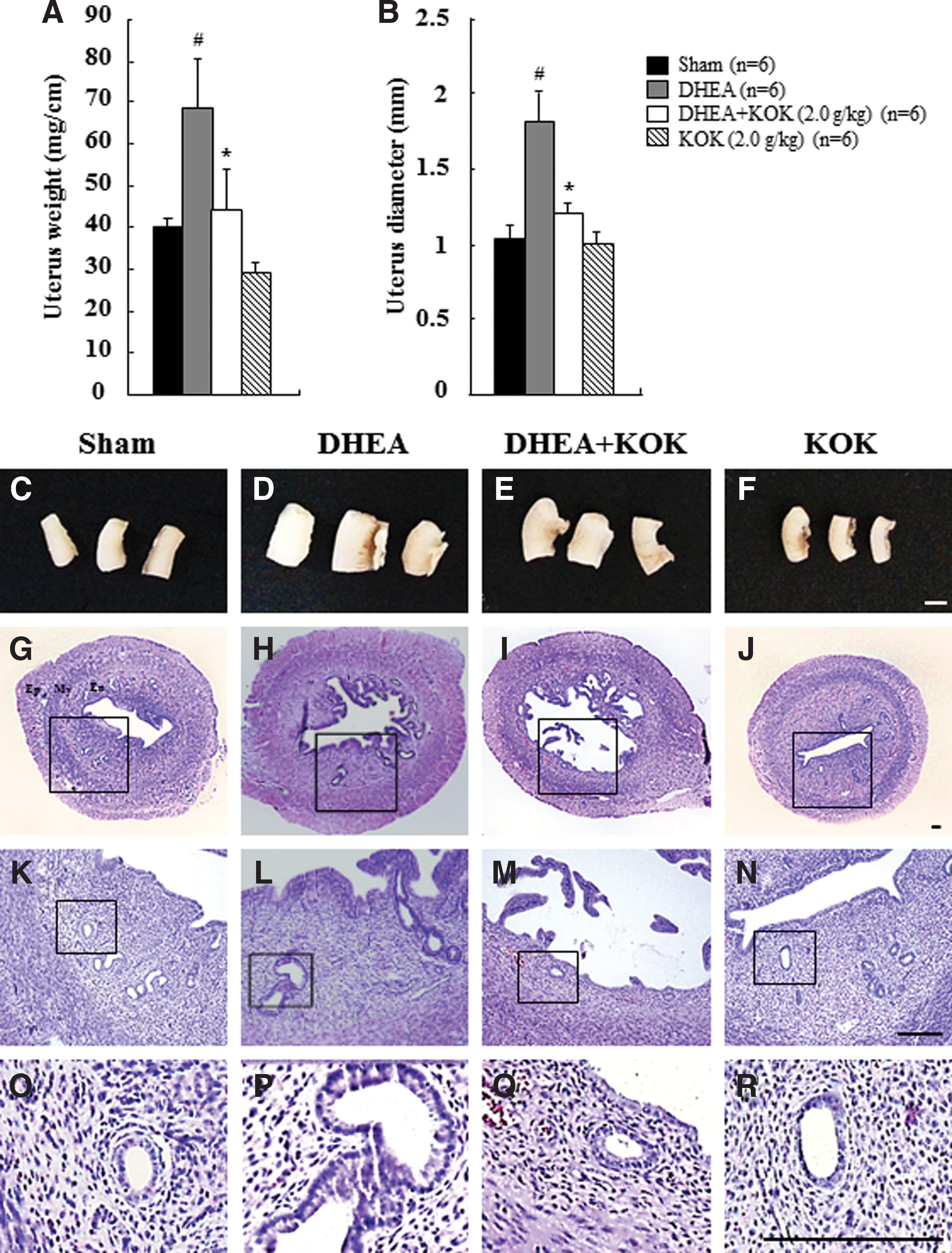
Effect of KOK on uterine weight and endometrial abnormality in rats with PCOS.
KOK ameliorates apoptosis in the endometria of PCOS rats
Because hyperandrogenism induced by DHEA is associated with a greater number of apoptotic cells in the endometria and metformin (insulinomimetic or insulin-sensitizing agent) is able to reduce the increased number of apoptotic cells, 36 we investigated the effect of KOK on apoptosis in utero. Apoptotic cells by TUNEL stain were rarely detected in uterine tissue of the sham or the KOK-alone group. However, the number of TUNEL-positive cells was increased in the endometria of the DHEA group. Interestingly, the increased number of apoptotic cells was significantly (p<0.05) reduced after administration with KOK (Fig. 3). These results indicated that preadministering KOK inhibited endometrial malformation induced by DHEA by reducing endometrial apoptosis.
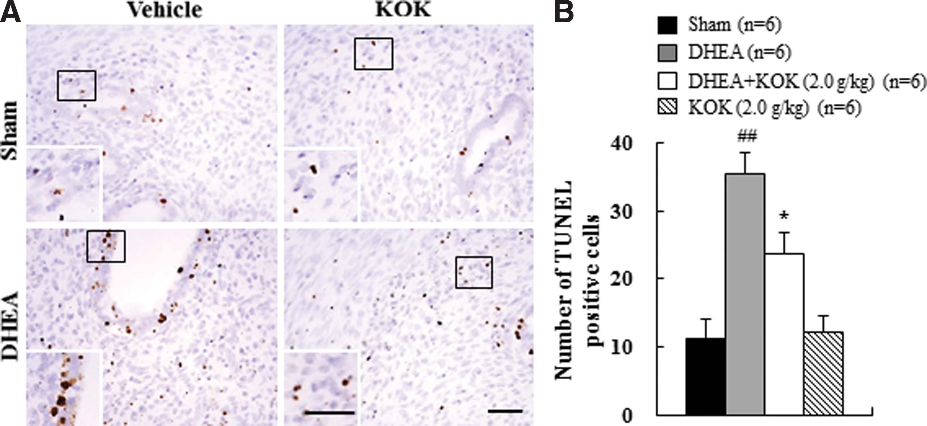
Effect of KOK on apoptotic cell death in the endometria of rats with PCOS.
KOK diminishes the infiltration of macrophages and T cells into the endometria of PCOS rats
Macrophages and T cells play important roles in the health of human endometrium by infiltrating or migrating to the endometrial inflammation sites. 37 –39 In our previous study, KOK has been found to be able to decrease the number of macrophages, CD4+ T cells, and CD8+ T cells in the retroperitoneal lymph nodes and ovaries from DHEA-induced PCOS rats by either immunohistochemistry or flow cytometry analysis. 20 Therefore, we further investigated the effect of KOK on the infiltration of macrophages and T cells in uterine tissue with PCOS by immunohistochemistry. Uterine sections from the sham and DHEA groups harbored Iba-1+ macrophages, CD4+ T cells, and CD8+ T cells at very low frequencies. These cells were mainly localized in the endometrium, rarely in the myometrium or epimetrium (Fig. 4A, D, E, H, I, L–O). In uterine tissue in the DHEA group, the numbers of each cell type were increased in the endometrium compared to those in the sham and KOK groups (Fig. 4B, F, J, M–O). However, these increased numbers in each cell type were significantly (p<0.01) decreased after administering KOK (Fig. 4C, G, K, M–O). These results suggested that preadministration with KOK could inhibit the infiltration and activation of macrophages, CD4+ T cells, and CD8+ T cells in the endometria of rats with PCOS.
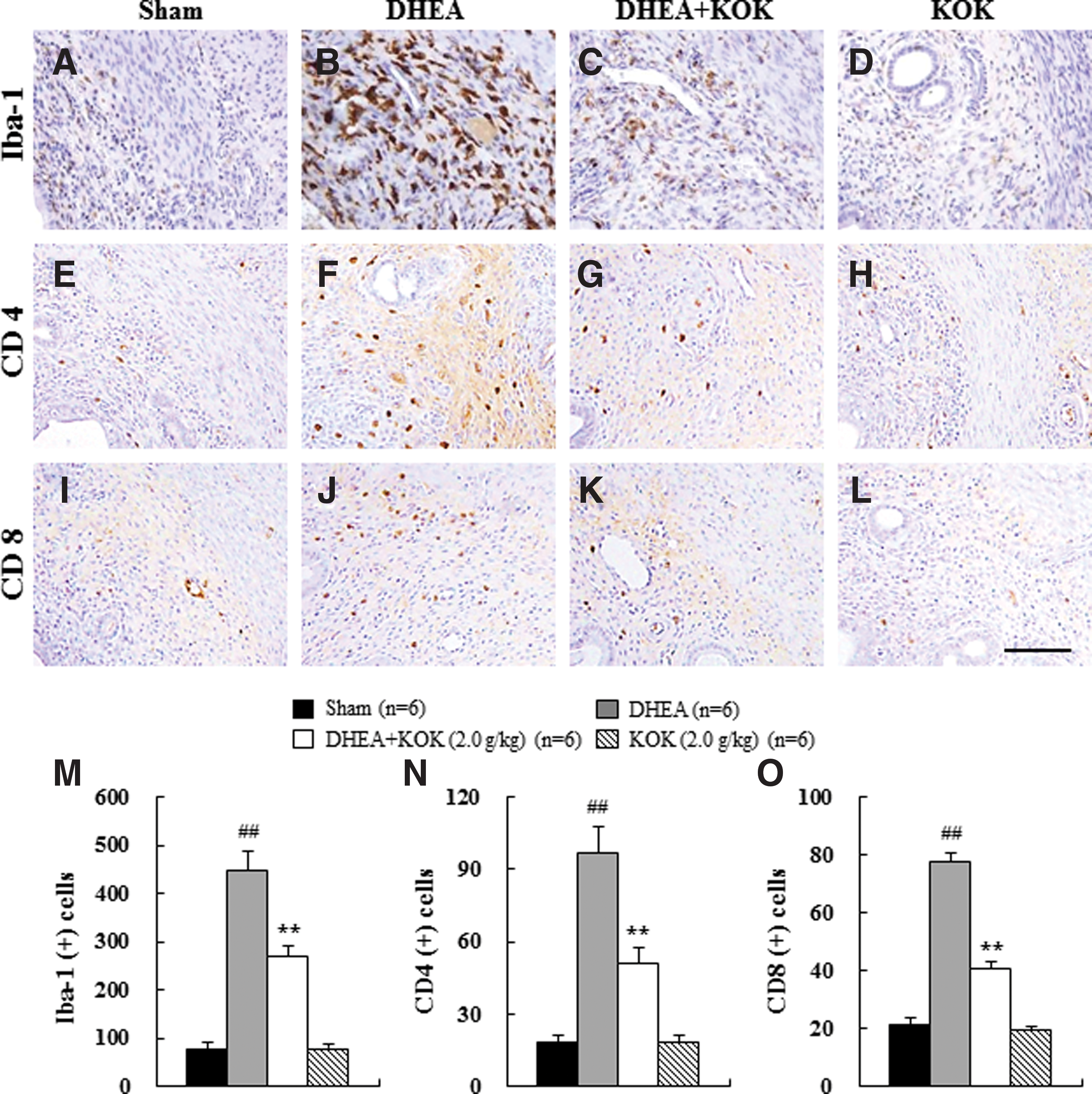
Effect of KOK on the infiltration of macrophages, CD4+ T cells, and CD8+ T cells into the endometria of rats with PCOS.
KOK reduces the expression of inflammatory mediators in the uteri of PCOS rats
PCOS in humans has been linked to chronic inflammation. 40 Endometrial cell types from women with PCOS display inflammatory and oncogenic potential. 41 Thus, we investigated whether administering DHEA and KOK could regulate the mRNA expression of proinflammatory cytokines (IL-1β and IL-6), chemokines (IL-8), damaging protease (MMP-3), and growth factors (IGF-β1, TGF-β, TGF-β1, and VEGF) in the uteri of each group at 20 days after DHEA injection and KOK administration. Results are shown in Figure 5. The uterine mRNA levels of IL-1β (by 7.1-fold), IL-6 (by 13.3-fold), IL-8 (by 54.5-fold), and MMP-3 (by 2.3-fold) were significantly (p<0.05) elevated in the uteri from the DHEA group compared to those from the sham or KOK group. However, the increased mRNA levels were decreased significantly (p<0.05) by 76.2%, 76.6%, 96.2%, and 41.5% in the DHEA+KOK group compared to those of the sham group (Fig. 5). In contrast, the mRNA expression levels of IGF-β (by 0.3-fold), TGF-β (by 0.5-fold), TGF-β1 (by 0.5-fold), and VEGF (by 0.3-fold) were significantly (p<0.05) reduced in the uteri of the DHEA group compared with those of the sham or KOK group. These decreased mRNA expression levels were significantly (p<0.05) increased by 112.7%, 55.4%, 43.6%, and 126.5%, respectively, by KOK administration. KOK itself had no effect on the mRNA expression level of these factors (Fig. 5). These results suggest that KOK might have inhibited the development of endometrial malformation by DHEA-induced hyperandrogenism.

Effect of KOK on the expression of inflammatory mediators in the uteri of rats with PCOS.
Discussion
KOK is a well-known herbal formula as a tonic in traditional Oriental medicine. It has been used for patients with age-associated disorders. Its efficacy determination has relied on empirical results. 21 However, its actual efficacy has not been systematically or scientifically demonstrated until recently. Moreover, the mechanism that underlies the effects of KOK on the female reproductive system has not been investigated yet. In the present study, we demonstrated that KOK could reduce the development of endometrial abnormalities in rats with PCOS by anti-inflammatory action, indicating that KOK might be used as a new and safe therapeutic strategy to control endometrial abnormalities. Further study is needed to clarify its exact molecular mechanisms.
The detrimental effects of hyperandrogenism on endometrial function contribute to the infertility of women with PCOS by inhibiting endometrial cell growth and secretory activity and inducing endometrial hyperplasia and endometrial cancer. 4,42 –44 However, the exact pathogenesis and specific therapeutic strategies for endometrial abnormalities, including endometrial hyperplasia and endometrial cancer by hyperandrogenism, have not yet been established. In humans, endometrial hyperplasia is a premalignant condition that usually heralds endometrial cancer. It shares identical risk factors with endometrial cancer. 4 Recent reports have indicated that there is a significant risk of endometrial cancer in women with PCOS, although further research is needed to clarify the exact molecular mechanisms. 4 Additionally, the use of animal models to study endometrial dysfunction related to human PCOS has only recently been well established. Elia et al. have reported that DHEA can alter uterine histology in ways closely related to the development of precancerous structures concomitantly with increased apoptosis and proinflammatory as well as pro-oxidant status in mice endometria. 36,45 These pathological features are similar to those of endometrial hyperplasia of patients with PCOS. 4,36,45 In the present study, DHEA-induced androgenization in rats induced the formation of follicular ovarian cysts and endometrial abnormalities, including increased endometrial thickness and abnormal endometrial glands (lined with pseudostratified and stratified epithelia with atypical cells and nuclei). The patterns were similar to those reported in a previous study when DHEA treatment was started earlier (at 23 days) and continued for 20 consecutive days (until 43 days of age). 36,45 However, DHEA-induced hyperandrogenism has not been broadly admitted in animal models for studying pathogenesis or therapeutic strategies for endometrial hyperplasia and endometrial cancer. Therefore, in the present study, we referred to any abnormal endometrial changes related to hyperandrogenism by DHEA in uterine function as endometrial abnormality or malformation.
The endometria of most PCOS patients are thick, exhibiting simple, complicated, atypical hyperplasia or malignant transformation that may be correlated with endometrial cell apoptosis. 46,47 DHEA-induced hyperandrogenism was found to increase the number of apoptotic cells in the endometria. However, administering metformin reduced the increased number of apoptotic cells, which was associated with prevention against some inflammatory and oxidative alterations produced by hyperandrogenism. 4,36,45 In this study, the number of TUNEL-positive apoptotic cells was increased in the endometria by DHEA-induced androgenism compared to those in the sham group. However, the increase in the number of apoptotic cells was decreased by KOK treatment (Fig. 3). These findings suggest that KOK can diminish the development of endometrial abnormalities through inhibiting the apoptosis produced by hyperandrogenism.
The immune system plays an active role in the female reproductive tract, and macrophages are one of the most abundant immune cells in uterine functioning. 39 Macrophages are scattered throughout the endometrium. They are found especially around glands. 48 They are significantly increased in inflamed endometria, such as in endometrial hyperplasia, endometrial cancer, the secretory phase (especially before menses), and implantation. 39,49 Uterine macrophages can produce cytokines, chemokines, and growth factors in normal, inflammatory, and disease processes of the uterus. Metformin regulates the nitric oxide synthase and cyclooxygenase-2 activity in uterine tissue and tumor necrosis factor-alpha activity in serum, corresponding to recovery from hyperandrogenism caused by DHEA. 36,45,50 Here, we demonstrated that KOK diminished the increased frequency of Iba-1-immunoreactive macrophages in the endometrium (Fig. 4). Our results suggest that KOK has an anti-inflammatory action by inhibiting the infiltration of macrophages into the endometria in DHEA-induced PCOS rat model. T cells are also important in the female reproductive tract. 39 CD4+ T (helper/inducer) cells and CD8+ T (cytotoxic/suppressor) cells are increased in the retroperitoneal or lumbar lymph nodes, 20,51,52 ovarian tissue, 51 and uterine tissue 36 of DHEA-induced PCOS rats and mice. Metformin can reduce the increased number of CD4+ T cells in uterine tissue from PCOS mice. 36 In the present study, KOK reduced the increased infiltration of CD4+ T cells in the endometria of rats with PCOS based on immunohistochemistry, supporting the inhibitory effect of metformin and KOK on the percentages of CD4+ T cells in lumbar lymph nodes from PCOS rats as reported in previous studies. 20,36 These data indicate that KOK could diminish endometrial abnormalities by blocking the infiltration of CD4+ T cells into the endometrial of rats with PCOS.
Macrophages, CD4+ T cells, and CD8+ T cells can release inflammatory mediators to regulate uterine function. 39 Here, we demonstrated that KOK significantly decreased the increased mRNA levels of IL-1β, IL-6, IL-8, and MMP-3 but increased the decreased mRNA expression levels of IGF-β, TGF-β, TGF-β1, and VEGF in uterine tissue with PCOS (Fig. 5). These results indicate that KOK has beneficial effects on uterine dysfunction from PCOS in rats by inhibiting the expression of proinflammatory cytokines and chemokines while inducing the expression of growth factors. Targeting this inflammatory process by inflammatory mediators might be a therapeutic alternative to the current treatment for uterine dysfunction caused by hyperandrogenism.
Among the major herbs that comprise KOK, Rehmannia glutinosa Liboschitz has been traditionally prescribed to reduce fever, regulate immunity, and improve various diabetic disorders. 53,54 Its dried root can prevent bone loss in ovariectomized rats. 55 The fruit of Lycium chinense has been used traditionally for antiaging purposes in East Asia. 21 It can ameliorate learning and memory deficits in attention-deficit mice. 56 It can also prevent or alleviate oxidative stress-induced hepatotoxicity. 57 Aquilaria agallocha Roxburgh contains 4-butyl-α-agarofuran as a major ingredient with significant antianxiety activity in animal models. 58 Poria cocos Wolf, a fungus that grows around the roots of pine trees in East Asia and North America, has been used to treat chronic gastritis, acute gastroenteric catarrh, gastric atony, edema, nephrosis, dizziness, nausea, and emesis. 59,60 Panax ginseng C.A. Meyer is a perennial herb of the family Araliaceae. It contains ginsenosides as a major active ingredient. 61,62 Its extract has been used to increase physical strength, prevent aging, and restore stamina in East Asia for thousands of years. 61 It has various pharmacological activities, such as immune, endocrine, cardiovascular, nervous, and cancer-related benefits. 62 Panax ginseng can antagonize estrogen decline in ovariectomized mice, 63 modulate ovarian function in immature rats, 64 and improve estradiol valerate-induced PCOS in a murine model. 65
As mentioned above, each ingredient of KOK can produce various pharmacological benefits in normal or abnormal states. Although it is currently unknown which herb in KOK can confer beneficial effect for uterine dysfunction related to PCOS, the present data offered the first step toward its identification. In addition to its positive effect for ovarian function following PCOS, KOK might be used as possibly a safe and effective strategy to improve endometrial abnormalities and treat infertility in women with PCOS. Although our findings suggest its beneficial effects on endometrial abnormalities in rats with PCOS, the underlying mechanisms remain unclear. More studies are needed in the future to further clarify the mechanisms of KOK and its potential uses for endometrial disorders related to PCOS.
In conclusion, our results for the first time suggest that KOK can regulate some inflammatory and immune parameters of hyperandrogenized uterine function. Its activity can prevent endometrial abnormalities, including the formation of precancerous structures and proapoptotic status. Our findings present novel evidence about the clinical implications of KOK treatment not only in normal ovarian function 20 but also in preventing uterine dysfunction, including the formation of precancerous structures.
Funding
This work was supported by a grant from the Kwang Dong Pharmaceutical Co., Ltd., the Kyung Hee University in 2013 (KHU-20130542), and the Cooperative Research Program for Agriculture Science & Technology Development, Rural Development Administration, Republic of Korea (project no. PJ011582042015). The funders had no role in study design, data collection, data analysis, decision to publish, or preparation of the article.
Footnotes
Author Contributions
C.S.B., K.S.P., Y.O.K., and I.H.C. conceived and designed the experiments. M.J.L., M.J., and I.H.C. performed the experiments. M.J.L., M.J., C.S.B., and I.H.C. analyzed the data. H.J.K., S.L., and S.W.L. contributed reagents/materials/analysis tools. I.H.C. wrote the article.
Author Disclosure Statement
No competing financial interests exist.