




Abstract
Baicalein is a flavonoid isolated from the roots of Scutellaria baicalensis Georgi. This study aimed to ascertain the effects and potential underlying mechanisms of baicalein in
Introduction
A
Baicalein is a flavonoid extracted from the roots of Scutellaria baicalensis Georgi. Baicalein has extensive pharmacological properties, including antivirus, 3 antibacteria, 4 antitumor, 5 and cardiovascular protection. 6 Particularly, baicalein protects against several cerebral diseases such as traumatic brain injury, 7 ischemic stroke, 8 Parkinson's disease, 9 –12 and Alzheimer's disease. 13,14 Interestingly, we found that 0.04, 0.2, and 1 mg/mL baicalein extended the mean, median, and maximum life spans of Drosophila melanogaster, 15 suggesting its potential antiaging roles. However, the antiaging effects of baicalein have not been deeply investigated.
Although the exact mechanism of aging remains a riddle, chronic inflammation plays important roles. 16 It is appreciated that chronic low-grade inflammation, termed “inflammaging,” positively correlates with age. 16 Accumulated data strongly suggest that continuous upregulation of proinflammatory mediators are induced during the aging process. 17 –19 Mitochondrial dysfunction, a hallmark of aging, 20 can accelerate aging in mammals. Mitochondria play a role in modulating the inflammatory response, and the production of proinflammatory mediators impairs mitochondrial function by increasing reactive oxygen species (ROS) production.
Metabolomics has significant potential in pharmaceutical research, including discovery of biomarkers for disease and the elucidation of the mechanism of action of drugs. 21 Metabolomics can comprehensively and quantitatively determine the metabolites, whose levels can be regarded as the ultimate response of biological systems to genetic and environmental changes. 22 With the utilization of a metabolomics approach, it has been discovered that the endogenous metabolites are correlated with aging. 23 –25
In the present work, the antiaging effects and the underlying mechanisms of baicalein were investigated in
Materials and Methods
Animals and drug administrations
Sixty male Sprague Dawley rats (180–220 g) of 8-week-old were commercially obtained from Beijing Vital River Laboratories (Beijing, China). The rats were adapted to the new environment for 1 week before experiments were carried out. All rats were accommodated in room temperature (20°C–25°C) and constant humidity (40%–50%) under a 12 hours light–dark cycle with food and water ad libitum. All recommendations in the Guide for the Care and Use of Laboratory Animals of the National Institutes of Health were stringently followed in all procedures of this study.
Sixty rats were randomly divided into five groups (n = 12 per group): control group,
Behavioral tests
Novel object recognition test
The object recognition test was carried out as method presented. 26 The apparatus consisted of a square open box (55 cm long, 20 cm high, and 35 cm wide). The objects to be discriminated were in two different shapes: block and ball, and they could not be displayed by rats. This test included periods of adaptation, familiarity, and test. During the adaptation, each rat was placed in an empty box for 6 minutes. On the familiarity day, two identical objects (AA) were put in the test case and the rat was put into the box from the same position to the object (6 minutes). On the test day, one of the other set of objects replaced an object used in the familiarity day in the box (AB) and the rat was put into the box (6 minutes).
The time spent exploring the objects during the experimental sessions was defined as follows: sniffing or touching the object with nose. The recognition index was used to evaluate the ability of object recognition of rats. The “recognition index” was calculated by equation TB/(TA + TB), where TA is the time spent exploring the familiar object, and TB is the time spent exploring the novel object.
Morris water maze test
The spatial learning and memory ability was evaluated by the Morris water maze test, which included a 5-day place navigation training and a test day. 27 The maze consisted of a black circular water tank (120 cm in diameter, 38 cm in height), containing water (24°C ± 1°C) to a depth of 30 cm and a hidden platform submerged 1 cm below the water surface. The water cistern was divided into four quadrants, one of which contained a circular escape platform (12 cm diameter) placed at a fixed position. Place navigation trials were executed twice a day for 5 days before the test day, and there was no intertrial time interval between the two trials of the same day. For each trial, the rat was placed in the water facing the pool wall at one of four starting quadrant points and the starting quadrant was varied randomly over the trials.
For all training trials, the time that rats spent to reach the fixed platform (escape latency) was recorded to assess spatial learning capacity. 28 If a rat did not find the platform within 60 seconds, it was placed on the platform for 20 seconds at the end of the trial, and the escape latency was recorded as 60 seconds. On the test day, the platform was removed. The numbers of target crossings were recorded as well as the escape latency before reaching the target place. Data were collected by the video tracking equipment and processed by a computer equipped with an analysis management system. 28
Inflammatory mediators detection
Serum samples were harvested for detection of inflammatory mediators. The content of nitric oxide (NO) was measured by NO assay kit (Nanjing Jiancheng Bioengineering Institute, Jiangsu, China) according to the manufacturer's instructions.
The concentrations of interleukin-6 (IL-6), interleukin-1 beta (IL-1β), and tumor necrosis factor-α (TNF-α) were determined using commercially available enzyme-linked immunosorbent assay (ELISA) kits obtained from Sangon Biotech (Shanghai, China). In brief, the samples were pipetted into the 96-well plates that contained rat-specific antibodies. After incubation and washing, the absorbance was determined by following the instructions specified in the kit.
Preparation of cerebral cortex extracts and acquisition of 1H NMR spectral data
Metabolite extraction from the cerebral cortex of rats was achieved using methanol/chloroform/water. 29,30 After weighing, ice-cold methanol (4 mL/g) and double-distilled water (0.85 mL/g) were added to homogenize the cerebral cortex. The sample was vortexed, and then chloroform (2 mL/g) was added, and the sample was vortexed again. Subsequently, 2 mL/g chloroform and 2 mL/g water were added to the sample. The homogenate was left on ice for 15 minutes and centrifuged at 1000 g for 15 minutes at 4°C. Transfer the upper methanol/water phase (with polar metabolites) into separate glass vials. Remove the solvents from the samples under a stream of nitrogen.
The tissue extracts were reconstituted in 600 μL of NMR buffer (100 mM sodium phosphate buffer, pH 7.4, in 10% D2O, containing 0.01% sodium 3-trimethlysilyl [2,2,3,3-d4] propionate [TSP] as an internal NMR chemical shift standard) and vortexed and then centrifuged at 12,000 g for 5 minutes. Then, 550 μL of the supernatant was transferred into a 5 mm NMR tube for NMR spectroscopy.
NMR analysis of cerebral cortex extracts
The 1H NMR spectra of cerebral cortex extracts were acquired on a Bruker 600 MHz AVANCE III NMR spectrometer (Bruker) equipped with a Bruker 5 mm double resonance BBI probe at a temperature of 25°C. Spectra were recorded using the noesygppr1d pulse sequence.
The 1H NMR spectra were processed using MestReNova software (version 10.0.1; Mestrelab Research, Santiago de Compostela, Spain). All the spectra were manually phased and baseline corrected. The spectra of cerebral cortex extracts were referenced to the chemical shift of TSP with a chemical shift at δ 0.00 ppm. The regions containing the resonance from residual water (δ 4.52–5.20) were excluded to avoid the influence of water. All spectra were divided and then the signal integral computed in 0.01 ppm across the region δ 0.09–8.90 for cerebral cortex extract samples. The remaining spectral segments for cerebral cortex extracts were normalized to a total sum of the spectral intensity.
Pathway analysis
MetaboAnalyst 3.0, 31 a web-based analysis module, can perform metabolic pathway analysis by integrating two analysis approaches, including pathway enrichment analysis and pathway topology analysis. The main metabolic pathways regulated by baicalein were analyzed using this tool.
Statistical analysis
All data are expressed as a mean ± standard error of the mean. Repeated measures analysis of variance (ANOVA) followed by Fisher's least significant difference test was used for analysis of the data in acquisition phase of Morris water maze test. One-way ANOVA followed by Dunnett post hoc test was used for statistical analysis to determine significant differences of other behavior and biochemical changes among control,
Results
Body weight and food intake
Compared with the rats in control group, rats treated with
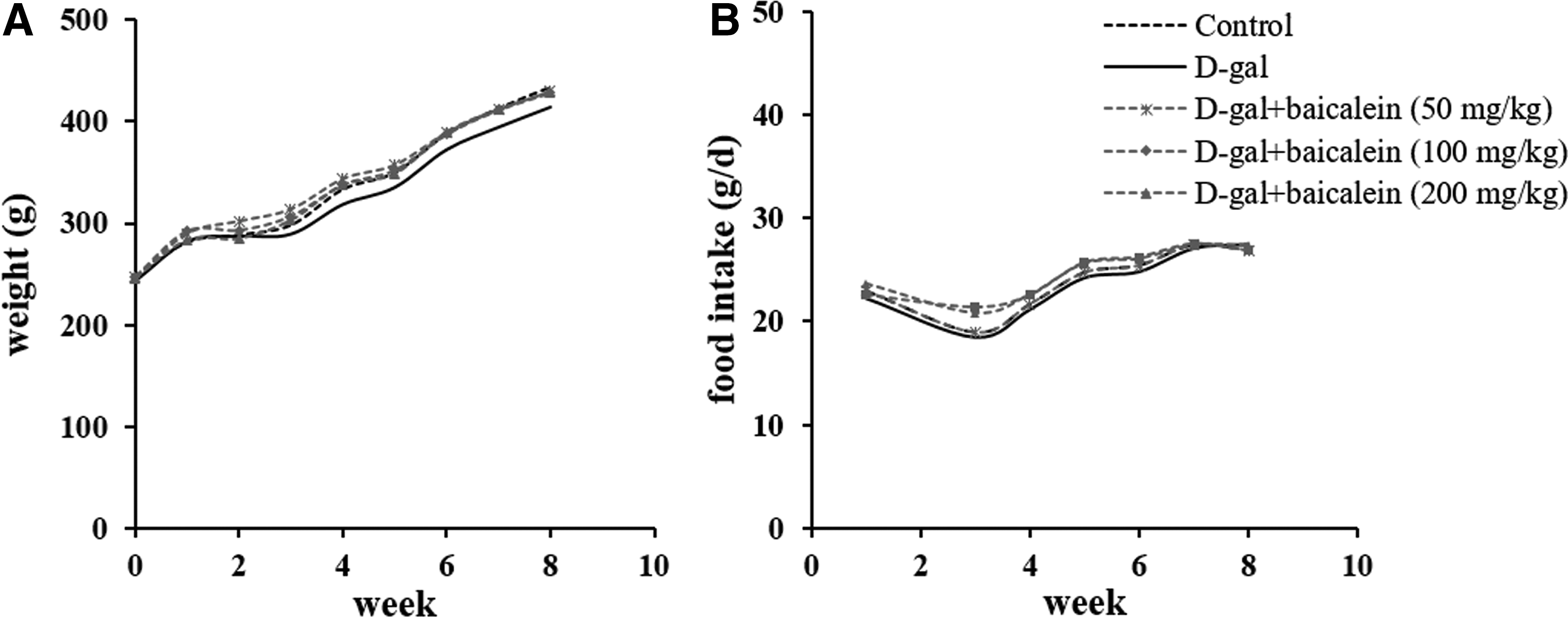
Effects of baicalein on body weight and food intake in
Effects of baicalein on the behavior of d -gal-treated rats
Novel object recognition test showed that recognition memory was significantly declined in the
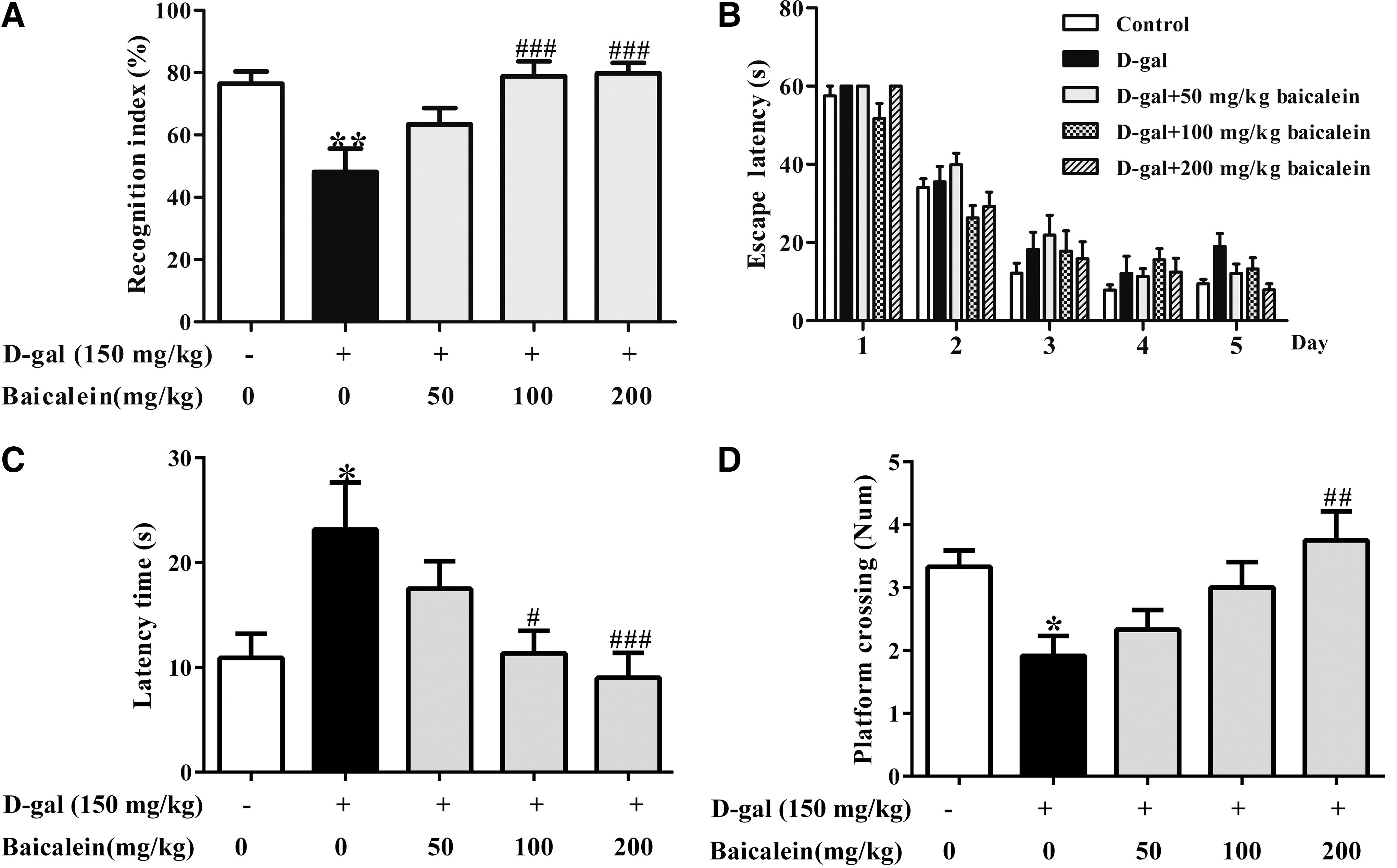
Effects of baicalein on the behaviors of
The results of repeated measures ANOVA showed that the escape latency was significantly higher in
On the test day, rats in
Effects of baicalein on the levels of inflammatory mediators
The NO level was significantly increased in rats of the

Effects of baicalein on the levels of inflammatory mediators in the serum.
Metabolomic study of cerebral cortex of rat brain
The typical1H NMR spectra of cerebral cortex obtained from the control rats are shown in Figure 4. The resonances in these spectra were assigned to individual metabolite by matching to the chemical shifts of compared standards, and those recorded in the literature, 30,32 and NMR databases such as the Human Metabolome Database (HMDB), Biological Magnetic, and Resonance Data Bank (BMRB). Identified endogenous metabolites in cerebral cortex of rats are listed in Table 1. Those detectable metabolites include numerous organic acids, amino acids, carbohydrates, tricarboxylic acid cycle intermediates, and nucleotide derivatives, together with some other metabolites.
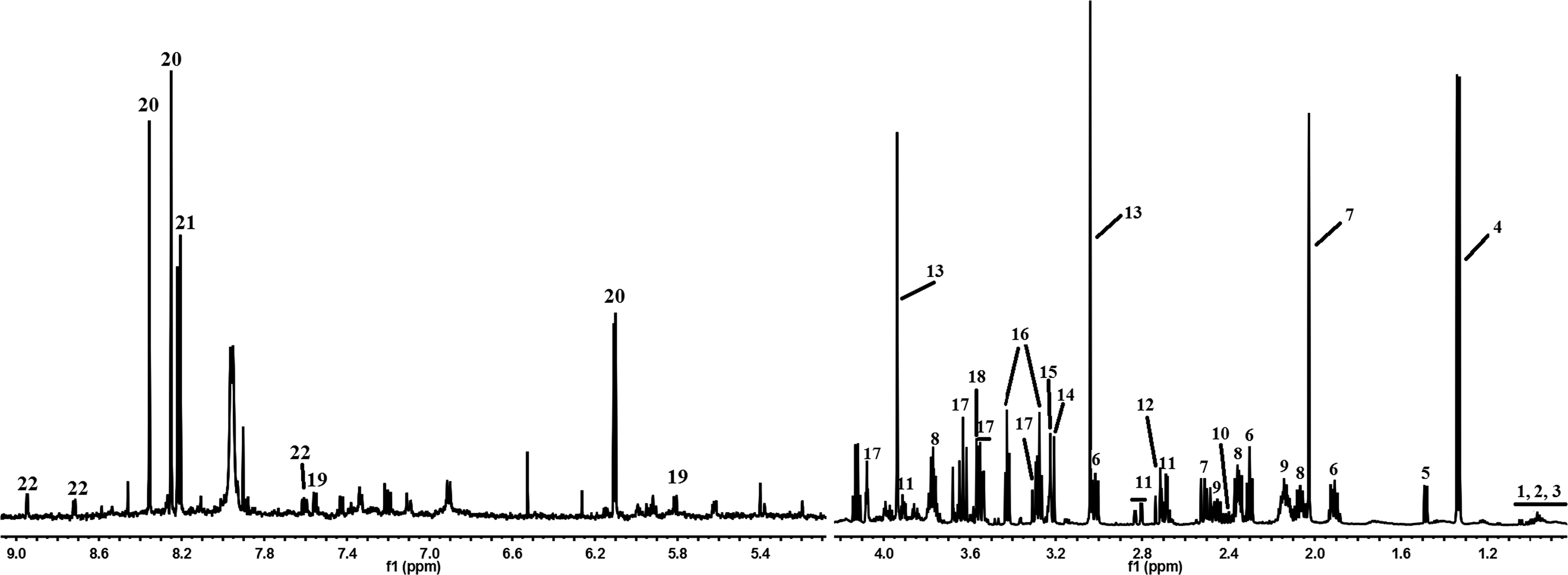
Typical 1H NMR spectrum of cerebral cortex in control rat. NMR, nuclear magnetic resonance.
1
Multivariate data analysis
To obtain the more detailed metabolic differences between the control,

Orthogonal projection to the latent structure with discriminant analysis (OPLS-DA) was further performed to find the differential metabolites between the control and
As shown in Figure 6, the principal component analysis (PCA) score plot showed good separations among the control,
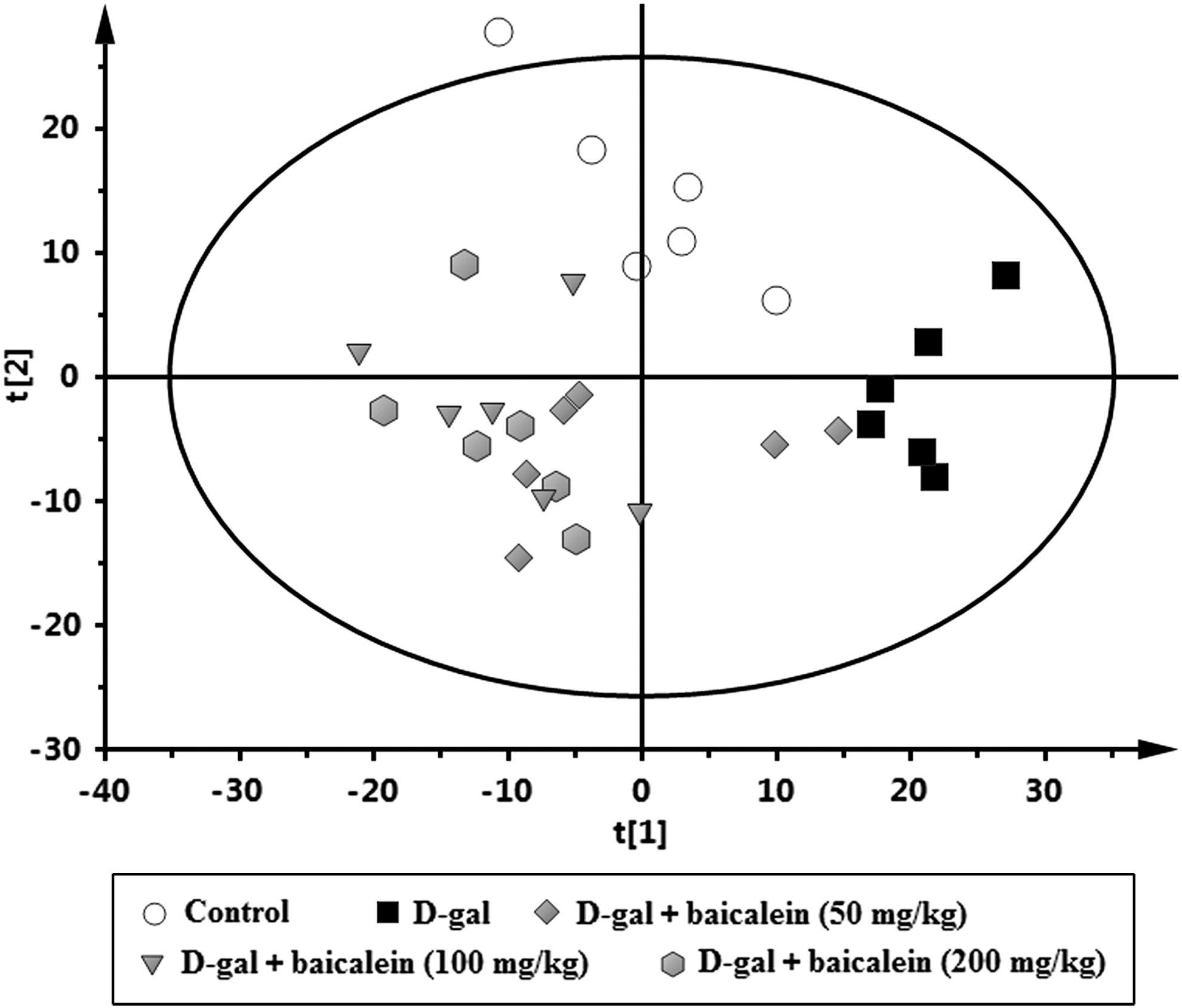
PCA score plot derived from 1H NMR spectra of cerebral cortex from rats in control group,

Comparison on relative peak areas of some potential differential metabolites in rat cerebral cortex. *p < 0.05, **p < 0.01, ***p < 0.001 versus control group. #
p < 0.05, ##
p < 0.01, ###
p < 0.001 versus
Metabolic pathways regulated by baicalein
Metabolites with significant abundance changes between sample groups were subjected to MetaboAnalyst 3.0 as well as the KEGG database (
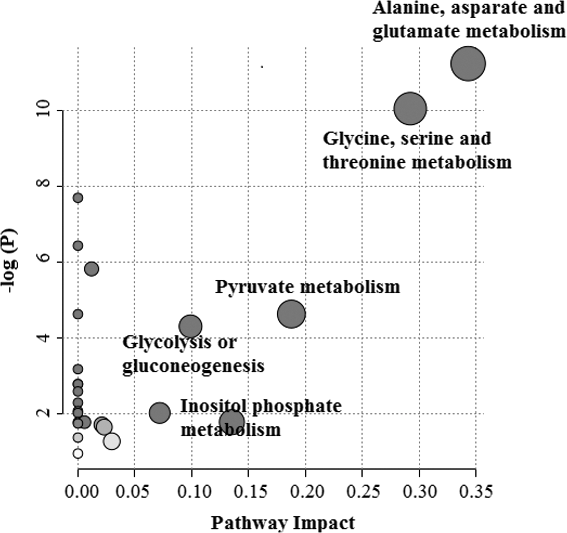
MetPA of metabolic pathway. The “metabolome view” presents all metabolic pathways arranged according to the scores based on enrichment analysis (y axis) and topology analysis (x axis).

Schematic diagram of the perturbed metabolic pathways regulated by baicalein for the beneficial effects in
Discussion
It was reported that caloric restriction or dietary restriction can prolong lifespan and improve the behavioral characteristics in later life of mice.
33,34
Our results demonstrate that the body weight and food consumption have not been significantly altered by baicalein in
In our research, we studied the effects of baicalein on the chronic
NO is a neurotransmitter and a marker of central nervous system inflammation. The higher concentrations of NO are neurotoxic, and it can induce defect in spatial memory,
43
inhibit mitochondrial respiration,
44
and diminish the ability of the cells to deal with oxidative stress.
45
Following
Glu, Gln, NAA, and Asp are involved in alanine, aspartate, and glutamate metabolism. Glu is the most abundant excitatory neurotransmitter in the human cortex, and its synthesis primarily occurs in astrocytes. Glu could potentially trigger excitotoxic cell injury and mitochondrial dysfunction. The high level of Glu is thought to be the primary contributor to cell damage. 45 Gln is synthesized from Glu and is vital in cerebral function. The level of Gln is correlated with cell density, and the reduced concentration might be related to cell loss with aging in cortical areas. 47 NAA is synthesized in the neuronal mitochondria, and is the marker of neuronal injury and cellular dysfunction. 48 NAA is related to neuronal integrity in many disorders, and it is involved in anti-inflammatory activity. 49
The decreases in Gln, NAA, and Asp concentrations were related to neuronal dysfunction and neuronal mitochondria deficit. Consistent with these changes, our results showed that the content of Glu was increased, and the levels of Gln, NAA, and Asp were decreased in
Gly, Cho, Cr, and Pyr participate in glycine, serine, and threonine metabolism. Gly could inhibit oxidative stress, neuroinflammation, and apoptotic neurodegeneration in postnatal rat brain. 50 Cho is an essential nutrient for all cells because it plays a role in the synthesis of the membrane phospholipid components of the cell membranes as well as in the synthesis of neurotransmitter acetylcholine. 51 The decrease of Cho level suggests a decreased turnover of cell membrane that may be related to hypometabolism or dysfunction. 52
Cr is a naturally occurring nitrogenous organic acid, which participates in metabolic reactions within cells and plays an important role in maintaining cellular energy homeostasis. Research has found that Cr has potential neuroprotective effects in various models of neurological disease. Cr supplementation can alleviate cerebral energy depletion, exacerbated oxidative stress and mitochondrial dysfunction in neurodegenerative disorders. 53 Furthermore, Cr supplementation can improve cognitive performance in elderly people 53 and improve memory in vegetarians and omnivores. 54
In accordance to these reports, our data found that the levels of Gly, Cho, and Cr in the
Furthermore, the energy metabolism, including pyruvate metabolism and glycolysis or gluconeogenesis, also might be involved in the beneficial effects of baicalein in
The above results suggest that baicalein exerts beneficial effects in
Conclusion
The behavior examination, biochemical detection, and 1H NMR-based metabolomic approach were combined to investigate the effects and molecular mechanisms of baicalein on
In short, baicalein improves learning and memory dysfunction induced by
Footnotes
Acknowledgments
This project was supported by the National Natural Science Foundation of China (81603319), and partly supported by Programs of Science and Technology and Higher Education of Shanxi Province (No. 2015118), Science and Technology Innovation Team of Shanxi Province (No. 201605D131045-18), and Key Laboratory of Effective Substances Research and Utilization in TCM of Shanxi province (201605D111004).
Author Disclosure Statement
No competing financial interests exist.