




Abstract
In this work, poly (
Introduction
Neuropathic pain arises due to nerve damage, injury, or lesion in nervous system, making this type of pain chronic and challenging to treat upon. 1 Although the exact etiology of this condition is unknown, major causes include any spinal or major nerve injuries, 2 diabetes, 3 cancer or chemotherapy, 4 postsurgical nerve injury, 5 and HIV infection. 6 Neuropathic pain associated with these types of multiple conditions leads to a large number of patients worldwide suffering from chronic pain syndromes.
The current line of treatment for neuropathic pain management uses tricyclic antidepressants and or nonsteroidal anti-inflammatory drugs. Since neuropathic pain is much intense than normal nociceptive pain, various other classes of drugs such as serotonin or norepinephrine uptake inhibitors and anticonvulsants are the choice for this pain management. 7,8 Even combinatorial therapies are given to patients in various combinations. Repetitive doses of these drugs result in higher peripheral organ toxicity.
Neuropathic pain is associated with multiple diseases, which are very common in elderly patients. Under such conditions, it is difficult to achieve prolonged pain relief as patients are already under certain treatment protocols and effective pain treatment is not achieved without multiple side effects. Under such conditions, nanoparticle-based drug delivery of antispasmodic agents such as baclofen (Bcf) for pain management could be helpful. The reduced doses achieved by Bcf nanoparticles could be helpful as drug therapy for treatment of various associated conditions such as painful diabetic neuropathy, pain associated with multiple sclerosis, poststroke syndromes, and postherpetic neuralgia commonly experienced with increasing age or in elderly patients. 9
Bcf is a GABA derivative, which is used as a muscle relaxant and central nervous system (CNS) depressant and offers positive results for strong opioid-resistant chronic pain conditions. Bcf suppresses the effect of excitatory neurotransmitters such as aspartate and glutamate, thus providing relief in painful conditions such as trigeminal neuralgia, multiple sclerosis, and severe spasticity. 10,11 Currently, patients taking this drug have to deal with multiple problems such as repetitive daily dosages and multiple side effects because of peripheral interactions such as neuropsychiatric problems, hypertension, gastrointestinal, and genitourinary problems.
Bcf is a structural analogue of an important neurotransmitter γ-aminobutyric acid and shows inhibitory effects on (GABA)-B receptors primarily found in CNS. It is reported to have rapid absorption in blood because of its hydrophilic nature (70%–80% oral bioavailability). 12 However, conventional oral administration of Bcf has a short half-life of around 3–4 hours that leads to high maintenance doses, which are associated with multiple side effects. 13 Currently, there is a limitation of short acting and instant relief for patients suffering from severe or chronic pain conditions limited to invasive intravenous (i.v.) approach. Intranasal (i.n.) delivery of Bcf provides transmucosal absorption directly to brain by bypassing the blood–brain barrier, providing a convenient option of administration, early and rapid onset of action, and bypassing hepatic presystemic elimination. Nanoparticles administered through the i.n. route can be absorbed through intracellular or paracellular pathways and transported to brain sites. 14 –18
In this work, Bcf-loaded PLGA nanoparticles for i.n. delivery were developed. The nanoparticles were prepared by the modified nanoprecipitation method and further characterized for particle size, polydispersity index (PDI), and zeta potential. Drug release profiles were studied in simulated media such as phosphate-buffered saline (PBS, pH 7.4) and simulated nasal fluid (SNF). In vitro cytotoxicity was evaluated on Neuro-2a cell line. As it is important to study the path of drug upon i.n. administration, in vivo experiments were planned through radiolabeling of Bcf so that upon administration the drug path can be observed using a gamma camera. Furthermore, to ascertain the amount of drug reaching the brain vis a vis blood following i.n. administration, biodistribution studies of Bcf-PLGA-NPs were conducted in Sprague Dawley rats to assess the distribution of Bcf to brain and blood.
Materials and Methods
Materials
Bcf was a generous gift from SNA Healthcare Pvt. Ltd., Mumbai, India. Poly (
Development and optimization of nanoparticles
Bcf-loaded PLGA nanoparticles were prepared using the nanoprecipitation method. In brief, PLGA was accurately weighed and dissolved in acetone as organic phase. The organic phase was added drop wise (1 mL/min) to aqueous phase (containing poloxamer 407 as surfactant and Bcf) under continuous stirring. This colloidal suspension was homogenized using high-speed homogenizer (Tissue Master, Omni International, Georgia) at 10,000 rpm for 10 minutes. Furthermore, the suspension was ultrasonicated using ultrasonic probe sonication (Model UP400S, Hielcher, Ultrasound Technology, Germany) at an amplitude of 40% for 150 seconds with 10 seconds on–off cycle. The nanoparticle suspension was then left on continuous stirring of 300 rpm for 3 hours for evaporating the organic phase. The prepared Bcf-PLGA-NPs were then centrifuged at 12,000 rpm at 4°C for 15 minutes to obtain nanoparticle pellets and washed twice using deionized water to remove any traces of unentrapped drug. The final Bcf-PLGA-NPs were finally dissolved in water for further analysis. 19,20
UV method development and RP-HPLC analysis of Bcf
For spectrophotometric determination of Bcf, double beam spectrophotometer (Shimadzu UV-Visible, UV-1800) was used that was equipped with UV-Probe Software. In brief, for standard plot preparation, Bcf was dissolved in distilled water (1 mg/mL) and was diluted to a concentration range of 2–20 μg/mL and absorbance was recorded at 220 nm. 21 Reversed phased HPLC method was developed using the isocratic RP-HPLC system (Waters, Vienna, Austria) equipped with nonpolar C-18 column (Sunfire 250 × 4.6 mm, 5 μm) with constant mobile phase of methanol–water (53:47 v/v) with a flow rate of 1 mL/min and injection volume of 20 μL. The peak analyses were done using UV-visible detector at wavelength of 228 nm. Bcf standard curve was developed by preparing the stock solution in mobile phase (1 mg/mL). 22
Encapsulation efficiency and drug loading
Bcf-loaded PLGA nanoparticles suspension was centrifuged at 12,000 rpm at 4°C (Remi, Mumbai, India) for 20 minutes and washed twice using HPLC water and supernatant was collected in a different vial. For determining the amount of free or unentrapped drug present in the supernatant solution, encapsulation efficiency and drug loading on nanoparticles were calculated using the following equations 23 :
Particle size and zeta potential
Average particle size of developed Bcf-PLGA-NPs was determined using Malvern Zetasizer (Malvern, United Kingdom), which works on the principle of dynamic light scattering. The prepared nanoparticles were mixed in the ratio of 1:50 v/v with HPLC water. PDI values were determined to know the homogeneity of prepared drug-loaded nanoparticles. PDI values with higher range show the mass aggregates of nanoparticles of variable ranges, thus leading to nonuniformity between the sample and poor stability of prepared nanoparticles. Similarly, zeta potential was also determined with similar dilution ratio for determining surface charge of developed particles. 24
In vitro permeation studies
In vitro permeation studies were carried out to study the release profile of freshly prepared Bcf-loaded PLGA nanoparticles. A known amount of Bcf-PLGA-NPs was loaded in a pre-treated dialysis membrane (cutoff 12,000 Da, Sigma-Aldrich) and sealed from both ends. The complete study was conducted in USP Type II apparatus (Veego, Mumbai, India) and drug release was studied on methanolic PBS (pH 7.4), simulated cerebrospinal fluid (CSF), and simulated SNF at 75 rpm. It is likely that after nasal administration, drug goes both to brain (through olfactory region) and to the systemic circulation (through respiratory region); therefore, permeation studies were conducted in various simulated media to mimic various body systems where PBS (pH 7.4) helps the distribution of prepared nanoparticles in systemic circulation as studied later in in vivo studies. In addition, release profile in simulated SNF helped in understanding the release of drug in nasal cavity and simulated CSF helped in understanding release of drug in brain. Sample aliquots of 2 mL were taken periodically and fresh dissolution medium of equal volume was replaced to maintain sink conditions. Bcf estimation was done using the spectrophotometric method developed as discussed in the previous sections.
In vitro cytotoxicity studies
In vitro cytotoxicity analysis was carried out using MTT (3-(4,5-dimethylthiazol-2-yl)-2,5-diphenyltetrazolium bromide) assay to assess the percentage cell viability on Neuro-2a cell line (neuroblastoma cell line). The cells were maintained using DMEM (Sigma-Aldrich) supplemented with 10% FBS at 5% CO2 at 37°C. Cells (1 × 10
4
) were initially seeded for around 24 hours followed by supplementation with aqueous Bcf drug, Bcf-PLGA-NPs, and the corresponding placebo. The developed formazan crystals were dissolved in dimethyl sulfoxide (DMSO) and quantified using scanning multiwall spectrophotometer at 570 nm.
25,26
The percentage viability was calculated using the formula
Radiolabeling of Bcf and Bcf-loaded nanoparticles
For radiolabeling of Bcf, 99mTc-pertechnate (99mTc) was used. Bcf was radiolabeled using the direct labeling method using stannous chloride (2 mg/mL in ethanol) as reducing agent followed by 99mTc-pertechnetate (200 μL, 5 mci) upon gradual mixing. The resultant mixture was incubated at room temperature for 30 minutes and volume was made up to 2 mL using normal saline (0.90% w/v sodium chloride). After incubation, the drug tagging was evaluated using thin layer chromatography using silica gel-coated sheets (Gelman Sciences, Inc., Ann Arbor, MI) and acetone as mobile phase. The reaction time and concentration of SnCl2 were optimized to achieve the final working concentrations. 27 The optimized conditions were further used for radiolabeling of Bcf-PLGA-NPs and stability studies were additionally performed on normal saline and rat blood plasma 28 before using for scintigraphy and biodistribution studies. Radiolabeling efficiency was calculated using the following equation. 29
Gamma scintigraphy
In vivo experiments were carried out at INMAS, DRDO, Delhi, India, upon proper ethical clearance obtained by Institutional Animal Ethics Committee (IAEC, New Delhi, IAEC vide number INM/IAEC/16/10) and their guidelines were followed throughout the study. Sprague Dawley rats (3–4 months old, 180–200 g) were used for the entire in vivo experiments. Animals were maintained in Central Animal House Facility, INMAS, Delhi, at room temperature (25°C ± 5°C). Animals were divided into five groups with three animals each for all the time points (0.5, 1.5, 3, 6, and 24 hours). Ketamine hydrochloride (50 mg/mL) was used for anesthetizing through intraperitonial injection (0.4 mL). Radiolabeled formulation (99mTc-Bcf-PLGA-NPs) was delivered orally, intranasally, and intravenously, whereas radiolabeled aqueous drug (99mTc-Bcf) was delivered intravenously and intranasally. Low-density polyethylene tube catheter (diameter 0.1 mm) was used for i.n. delivery of nanoparticles. Periodic images (0.5, 1.5, 3, 6, and 24 hours) were taken by placing anesthetized rats on imaging platform under single photon emission CT gamma camera (SPECT, LC 75-005, Siemens, Germany). 27
Group 1: 99mTc-Bcf-aqueous (i.v.)
Group 2: 99mTc-Bcf-PLGA-NPs (i.v.)
Group 3: 99mTc-Bcf-PLGA-NPs (oral)
Group 4: 99mTc-Bcf-PLGA-NPs (i.n.)
Group 5: 99mTc-Bcf-aqueous (i.n.)
Each group contains three animals for each time point.
Biodistribution studies
Biodistribution studies were also carried out using Sprague Dawley rats (3–4 months old, 180–200g). Animals were divided into four groups with three animals each for all the time points (0.5, 1.5, 3, 6, and 24 hours). Rats were anesthetized using intraperitoneal injections of ketamine hydrochloride (50 mg/mL). Radiolabeled formulation (99mTc-Bcf-PLGA-NPs) and radiolabeled aqueous drug (99mTc-Bcf) were delivered intranasally and intravenously. Blood samples were collected for various time points by puncturing of retro-orbital vein followed by sacrificing of rats by spinal dislocation. Brain was then dissected out, washed using normal saline to remove any unwanted tissues or fluid, and properly weighed. Radioactivity present in brain tissues was measured using shielded well-type gamma counter (SPECT; LC 75–005, Diacam, Siemens AG; Erlanger, Germany). Furthermore, percentage radioactivity per gram of brain tissue, DTE %, and DTP % were calculated using the following equations 27,30,31
For assessing various pharmacokinetic parameters, drug targeting efficiency (DTE %) and direct transport percentage (DTP %) were calculated using the following equations:
where Bx is the AUC of brain through i.n. administration, Bi.v. is AUC(0–24) of brain through i.v. administration, Pi.v. is AUC(0–24h) of blood through i.v. administration, Bi.n. is AUC(0–24h) of brain through i.n. administration, and Pi.n. is AUC(0–24h) of blood through i.n. administration. Group 1: 99mTc-Bcf-PLGA-NPs (i.n.), Group 2: 99mTc-Bcf aq. (i.n.),Group 3: 99mTc-Bcf-PLGA-NPs (i.v.), and Group 3: 99mTc-Bcf aq. (i.v.).
Each group contains three animals for each time point.
Results and Discussion
Preparation and characterization of Bcf-PLGA-NPs
Bcf-loaded PLGA nanoparticles were prepared using nanoprecipitation method followed by homogenization and ultrasonication. The developed particles were centrifuged and pelleted particles were suspended in water for further analysis. Entrapment efficiency was calculated to know the percentage amount of drug that is entrapped in the form of nanoparticles. It was observed that on preparing Bcf-PLGA-NPs, around 86.45% ± 1.65% entrapment efficiency was achieved and drug loading was observed to be 9.48% ± 0.7%. Particle size of prepared Bcf-PLGA-NPs was observed to be 124.8 (d.nm), showing the prepared particles to be of nanometric range. For a drug nanoparticle, smaller size enhances its uptake through nasal mucosa and would lead to higher availability of drug in brain and thus reduces the side effects arising due to peripheral circulation of drugs. Furthermore, polydispersity index of prepared particles was observed to be 0.225, implying homogeneity among particles (Figs. 1 and 2). The range of PDI varies from 0 (monodispersed system) to 1 (polydispersed system). Another important parameter is zeta potential, which depicts the charge and stability of prepared nanoparticles. It has been widely reported that higher value of surface charge confers reduced aggregation of particles. 32 Zeta potential was observed to be −20.4 mV, where the negative charge is imparted because of PLGA polymer (Figs. 3 and 4).
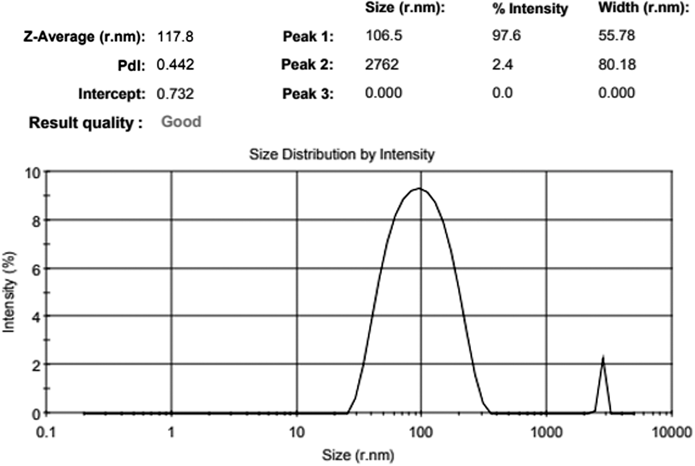
Particle size distribution of PLGA-NPs. PLGA-NP, poly (
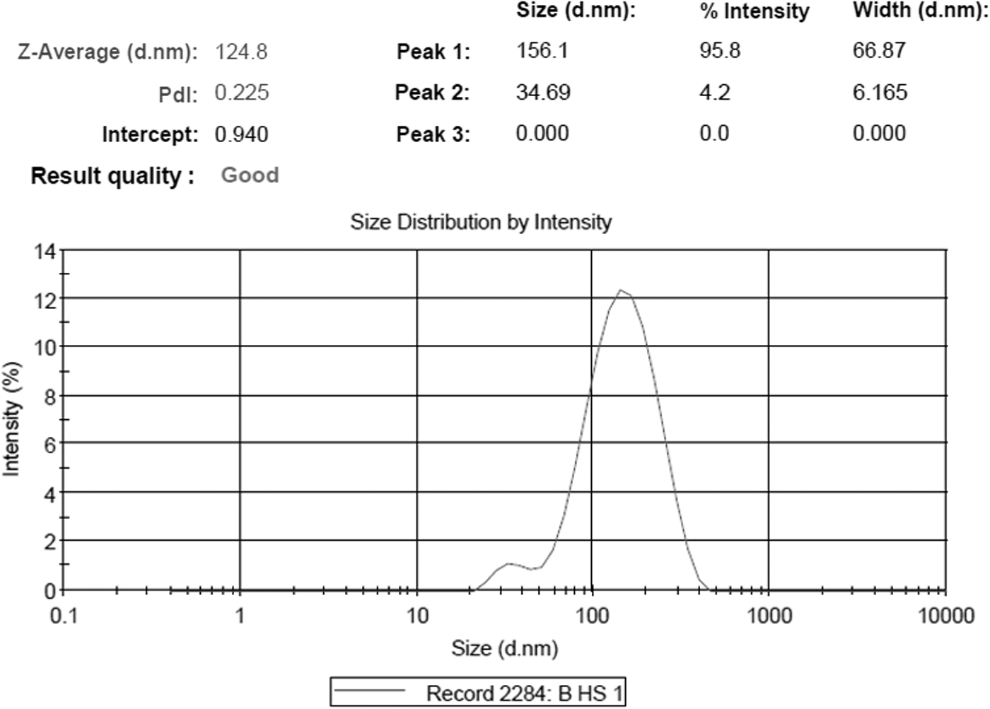
Particle size distribution of Bcf-PLGA-NPs. Bcf, baclofen.
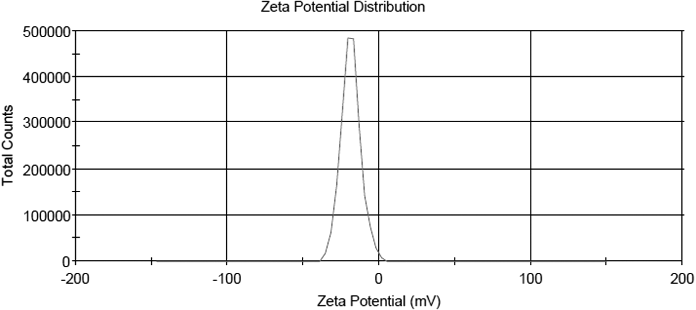
Zeta potential of PLGA-NPs.

Zeta potential of Bcf-PLGA-NPs.
In vitro permeation studies
In vitro release for Bcf-PLGA-NPs was done through dialysis membrane bags using methanolic PBS (pH 7.4), simulated CSF, and SNF. It was observed that within 30 min, around 38% drug release was seen in PBS, whereas very low initial release profile was seen in SNF. In PBS (pH 7.4), around 50% release was achieved within 2.5–3 hours, which increased to around 90% within 24 hours. On the contrary, comparably slow release was seen in SNF, achieving around 40% release up to 5 hours, which increased to around 73% in 24 hours (Fig. 5). Similarly, a very slow initial release was seen in CSF with around 26% release in initial 5 hours, which, however, increased to 98% in 24 hours. The initial drug release seen in Bcf-PLGA-NPs could be because of surface-adsorbed drug particles on nanoparticle surfaces, which were desorbed in the methanolic PBS faster than SNF, this could be due to high solubility of Bcf in PBS. 33,34 The rate of drug release reduced after initial release because of slow diffusion of drug from the PLGA polymer matrix, which slowly diffuses out in the dissolution media. This leads to a controlled release pattern with gradual release of Bcf throughout the 24 hours period. 20,35
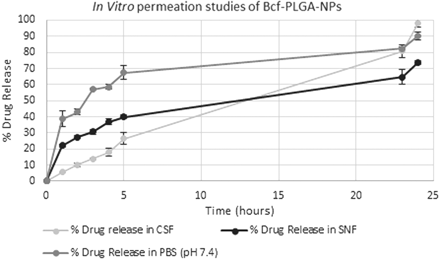
In vitro release of Bcf-PLGA-NPs in PBS (pH 7.4) and SNF. PBS, phosphate-buffered saline; SNF, simulated nasal fluid.
To assess the mechanism of drug release from the PLGA nanoparticle matrix, various kinetic models were applied: zero order release, first order release, Higuchi model, and Korsmeyer–Peppas model. The R2 values, that is, the coefficient of correlation was determined for all the models 36 (Table 1). It was concluded that the Bcf-PLGA-NPs followed Higuchi and Korsmeyer–Peppas model in all the simulated media (where R 2 values were exceeding 0.9). Korsmeyer–Peppas model suggested a two-step release profile (which corresponds to the profiles seen in Fig. 5 as well), where there is initial swelling of polymeric matrix followed by release or diffusion of the drug. Based upon the release component (n value) in Korsmeyer–Peppas model, it was seen that in PBS and SNF, non-Fickian diffusion occurred, whereas in CSF, Fickian diffusion occurred. Higuchi release profiles suggest that a homogenously dispersed Bcf in polymeric matrix is diffused out without any erosion of matrix system. These release systems suggested a constant controlled release of drug from nanoparticles. 37,38
R 2 Values for Various Simulated Media for Model Selection
CSF, cerebrospinal fluid; PBS, phosphate-buffered saline; SNF, simulated nasal fluid.
In vitro cytotoxicity studies
In vitro cytotoxicity of Bcf-PLGA-NPs, Bcf aqueous drug solution, and the corresponding placebo nanoparticles was evaluated using MTT assay on Neuro-2a neuroblastoma cell line. The chosen concentration range of Bcf from 0.625–50 μg/mL covered the reported Cmax value of 737.6 ng/mL. The cytotoxicity was examined for various drug concentration levels (Cmax, 2 × Cmax, 4 × Cmax, and 40 × Cmax). Neuro-2a cells exhibited dose-dependent cytotoxicity, nevertheless, ∼75% cells were viable upon treatment with the highest concentration of aqueous drug and prepared nanoparticles. Bcf-PLGA-NPs showed higher cell viability (∼90%–100%) on N2a cells than Bcf aqueous suspension (81%–90.77%) for the respective concentration range of 25–0.625 μg/mL. Bcf-PLGA-NPs elicited >90% cell viability on mammalian cells at approximate four times Cmax value and reached almost 100% cell viability at near twice of Cmax value (Fig. 6). These low-toxicity values in placebo and in formulations suggest PLGA as suitable carrier system for Bcf nanoparticles.

In vitro cytotoxicity assay of prepared nanoparticles with corresponding placebo and aqueous drug.
In vivo studies
Bcf was radiolabeled using 99mTc as radiolabeled isotope by direct labeling method as already discussed above. Stannous chloride (dihydrated) was used as a reducing agent and the samples were incubated for 30 minutes at room temperature. The percentage of radiolabeling efficiency was observed through instant thin layer chromatography and found to be 96.61% ± 0.8%. After radiolabeling, the radiolabeled drug was used for the preparation of PLGA nanoparticles for their administration to the animals. To check the in vitro stability of radiolabeled NPs, radiolabeled formulation was mixed with blood serum and normal saline stability was observed for 24 hours and the radiolabeled conjugates showed fairly good radiolabeling efficiency of 94.94% ± 1.2%, which was seen to be well correlated with the observed percentage radiolabeling efficiency (96.61% ± 0.8%). To explore the nose to brain drug uptake pathway, gamma images of Sprague Dawley rats at various time points (0.5, 1.5, 3, 6, and 24 hours) were taken after oral, i.n., and i.v. administration of 99mTc-Bcf-PLGA-NPs. The aqueous drug solution of radiolabeled Bcf (99mTc-Bcf-Aq) was also administered through i.v. and i.n. route to compare the scintigrams. From the scintigrams obtained, it was observed that there was slight distribution of drug in the brain of rats administered with 99mTc-Bcf-PLGA-NPs i.v. and oral route (Fig. 7b, c). Whereas the images of rats administered with 99mTc-Bcf-PLGA-NPs i.n. showed maximum distribution of drug in the brain (Fig. 8a) as compared with that in peripheral organs. The scintigram images clearly showed that maximum distribution was seen in the target organ at 3 hours and remained there for 24 hours post-administration of radiolabeled NPs through the i.n. route. The radiolabeled formulation administered through the i.v. route also showed higher uptake in brain at 3 hours (Fig. 7b) than the 99mTc-Bcf-aqueous solution administered group in which very low intensity was observed in brain, and majority of drug was distributed in abdomen and tail area (Fig. 7a). On the contrary, very slight distribution was seen in the brain of rats administered with 99mTc-Bcf-PLGA-NPs orally (Fig. 7c). The maximum uptake of radiolabeled NPs in the brain through the i.n. route could be due to polymer matrix formed by PLGA nanoparticles, which would act as a reservoir for drug 39 and direct administration of drug through olfactory lobe bypassing blood–brain barrier. 40,41 However, the minimum distribution of the 99mTc-Bcf-aqueous in the brain may be due to the hydrophilic nature of the Bcf and its majority absorption through blood vessels and distributed through systemic circulation. In the case of orally administered radiolabeled formulation, blood–brain barrier acts as a major hurdle in the route of distribution in the brain. 42,43 Gamma scintigrams gave a preliminary picture about the distribution patterns of nanoparticles and aqueous drug through different routes. Further biodistribution studies were carried out to know the exact levels of drug and drug-loaded nanoparticles in the brain.

Gamma scintigraphy image of

Gamma scintigraphy image of
Biodistribution studies were performed on Sprague Dawley rats after administration of 99mTc-Bcf-aqueous and 99mTc-Bcf-PLGA-NPs through i.n. and i.v. routes and percentage radioactivity per gram of tissue was calculated for brain and blood samples at different time points up to 24 hours (Table 2). The maximum percentage radioactivity (3.5% ± 0.14%) was seen at 3 hours (Fig. 9) in brains of rats administered with 99mTc-Bcf-PLGA-NPs through the i.n. route followed by (3.08% ± 0.19%) of radioactivity in brains of rats administered with 99mTc-Bcf-PLGA-NPs through the i.v. route. In the tissue samples of rats administered with 99mTc-Bcf-aqueous, the levels were quite low in brain with 2% ± 0.12% through the i.n. route and 1.5% ± 0.17% through the i.v. route. It was also seen that within 1.5 hours, 99mTc-Bcf-PLGA-NP levels spike in the brain (2.8% ± 0.19%), suggesting possibility of early onset of action. Similarly, percentage radioactivity levels were also checked in blood samples at different time intervals from 0.5 to 24 hours (Fig. 10). From the results, maximum radioactivity efficiency was observed in the rats at 1.5 hours (3.68% ± 0.22%) administered with 99mTc-Bcf-PLGA-NPs through i.v. followed by 99mTc-Bcf-aqueous (3.2% ± 0.19%) through i.v. This early and high concentration was expected because of hydrophilic nature of Bcf drug. However, upon i.n. administration, the maximum levels were achieved by 3 hours for both 99mTc-Bcf-PLGA-NPs (2.4% ± 0.13%) and 99mTc-Bcf-aqueous (1.5% ± 0.21%) as they were slowly absorbed to the blood stream.
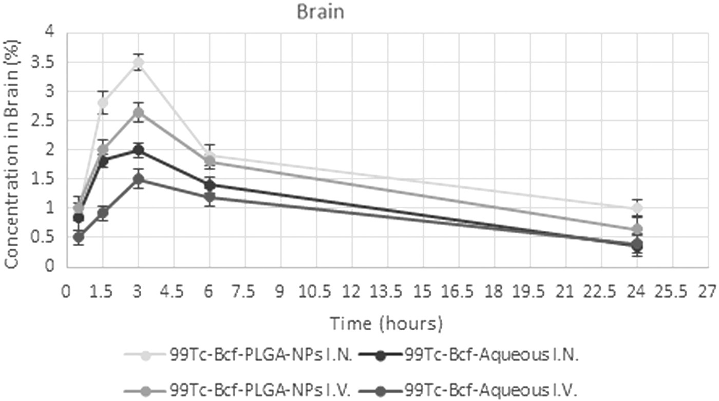
Drug concentration in brain samples of rat showing 99mTc-Bcf-PLGA-NPs and 99mTc-Bcf-aqueous (intravenous and intranasal).
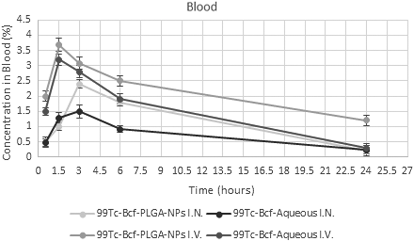
Drug concentration in blood samples of rat showing 99mTc-Bcf-PLGA-NPs and 99mTc-Bcf-aqueous (intravenous and intranasal).
Distribution of 99Tc-Bcf-PLGA-NPs and 99Tc-Bcf-Aqueous (Intranasal and Intravenous) in Sprague Dawley Rats in Percentage per Gram
Pharmacokinetic parameters were evaluated and Cmax (3.5%/g) at 3 hours for 99Tc-Bcf-PLGA-NPs administered i.n. was found higher in brain than Cmax (2.65%/g) at 3 hours administered i.v. (Table 3). The area under curve (AUC) was also calculated and AUCbrain (41%.h/g) of rats administered with 99mTc-Bcf-PLGA-NPs intranasally was observed significantly higher than AUCbrain (33.52%.h/g) of rats administered with 99mTc-Bcf-PLGA-NPs i.v. A similar profile of Cmax (2.4%/g) was seen in blood too in the rats administered with 99mTc-Bcf-PLGA-NPs intranasally with AUC0–24h of 33.12%.h/g, which was significantly higher than Cmax (1.5%/g) and AUCblood (17.13%.h/g) of rats administered with 99mTc-Bcf-aqueous drug solution. These lower levels seen in aqueous form are because of poor absorption of hydrophilic drug in the brain. On the contrary, the i.n. route bypasses the blood–brain barrier reaching directly to site of action. 44,45 In addition, the direct nose to brain transport was observed by calculating DTE% and DTP% for 99mTc-Bcf-PLGA-NPs as 183.85% and 45.92%, respectively, for 99mTc-Bcf-PLGA-NPs administered intranasally (Table 4). The DTE% and DTP% showed that the developed nanoparticles were able to reach the target organ (brain) efficiently. The higher DTE% (>100%) showed that upon i.n. application, the 99mTc-Bcf-PLGA-NPs were accumulated in brain region (target organ) as compared with that upon i.v. administration. This higher uptake through i.n. route could be because of bypassing blood–brain barrier. Our results were in agreement with the gamma scintigrams obtained at different time points and were significant with p value <0.05. From the observed results, it was suggestive that i.n. route of administration of PLGA nanoparticles loaded with Bcf enhances the absorption and distribution of drug at target site through bypassing the blood–brain barrier.
Pharmacokinetics of 99mTc-Bcf-PLGA-NPs and 99mTc-Bcf-Aqueous (Intranasal and Intravenous) in Sprague Dawley Rats
Drug Targeting Efficiency and Direct Target Organ Transport After Intranasal Administration of 99mTc-Bcf-PLGA-NPs
The blood and brain levels helped in understanding the amount and rate of permeation of prepared Bcf nanoparticles to brain and to systemic circulation. It is widely accepted to understand these levels in rodent models so as to establish the brain–plasma concentration, the actual amount of uptake in brain when administered through various routes (such as i.v. and i.n.).
Conclusion
In this work, Bcf-loaded PLGA nanoparticles were developed using the nanoprecipitation method followed by high-speed homogenization and ultrasonication. Developed nanoparticles were of nanometric range and their in vitro release profiles were studied. In PBS, there was an immediate burst release within 30 minutes, whereas in SNF, the release was quite slow. In in vitro cytotoxicity assay, dose-dependent cytotoxicity was seen on Neuro-2a cell line where prepared formulation (Bcf-PLGA-NPs) showed comparative lower toxicity and higher cell viability. In vivo studies involving gamma scintigraphy showed prolonged retention of Bcf-PLGA-NPs in brain unlike aqueous drugs, which were confirmed by biodistribution studies suggesting PLGA as suitable carrier of Bcf in the form of nanoparticles for combating neuropathic pain conditions.
Footnotes
Acknowledgments
The authors thank Jaypee Institute of Information Technology, Noida, and INMAS, Delhi, for providing basic infrastructural support to carry out the project. The authors thank Dr. A. Panda, National Institute of Immunology, Delhi, and Dr. M. Kalia, THSTI, Faridabad, for providing required resources for completion of this work.
Author Disclosure Statement
No competing financial interests exist.