




Abstract
The incidence of type 2 diabetes mellitus (T2DM) has been increasing in recent years and has become a serious threat to human health. Zengye Decoction (ZYD), a well-known traditional Chinese medicinal formula, has been used in the treatment of T2DM with yin asthenia and extreme heat since Qing Dynasty. However, the characteristics of antidiabetic activities of ZYD have not been fully elucidated. In our study, high-fat diet and streptozotocin were used to establish the T2DM rat model. After 3 weeks of treatment with ZYD, the fasting blood glucose (FBG), oral glucose tolerance, the fasting serum insulin concentration, insulin sensitivity index (ISI), serum lipid profiles, and pancreas histopathology were measured. Then, under circumstance of insulin-resistant glucose consumption, 2-(N-(7-nitrobenz-2-oxa-l,3-diazol-4-yl) amino)-2-deoxyglucose (2-NBDG) uptake and glycogen content in C2C12 myotubes, 3T3-L1 adipocytes, and HepG2 cells were determined, respectively. Finally, the expressions of key targets in the insulin signaling pathway were measured to explain the potential mechanism underlying these activities. After administration with ZYD, a notable reduction in FBG levels, oral glucose tolerance test-area under the curve, blood lipid metabolism, and ISI values were observed compared with the diabetic control group. Moreover, ZYD restored the damaged islet cells in T2DM rats. Significant increases in glucose consumption, glucose uptake, glycogen content, expression of glucose transporter type 4, and the ratio of p-Akt/Akt were observed in the ZYD groups. According to the above results, ZYD exhibited glucose disposal, including glucose consumption, glucose uptake, and glycogen content and promoted the Akt signal pathway, which indicates that ZYD exerts significant hypoglycemic effect in T2DM.
Introduction
Diabetes mellitus is one of the most common metabolic disorders in the world. Type 2 diabetes mellitus (T2DM) is the primary type of diabetes, and according to epidemiological surveys, the prevalence of T2DM is increasing dramatically. 1 The number of T2DM patients is estimated to increase to 642 million over the next 20 years, 2,3 and T2DM has gradually become one of the ten leading causes of death and the world's most expensive chronic disease. 4 Under these circumstances, it is crucial to put more effort into developing antidiabetic drugs that are safer and more effective.
T2DM is a heterogeneous disorder characterized by insulin resistance (IR), relative pancreatic beta cell dysfunction, hyperglycemia, dyslipidemia, and uncontrollable glucose and protein metabolism. 1,5 T2DM leads to many macrovascular, microvascular, and neurological complications, including cardiac dysfunction, atherosclerosis, nephropathy, diabetic foot, and neuropathy. 6 Therefore, it is crucial to discover new drugs to alleviate T2DM and its complications. Drugs currently available for the treatment of T2DM include biguanide, thiazolidinedione, sulfonylurea, α-glycosidase inhibitors, and insulin. 7 However, the use of these drugs is limited due to several rather large side effects. The success of metformin, the current first-line drug for the treatment of T2DM, which is isolated from extracts of Galega officinalis, 8 motivated the search for other hypoglycemic Traditional Chinese Medicines (TCMs).
The regulation of glucose transport into adipose tissue, skeletal muscle, and the liver is essential for maintaining systemic glucose homeostasis. 9 Liver helps to maintain blood glucose levels through gluconeogenesis, glycogenolysis, and glycogen synthesis. 10 Aside from the regulation of glucose in liver, glucose uptake by muscle and adipose tissue also significantly contributes to the insulin-dependent reduction in circulating glucose levels. In humans, ∼75% of whole-body glucose is uptake by skeletal muscle and ultimately stored as glycogen in muscle. 11 Therefore, it is essential to investigate glucose disposal in skeletal muscle and adipose tissue.
The insulin signaling pathway is thought to be the primary mechanism involved in the regulation of blood glucose metabolism. First, glucose in the blood can be absorbed into cells by glucose transporters such as glucose transporter type 4 (GLUT4), which is an insulin-sensitive glucose transporter and is mainly distributed in adipose tissue, skeletal muscle, and myocardial tissue. Abnormality in GLUT4, the main transporter of glucose in the signal transduction pathway, is considered the crucial cause of IR. 6 Akt is an important target protein involved in the insulin signaling pathway, which is activated by phosphorylation as a regulator of glucose transport, glycolysis, protein synthesis, lipogenesis, glycogen synthesis, and suppression of gluconeogenesis. 10 In addition, Akt directly regulates the GLUT4 vesicles in cytoplasm translocated to the plasma membrane and thus promotes glucose transport. 12
TCMs and its formula have been used in China for thousands of years and have recently attracted increasing attention. 13 Zengye Decoction (ZYD), a well-known formula, consists of three yin-nourishing herbs as follows: Radix Scrophulariae (Xuanshen), Radix Rehmanniae (Shengdi), and Radix Ophiopogonis (Maidong) and was first recorded in WenBingTiaoBian (China's Qing Dynasty, written by Ju-Tong Wu).
The monarch drug Xuanshen, which has numbers of constituents, including catalpol, acteoside, angoroside C, cinnamic acid, harpagide, and harpagoside, possesses various bioactivities, including antichronic inflammatory, antihypertensive, abirritative, antispasmodic, antihepatitis B virus, and immunological enhancement. 14 The minister drug Maidong mainly contains Ophiopojaponin and homoisoflavones and possesses various bioactivities, including antimyocardial ischemia, hypoglycemia, and antiaging. 15 The assistant and messenger drug Shengdi mainly contains iridoid glycosides and phenethyl alcohol glycosides, which have various bioactivities, including alleviating endothelial dysfunction, anti-inflammatory, antidiabetic, and so on. 16
ZYD has long been used for treating constipation and treating diseases related to “Yin deficiency.” In modern clinical practice, ZYD is used to treat type 2 diabetes with yin asthenia and extreme heat.
In recent years, several studies have been performed on ZYD. A hypoglycemic effect and potential active constituents were confirmed in ZYD in mice with T2DM. 15 Liu reported that ZYD could markedly upregulate the p-AMPKα levels and p-AMPKα/AMPKα ratios and promote glucose consumption and glucose uptake and decrease the lipid content in IR-HepG2 cells, and the mechanisms may be associated with AMPK activation. 17 However, the regulation of blood glucose concentration is affected by many factors, such as glycogen synthesis, not just glucose uptake in the liver. In addition, other target organs such as skeletal muscle and adipose tissue are also involved in the regulation of blood glucose.
Until now, the mechanisms of ZYD have not been fully elucidated. Therefore, in this study, we aimed to explore the regulation of glucose in skeletal muscle and adipose tissue by ZYD and the mechanism of improving IR, to learn more about ZYD and provide scientific evidence for the beneficial actions and traditional use of ZYD in the treatment of T2DM.
In this study, the antidiabetic effect of ZYD was evaluated, including its hypoglycemic, antihyperlipidemic, and pancreatic protective effects in high-fat diet (HFD)-fed and streptozotocin (STZ)-induced T2DM rats. ZYD was found to improve glucose consumption, glycogen synthesis, and glucose uptake in HepG2 cells, C2C12 myotubes, and 3T3-L1 adipocytes, respectively, as well as promote Akt activation and GLUT4 expression, which will lay a foundation for research into antidiabetic mechanisms and further development of T2DM drugs.
Materials and Methods
Chemicals and reagents
STZ was purchased from Sigma (St. Louis, MO). Metformin and rosiglitazone were obtained from Aladdin (Shanghai, China). Insulin was from Novo Nordisk (Bagsvaerd, Denmark). Fetal bovine serum (FBS) and Dulbecco's modified Eagle's medium (DMEM) were purchased from Gibco (California, CA). 2-NBD-glucose (2-NBDG) was purchased from Invitrogen (Eugene, OR).
Preparation of ZYD extract
The crude drugs of Radix Scrophulariae, Radix Ophiopogonis, and Radix Rehmannia were purchased from Nanjing TCM Clinic (Nanjing, China). All three plants were identified by Professor Jin-Qi of School of Traditional Chinese Pharmacy, China Pharmaceutical University, Nanjing. The voucher specimen (20170301-20170303) was deposited in Jiangsu Key Laboratory of TCM Evaluation and Translational Development of the University. ZYD was obtained by decocting the crude drugs of Radix Scrophulariae, Radix Rehmanniae, and Radix Ophiopogonis at a proportion of 5:4:4 according to traditional use. The crude drugs were extracted with 10, 8, and 6 times the volume of distilled water for 1 hour, respectively. After filtering, the supernatant was collected, concentrated, and lyophilized to freeze-dried powder.
Animals
Six-week-old, 180 ± 10 g male Wistar rats (n = 50) were obtained from the experimental animal center of Yangzhou University. All the animals were housed at a constant temperature (25 ± 1°C) on a 12-h light/12-h dark cycle with free access to food and water. All the operations were approved by the Institutional Animal Care and Use Committee of the Jiangsu Province Academy.
Induction of T2DM and experimental design
All the rats were housed for 1 week before the study. Eight rats were randomly assigned to the normal control (NC) group and fed on a normal diet. The other rats were fed with a HFD (78.8% normal diet, 10% lard oil, 10% egg yolk powder, 1% cholesterol, and 0.2% sodium cholate) for 3 weeks. 18 The animals were fasted for 12 hours and then received an intraperitoneal injection of STZ (35 mg/kg, 0.1 M citric acid/sodium citrate buffer, pH 4.4). 19 After 72 hours, rats were fasted for 12 hours, and the fasting blood glucose (FBG) levels of the rats were measured. The rats with an FBG of >11.1 mmol/L were used as T2DM animals in subsequent experiments.
The T2DM animals were divided randomly into five groups: the Diabetic Control (DC) group, ZYD-L group (4 g/kg), ZYD-M (8 g/kg), ZYD-H group (16 g/kg), and metformin group (250 mg/kg), 20 which were in a dose volume of 1 mL/100 g. The model group was given the same volume of double distilled water.
Measurement of FBG levels and oral glucose tolerance test
All rats were fasted for 12 hours and their FBG levels were measured using a glucometer (Accu-Chek Performa Nano, Roche, Germany).
After 3 weeks of intragastric administration of ZYD, the 12-hour fasted rats in all groups were given glucose orally (2.0 g/kg). Blood glucose levels were measured at 0, 30, 60, and 120 minutes after glucose administration. Blood glucose area under the curve (AUC) was calculated as follows: 0.5 × (Bg0 + Bg30)/2 + 0.5 × (Bg30 + Bg60)/2 + 1 × (Bg60 + Bg120)/2. 21
Biochemical analysis
After 3 weeks of treatment with ZYD and metformin, the rats were sacrificed and blood samples were obtained from orbital sinus and centrifuged (3500 rpm for 10 minutes) to isolate the serum. Fasting serum insulin (FINS) concentrations were measured using an Enzyme-Linked Immunosorbent Assay (ELISA) Kit according to the manufacturer's instructions (ExCell Bio, China). The level of the FBG was measured with a Glucose Assay Kit (RongSheng, Shanghai China), and total cholesterol (TC), triglyceride (TG), low-density lipoprotein cholesterol (LDL-c), and high-density lipoprotein cholesterol (HDL-c) were measured using a biochemical analyzer (Jiancheng Bioengineering Institute, Nanjing, China). The insulin sensitivity index (ISI) was calculated according to the formula: ISI = 1/(blood glucose [mM/L] × blood insulin [μIU/mL]). 21
Histopathology
At the end of the experiment, the pancreatic tissues were removed and fixed in 10% formalin solution. Then, samples were dehydrated, embedded in paraffin, and prepared for observation under a light microscope by slicing to a thickness of 4 μm and staining with hematoxylin and eosin (H&E).
Anesthesia Procedure
The animals were fasted for 12 hours and weighed before anesthesia administration to ensure appropriate dosage. Then each rat received an intraperitoneal injection of a dose of 200 mg/kg sodium pentobarbitone. Before sacrificing, we waited until each rat appeared to be fully unconscious.
Cell culture and differentiation
HepG2 cells, C2C12 myoblasts, and 3T3-L1 preadipocytes were cultured in DMEM supplemented with antibiotics (100 U/mL penicillin A and 100 U/mL streptomycin) and 10% FBS and were maintained at 37°C in a humidified incubator containing 5% CO2.
Differentiation of myoblasts into myotubes was induced by switching the medium from 10% FBS DMEM to a differentiation medium consisting of DMEM supplemented with 2% horse serum (5 days) when C2C12 myoblasts had grown to 90% confluence.
When 3T3-L1 preadipocytes reached confluence, the cells were cultured in a differentiation medium containing 10% FBS DMEM, 10 μg/mL insulin, 1.0 μM dexamethasone, and 0.5 mM 3-lsobutyl-1-methylxanthine (IBMX) for 48 hours. Cells were then incubated in 10% FBS DMEM with 10 μg/mL insulin for another 48 hours. Then, the culture medium was changed to 10% FBS DMEM every 2 days until cells achieved full adipocyte morphology (∼10 days).
Induction of IR-HepG2, IR-C2C12 myotubes, IR-3T3-L1 adipocytes and treatments
Palmitic acid (PA) was used to induce IR. 22,23 After cells reached 70%–80% confluence, the cells were then cultured in DMEM supplemented with 100 μM PA (PA was first dissolved in ethanol at 200 mM and then combined with 10% BSA to obtain the final concentration of 10 mM) in the absence or presence of different concentrations of ZYD (25, 50, and 100 μg/mL) and positive drugs for 24 hours.
HepG2 cell glucose consumption assay
HepG2 cells were seeded into a 96-well plate at a density of 1.5 × 10⁴ cells/well. Following 24 hours of stabilization, the cells were incubated with ZYD and metformin for 24 hours. Then, the medium was removed and the cells were washed once with PBS, followed by 4-hour incubation with 100 μL phenol red-free medium containing 100 nM insulin and 5.6 mM glucose, and the cell supernatant was collected. The glucose content was measured using a Glucose Assay Kit, and the consumption of glucose was calculated.
Glucose uptake assay
Cells were seeded into a 24-well plate and incubated with ZYD and rosiglitazone for 24 hours. Then, the medium was removed and the cells were washed once with PBS, followed by incubation with 100 nM insulin for 30 minutes. After incubation, cells were exposed to 50 μM 2-NBDG for another 30 minutes. Then, the supernatant was discarded and washed twice with ice-cold PBS, and 2-NBDG fluorescence intensity was measured with a fluorescence microplate reader (Varioskan LUX, Thermo Fisher) at an excitation wavelength of 485 nm and emission wavelength of 538 nm.
Glycogen content assay
HepG2 cells and C2C12 myotubes were seeded into 24-well cell culture plates. Following 24 hours of stabilization, the cells were incubated with different drugs for 24 hours. Then, the medium was removed and replaced with DMEM containing 100 nM insulin. After 4 hours of incubation, the incubation medium was discarded and cells were washed to remove extracellular glucose and collected. Glycogen content was determined with a Glycogen Assay Kit (Jiancheng Bioengineering Institute, Nanjing, China). The protein content was quantified with a bicinchonininc acid (BCA) Protein Assay Kit (Jiancheng Bioengineering Institute, Nanjing, China). Glycogen content was normalized to protein level, and values are presented as the ratio of glycogen to protein.
Western blot
After pretreatment, cells were washed with ice-cold PBS, lysed with lysis buffer, and centrifuged at 12,000 rpm for 10 minutes at 4°C. The protein concentration was determined by BCA Protein Assay Reagent. Total proteins (30–50 μg) were separated by 10.4% SDS-PAGE gel and then transferred to polyvinylidene fluoride membranes. Membranes were blocked in a blocking buffer, composed of 5% skim milk powder dissolved in Tris-buffered saline containing 0.1% Tween 20 (TBST) for 2.5 hours at room temperature. Subsequently, blots were washed and incubated overnight at 4°C with primary antibody (p-Akt [Ser473]: Bioworld, China, Akt: Abcam, United Kingdom, GLUT4: Bioworld, China). After washing thrice with TBST, membranes were incubated with secondary antibody for 2 hours at room temperature. The membranes were then washed thrice with TBST. After the chemiluminescence reaction, bound antibodies were detected with peroxidase-conjugated antibody. For quantitative analysis, bands were detected and densities evaluated using ImageJ software and normalized for GAPDH density.
Statistical analyses
All data are presented as the mean ± standard error of the mean. Statistical significance between the data from different groups was performed by one-way analysis of variance (ANOVA) followed by Dunnett's post hoc test. Values of p < 0.05 were considered statistically significant. Statistical analyses were performed using GraphPad Prism 8.0 (GraphPad Software, Inc., La Jolla, CA).
Results
ZYD reduced blood glucose and improved oral glucose tolerance of T2DM in rats
According to tian s method, 15 the extract of ZYD was prepared and analyzed (Supplementary material). After treatment with ZYD, the FBG level of each group was showed in Figure 1A. Compared with the NC group, the FBG in the DC group increased significantly. After oral administration of ZYD for 3 weeks, compared to the DC group, the FBG in ZYD-L, ZYD-M, and ZYD-H groups showed a 36.84% (p < 0.01), 47.32% (p < 0.01), and 51.60% (p < 0.001) decrease in FBG levels, respectively. The oral glucose tolerance test (OGTT)-AUC and the OGTT levels are illustrated in Figure 1B and C. The values of OGTT-AUC in the ZYD-M and ZYD-H groups were 50.33 ± 2.90 and 48.1 ± 3.43, respectively, which were significantly lower compared with the DC group (55.18 ± 3.94). Metformin significantly decreased FBG levels and OGTT-AUC.
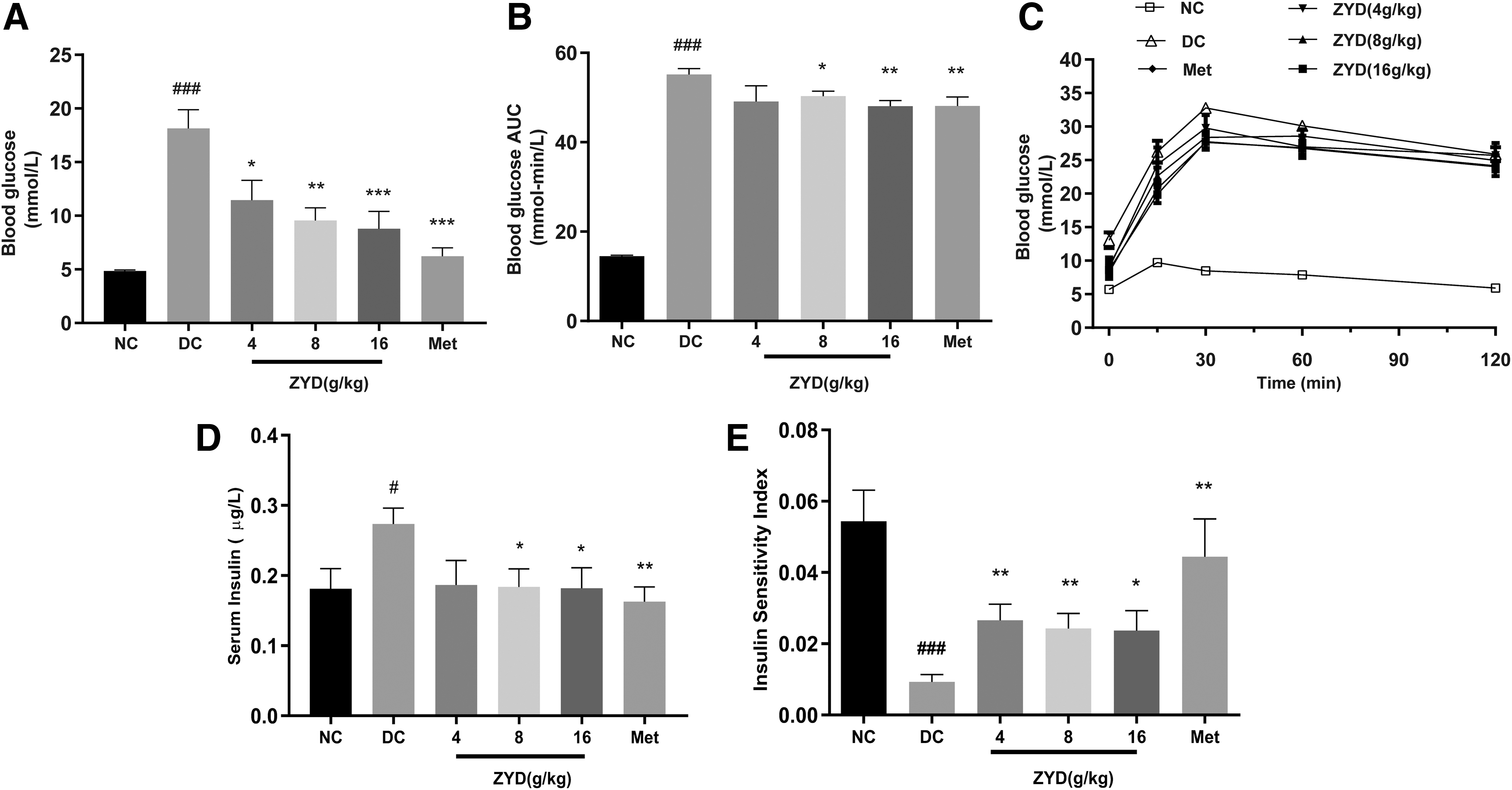
The effects of ZYD on blood glucose levels, FINS, and insulin sensitivity in T2DM rats. Fasting blood glucose level
ZYD improved the ISI of T2DM rats
As shown in Figure 1D, the FINS level of the DC group (0.273 ± 0.051 μg/L) was significantly higher than that in the NC group (0.181 ± 0.064 μg/L) (p < 0.05). However, after 3 weeks of treatment, compared to the model group, the FINS of both ZYD-M (0.184 ± 0.063 μg/L, p < 0.05) and ZYD-H (0.182 ± 0.065 μg/L, p < 0.05) groups was significantly reduced. The FINS level significantly decreased in the metformin group (0.163 ± 0.051 μg/L, p < 0.01). Figure 1F shows that the ISI value of the DC group was significantly lower than that in the NC group (p < 0.01). The values of ISI of ZYD groups were, respectively, higher than the DC group and the metformin group, which indicate that ZYD could effectively increase insulin sensitivity.
Antihyperlipidemic effects of ZYD
Figure 2 shows the lipid profiles of each group. At the end of study, the TC, TG, and LDL-c levels of the DC group significantly increased (p < 0.001) and HDL-c levels of the DC group decreased. However, after 3 weeks of treatment, the levels of TC, TG, and LDL-c of the ZYD groups significantly decreased and HDL-c levels significantly increased compared to the DC group. Those findings were similar with that of the metformin group, which provides clear evidence of an antihyperlipidemic effect.

The effects of ZYD on serum lipid profiles: TG, TC, LDL-c, and HDL-c. Values are expressed as mean ± SEM. ###
p < 0.001 versus control group, **p < 0.01, ***p < 0.001 versus model group. TG, triglyceride
Histopathological changes in pancreas and liver tissues
Figure 3A–F shows the histology pancreatic tissue excised from experimental rats under 400 × magnification. The pancreas islets of the NC group were round and cord like, and the edges were neat and of normal size. H&E staining of islet cells are lightly colored and showed no obvious degeneration (Fig. 3A). However, the islet cells of the DC group were irregular in shape and unevenly distributed, showing significant decreases in size and number. The islet cells were moderately degenerated (Fig. 3B). After drug application, the degree of degeneration and atrophy of the islets in each drug-administered group was alleviated (Fig. 3C–F).

Histopathology of pancreatic tissues in each group. Conventional H&E staining was performed (magnification: 400 × ). Control group
ZYD prevented glucose consumption reduction in HepG2 cells, and 2-NBDG uptake decreased in C2C12 myotubes, 3T3-L1 adipocytes, and HepG2 cells
The glucose concentration and the fluorescence intensity of 2-NBDG are presented in Figure 4A–D. HepG2 cells were exposed to 100 μM PA for 24 hours followed by insulin (100 nM) incubation for 4 hours. Compared to the NC cells, the glucose consumption of IR model cells exhibited an obvious decrease (21.06%), indicating that cells of the IR treatment were successfully induced. However, the glucose consumption was significantly increased by 11.05% and 15.75% at 50 and 100 μg/mL of ZYD group (p < 0.05) (Fig. 4A). Compared to the NC cells, an obvious decrease of 2-NBDG uptake was exhibited in IR model cells. The uptake of HepG2 cells was enhanced in each ZYD treatment group (Fig. 4B). In addition, the uptake of 3T3-L1 adipocytes (Fig. 4C) and C2C12 myotubes (Fig. 4D) was also enhanced in the 50 and 100 μg/mL ZYD treatments. Rosiglitazone, as a positive control, showed a significantly protective effect on 2-NBDG uptake. Besides, the 50 and 100 μg/mL ZYD treatment of HepG2 cells and the 100 μg/mL ZYD treatment of C2C12 myotubes were more effective than rosiglitazone treatment in enhancing 2-NBDG uptake.
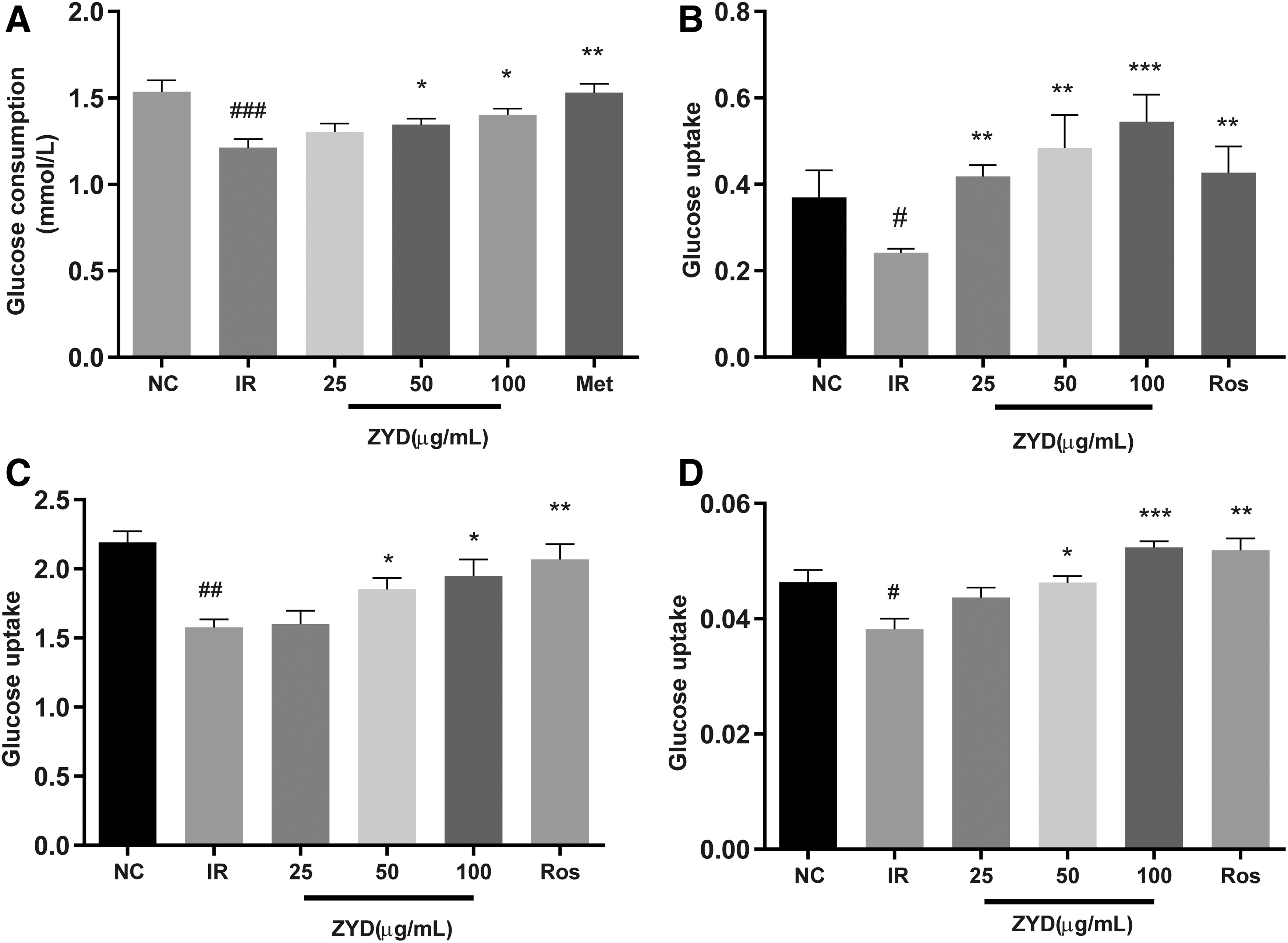
Effects of ZYD on glucose consumption in IR-HepG2 cells and glucose uptake in IR-HepG2 cells, IR-C2C12 myotubes, and IR-3T3-L1 adipocytes. Glucose consumption of HepG2 cells
ZYD enhanced glycogen synthesis in C2C12 myotubes and HepG2 cells
As shown in Figure 5, PA treatment significantly decreased glycogen content in the IR group. Compared to the IR group, we observed significantly increased glycogen content in HepG2 cells after treatment with 50 and 100 μg/mL ZYD (Fig. 5A) and in C2C12 myotubes after treatment with 25 and 100 μg/mL ZYD (Fig. 5B).
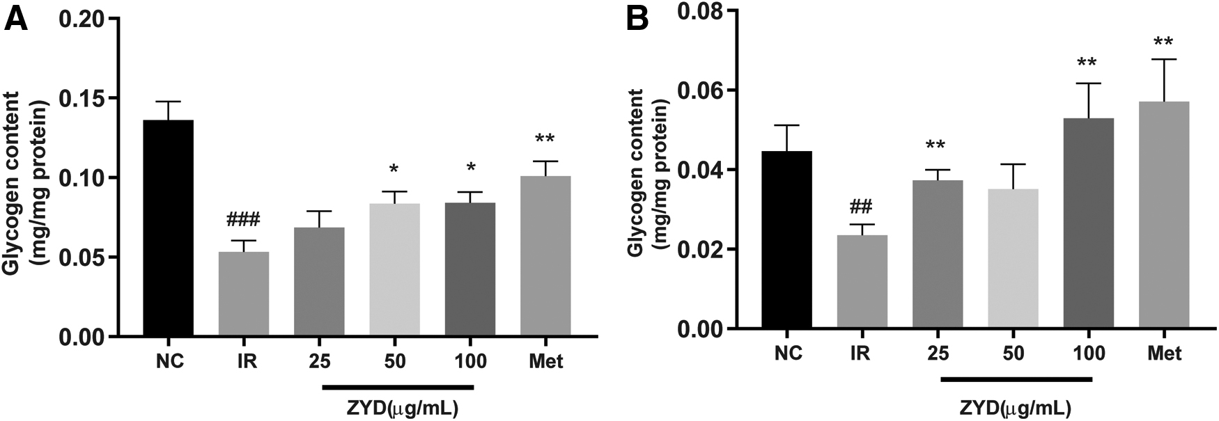
Effects of ZYD on glycogen content in IR-HepG2 cells and IR-C2C12 myotubes. Glycogen content in HepG2 cells
ZYD upregulated Akt phosphorylation level and the expression of GLUT4 in the insulin signaling pathway
Figure 6 shows the alterations of p-Akt/total Akt ratio and GLUT4 in 3T3-L1 adipocytes and C2C12 myotubes. A significant decrease was observed in p-Akt/total Akt ratio and GLUT4 expression in the IR group compared to that in the NC group. The ratio of p-Akt/total Akt and GLUT4 expression of the IR group decreased by 16.0% and 30.25% in 3T3-L1 adipocytes (Fig. 6A, B) and 37.0% and 29.4% in C2C12 myotubes (Fig. 6C, D), respectively. In 3T3-L1 adipocytes, compared to the IR group, rosiglitazone upregulated the ratio of p-Akt/total Akt (by 42.32%) and GLUT4 expression (by 84.59%) and ZYD upregulated the ratio of p-Akt/total Akt by 33.09%, 28.62%, and 36.02% and upregulated GLUT4 expression by 43.73%, 56.63%, and 73.19%, respectively, at 25, 50, and 100 μg/mL.
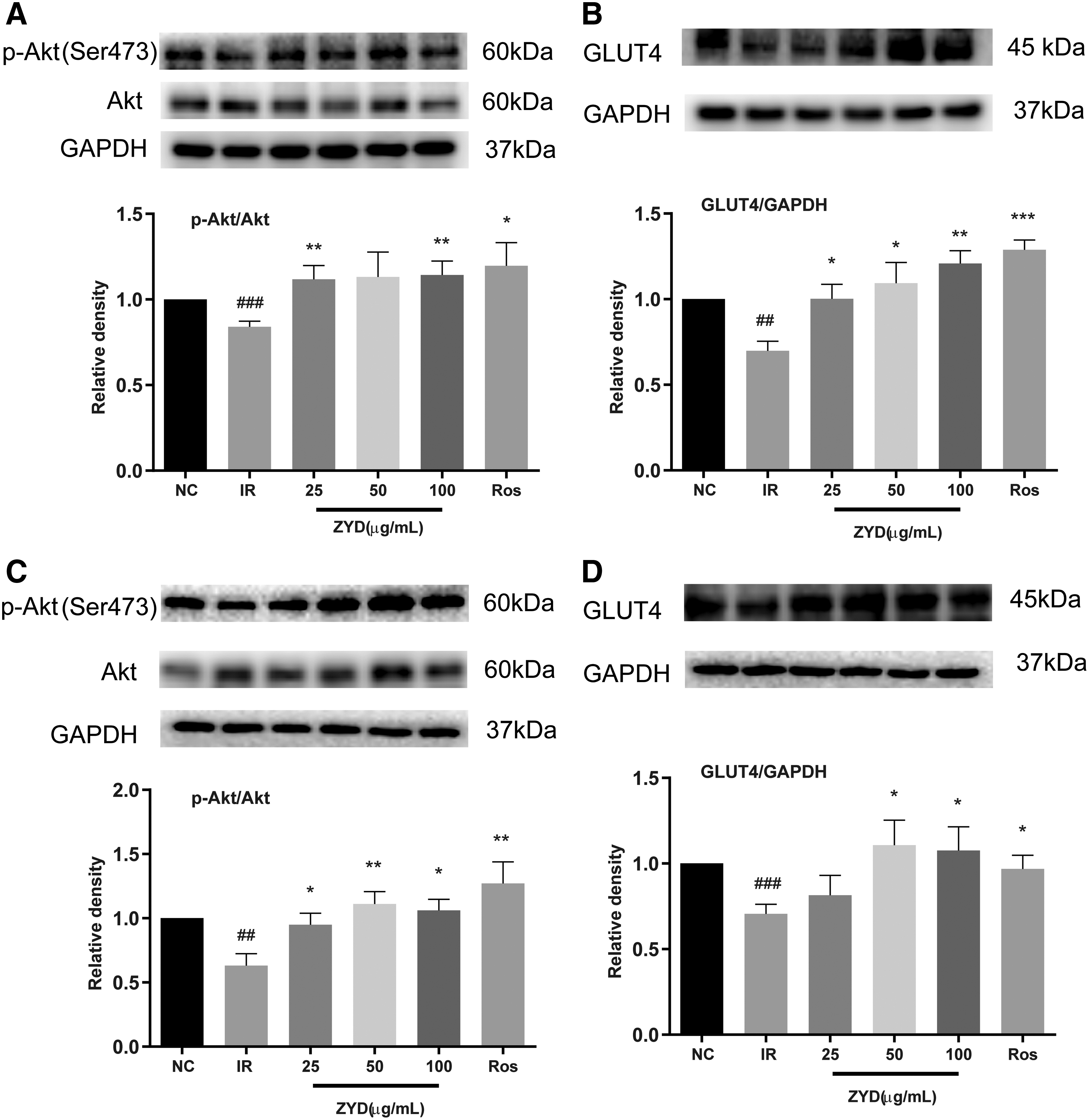
Effects of ZYD on the phosphorylated levels of Akt and the expression of GLUT4 in IR-C2C12 myotubes and IR-3T3-L1 adipocytes. The ratio of p-Akt/total Akt and GLUT4 expression in 3T3-L1 adipocyte cells
In C2C12 myotubes, compared to the IR group, rosiglitazone upregulated the ratio of p-Akt/total Akt and GLUT4 expression by 101.59% and 37.04%, and ZYD upregulated the ratio of p-Akt/total Akt by 50.79%, 76.19%, and 68.25% and upregulated GLUT4 expression by 15.30%, 56.87%, and 52.27%, respectively, at 25, 50, and 100 μg/mL.
Discussion
T2DM is mainly characterized by a progressive decline in insulin action (IR), followed by the inability of beta cells to compensate for IR (pancreatic beta cell dysfunction). 24,25 Many studies demonstrate that a combination of HFD and low dose of STZ treatment can be effectively used to generate a rat model that mimics the natural history and metabolic characteristics of the common type 2 diabetes in humans. 19 It becomes a useful model for studying the antidiabetic effects of new therapeutic drugs. 26 In our study, we used this method to induce T2DM. Compared with NC rats, T2DM rats showed significantly higher FBG levels, impaired glucose tolerance, compensatory hyperinsulinemia syndrome, more severe IR, severe pancreas damage, and higher levels of dyslipidemia, which indicated the successful establishment of T2DM model.
Lowering blood glucose levels and improving glucose tolerance are the primary treatment targets for diabetic patients. After 3 weeks of treatment, compared to the T2DM group, all ZYD treatments showed a significant decrease in FBG level. Moreover, the ZYD-M and ZYD-H treatments significantly improved glucose tolerance. After 3 weeks of treatment, the hypoglycemic effects of ZYD were relatively similar to those of metformin. These results suggest that the hypoglycemic activity of ZYD is reliable in treating T2DM.
These insulin-resistant animals also exhibited abnormal lipid metabolism, as evidenced by increased TC, TG, and LDL-c and decreased levels of HDL-c. In human T2DM patients, these factors may cause various cardiovascular complications. The administration of ZYD for 3 weeks increased HDL-c and reduced the TC, TG, and LDL-c levels, indicating its potential hypolipidemic activity. Moreover, ZYD repaired degeneration and atrophy of islets, and the ZYD-H group was even superior to the metformin group.
IR is the most important feature of T2DM. Feeding rats with HFD led to rat IR characterized by increased body weight (obesity), mild hyperglycemia, lipid metabolism disorder, and compensatory hyperinsulinemia. 19,27 The decreasing of IR is an important method to treat T2DM. The ISI, revealing insulin sensitivity in clinical and animal studies, was significantly decreased in the untreated diabetic rats, while ZYD considerably increased ISI values in T2DM rats, indicating the beneficial actions of ZYD. Moreover, our findings showed that ZYD caused a significant increase in glucose uptake in these three insulin-sensitive cells: HepG2 cells, C2C12 myotubes, and 3T3-L1 adipocytes, which also proved the evidence that ZYD can alleviate IR.
Glucose transport into Liver and skeletal muscle storage as glycogen also contributes to maintaining systemic glucose homeostasis. 10 The preliminary evaluation of ZYD on regulating glycogen synthesis is mainly focused on the two insulin-sensitive cells: HepG2 cells and C2C12 myotubes. In this study, we first chose PA to develop an IR model in C2C12 myotubes and HepG2 cells. Our findings showed that ZYD treatment caused a significant increase in glycogen content compared to model cells.
Considering the importance of the insulin signaling pathway in the pathogenesis of T2DM, we first measured the effects of ZYD on the insulin signaling pathway to further explore the underlying mechanism of ZYD on ameliorating IR. Insulin regulates glucose metabolism in skeletal muscle and adipose tissue by promoting glucose uptake through GLUT4 translocation. 21 Abnormality in GLUT4, the main transporter of glucose and the signal transduction, was considered as the crucial cause of IR. 6 Therefore, investigating its expression level is also crucial. It has been reported that the decrease of Akt could lead to downregulation of GLUT4. Moreover, the activation of Akt could promote the generation of glycogen by adjusting a series of downstream molecules. 10 In this study, treatment of 3T3-L1 adipocytes and C2C12 myotubes with PA induced the impairment in GLUT4 expression and the phosphorylated levels of Akt. In this study, ZYD significantly improved the phosphorylated levels of Akt and upregulated the expression of GLUT4, explaining the increase in glucose uptake.
Due to the complex composition of multiherb prescriptions and the large number of possible target interactions, ZYD may ameliorate T2DM through multiple mechanisms. However, these mechanisms have not yet been elucidated clearly, so further studies to clarify the mechanism of ZYD on treatment of T2DM are needed.
Conclusions
In summary, using in vivo studies, we present evidence that ZYD has good hypoglycemic activity and improves dyslipidemia, contributing to the amelioration of IR in T2DM rats. In our in vitro studies, we found that ZYD significantly regulated glucose metabolism, as evidenced by an increase in glucose consumption, glucose uptake, and glycogen content in IR cells. Moreover, ZYD also activated Akt phosphorylation and GLUT4 expression, which indicated that ZYD may ameliorate T2DM through the Akt pathway. This study lays the foundation for further research of ZYD in the management and treatment of T2DM patients.
Footnotes
Authors' Contributions
J.Q. conceived and designed all the experiments; M.W., S.Q.-C., Y.S.-T., and G.Q.-Z. carried out the experiments; M.W., G.Q.-Z. analyzed the data; J.Q. and M.W. performed the data analyses and wrote the article. All the authors approved the final version of the article before submission.
Author Disclosure Statement
No competing financial interests exist.
Funding Information
This work was financially supported by the National Natural Science Foundation of China (No. 81673555) and “Double First-Class” University project (CPU2018GF06 and CPU2018GY32).
Supplementary Material
Supplementary Data
Supplementary Figure S1
Supplementary Table S1
References
Supplementary Material
Please find the following supplemental material available below.
For Open Access articles published under a Creative Commons License, all supplemental material carries the same license as the article it is associated with.
For non-Open Access articles published, all supplemental material carries a non-exclusive license, and permission requests for re-use of supplemental material or any part of supplemental material shall be sent directly to the copyright owner as specified in the copyright notice associated with the article.
