




Abstract
Huntington's disease (HD) is an inherited neurodegenerative disease. 3-Nitropropionic acid (3-NP) causes increased reactive oxygen species production and neuroinflammation. Centella asiatica (CA) is a strong antioxidant. The aim of this study is to investigate the effect of hydroalcoholic extract of C. asiatica (HA-CA) on 3-NP-induced HD in adult zebrafish. Adult zebrafish (∼5–6 months old) weighing 470 to 530 mg was used and treated with 3-NP (5 mg/kg intraperitoneal [i.p.]). The animals received HA-CA (80 and 100 mg/L) daily for up to 28 days in water. Tetrabenazine (3 mg/kg i.p.) was used as a standard drug. We have done an open field test (for locomotor activity), a novel tank diving test (for anxiety), and a light and dark tank test (for memory), followed by biochemical analysis (acetyl-cholinesterase [AchEs], nitrite, lipid peroxidation [LPO], and glutathione [GSH]) and histopathology to further confirm memory dysfunctions. 3-NP-treated zebrafish exhibit reductions in body weight, progressive neuronal damage, cognition, and locomotor activity. The HA-CA group significantly reduced the 3-NP-induced increase in LPO, AchEs, and nitrite levels while decreasing GSH levels. Oral administration of HA-CA (80 or 100 mg/L) significantly reduces 3-NP-induced changes in body weight and behaviors, in addition to neuroinflammation in the brain by lowering tumor necrosis factor-α and interleukin-1β levels. Moreover, HA-CA significantly decreases the 3-NP-induced neuronal damage in the brain. HA-CA ameliorates neurotoxicity and neurobehavioral deficits in 3-NP-induced HD-like symptoms in adult zebrafish.
Introduction
Huntington's disease (HD) inherited fatal neurodegenerative disorder associated with striatal specific GABA-ergic medium spiny neurons delineated with choreiform movements, psychiatric, cognitive, and motor dysfunctions. 1 The mean HD prevalence is 5 in 100,000 people in the western world; small frequencies are known in Africa, Japan, China, and Finland. 2 HD is caused by cytosine, adenine, guanine (CAG) repeat expansion in exon 1 of the huntingtin gene (Htt). 3,4 CAG triplet repeats in exon 1 responsible for the enlargement of polyglutamine (poly Q) in HTT protein in the HD patients. The normal HTT has 35 CAG repeats whereas mutant HTT >40 CAGs, the patients having 36–39 CAG repeat expansion are at risk of HD. 5 The exact mechanism of HD is still unknown, apart from this neuroinflammation, mitochondrial dysfunction, oxidative stress, excitotoxicity, loss of brain-derived neurotrophic factor, and apoptosis are well-accepted mechanisms in HD. 6 –8 There is no effective therapeutics available for the management of HD.
However, the current therapies are mainly focused on psychiatric and neurological symptoms to improve the quality of life. 9 In 2008, the FDA approved tetrabenazine (TBZ) for the management of choreatic symptoms associated with HD.
In the current study, we employ 3-nitropropionic acid (3-NP) a natural neurotoxin produced by several plant and fungal species nowadays, it is widely used as a pre-clinical model agent that impersonates characteristics associated with HD. 10,11 It is evidenced that 3-NP causes decrease in calcium, adenosine triphosphate production, mitochondrial complex II, III, and IV activity, and increases mitochondrial damage in the striatal neurons of HD patients 12 via the irreversible inhibition of mitochondrial succinate dehydrogenase enzyme (SDH) that is involved in molecular mechanisms associated with neurodegeneration and mitochondrial dysfunction in HD. 13 According to a study, both short-term and long-term exposure to 3-NP in zebrafish causes changes in cells and motor problems, such as choreiform movements, oxidative stress, weight loss, and SDH inhibition. 14
In this context, zebrafish is an outstanding model system to evaluate new drug entity against various neurodegenerative disorders due to several advantages over the other experimental animals that include, ease of genetic manipulation, low cost, fast development, and large progeny. 15 It is well reported that, zebrafish shows 71% physiological similarity with human beings. Evidence suggested that zebrafish is an excellent animal model for the study of several brain disorders such as Alzheimer's disease (AD), 16 Parkinson's disease, 17 stress, 18 epilepsy, 19 dementia, 20 cognition, 21 multiple sclerosis (MS), 22 amyotrophic lateral sclerosis, 23 and traumatic brain injury. 24 However, there is no effective treatment available for the management of HD. As a result, new drugs that not only provide symptomatic relief but also modify the disease pathology of HD patients are required.
Hydroalcoholic extract of Centella asiatica (HA-CA) possesses a nootropic effect that prevents the brain from oxidative damage. This nootropic effect also stimulates the growth of nerves and neuronal dendritic arrangements. 25 It is well known to revitalize the brain and nervous system, increase attention span, concentration, and combat aging. 26 The study revealed the cognition enhancing and antioxidant property of HA-CA against streptozotocin (STZ) induced cognitive impairments and oxidative stress in rat model of AD. 27 Evidenced showed that HA-CA increases body length in rotenone-induced zebrafish larvae via the modulation of its antioxidant potential. 28 Similarly, Naß et al. reported that HA-CA decreases reactive oxygen species (ROS) production, increases resistance to osmotic stress, increases life span, and reduces heat stress due to its antioxidant potential in Caenorhabditis elegans wild-type and knockout mutants. 29
With this background, the present study is designed to investigate the effect of HA-CA against 3-NP-induced behavioural, biochemical, and molecular alterations and neurodegeneration in adult zebrafish.
Materials and Methods
Animal and housing
In the present study, zebrafish (Danio rerio) either-sex wild-type strain of ∼6 months old and weighing from 470 to 530 mg were procured from Aquarts (Kolkata, India). Zebrafish were kept in an aquarium (94.7 L) having temperature 26°C–27°C with constant aeration and pH (6–7) subjected to light and dark cycle 14 and 10 hours respectively. The temperature of the system was maintained at 26°C–27°C by an automatic thermostat. All other water quality parameters including nitrite, nitrate, alkalinity, hardness, and conductivity were maintained in accordance with Crim et al. 30 Fish were fed twice daily with a commercially available diet (Tetrabits Complete Bioactive Formula Fish Food). All of the experiments were done according to the rules set out by the Institutional Biosafety Committee (IBSC), which were approved by the number ISFCP/IBSC/M1/2020/12.
Chemical and drug
Sigma (St. Louis, MO) provided 3-NP, Tricaine MS-222, fish tumor necrosis factor-α (TNF-α) (ELK8512), and fish interleukin-1β (IL-1β) (ELK8342). ELISA kits were purchased from ELK Biotechnology (Wuhan, China). HA-CA was purchased from Natural Remedies (Bangalore, India), and TBZ was purchased from Sun Pharmaceutical Industries Ltd. (India), for commercial purposes. All other chemicals for biochemical analysis were procured from Himedia and SRL Limited, which were of analytical grade. Drugs and chemical solutions were freshly prepared before use.
Study design
A total of 48 zebrafish of either sex was selected randomly and experimenters are partially blinded to the treatment given to zebrafish with weight variation of not >30% (470–530 mg). Before the experiment, each fish acclimated to about 10 days of prior experimentation in laboratory conditions. After the completion of the acclimatization period, the fish were categorized into different groups (n = 6) as shown in Table 1. The HA-CA dose was adapted from a previous published study on rats 31 and administered at two concentrations (80 and 100 mg/L) daily for 28 days. The medium of the experimental tank was changed each day, 1 hour after feeding. HA-CA was dissolved in distilled water and administered for a period of 28 days. 3-NP (5 mg/kg) was dissolved in phosphate-buffer solution (PBS). Interestingly, we have tested a total of five doses of 3-NP, that is, 60, 40, 20, 10, and 5 mg/kg in zebrafish. But we see death at all doses except 5 mg/kg. Because of this, we chose the 5 mg/kg dose for further study (Supplementary Fig. S1).
Experimental Grouping of Animals
HA-CA, hydroalcoholic extract of Centella asiatica; i.p. intraperitoneal; 3-NP, 3-nitropropionic acid.
Behavioral parameters such as the novel tank diving test (NTD), light-dark test (LDT), and open field test (OFT) were assessed on days 1, 4, 8, 12, 16, 20, 24, and 28. Terminally, on day 29, animals were sacrificed by inducing anesthesia by placing the animal in a Tricaine MS-22 solution (100 mg/L). 32 The brain was taken out, mixed up, and put in the deep freeze at −80°C so that biochemical and inflammatory markers could be studied more (Fig. 1).
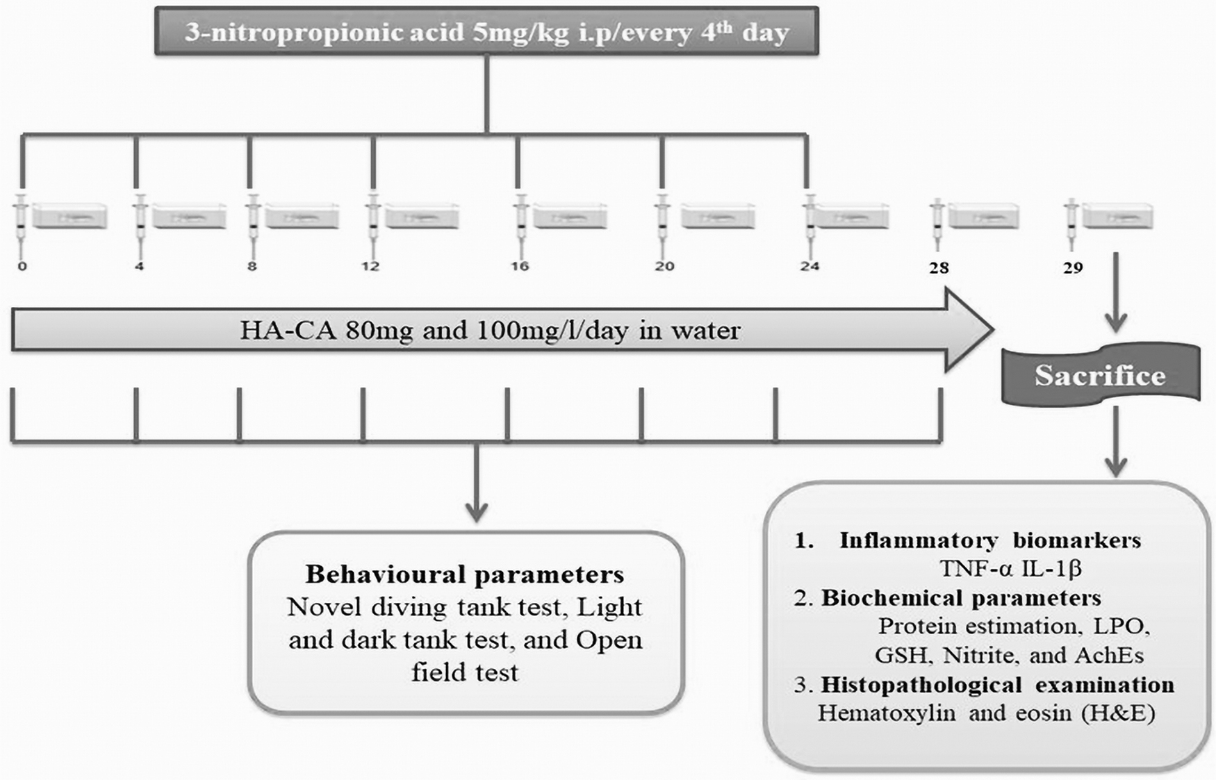
Experimental protocol. AchEs, acetylcholinesterase; GSH, glutathione; HA-CA, hydroalcoholic extract of Centella asiatica; IL-1β, interleukin-1β; LPO, lipid peroxidation; 3-NP, 3-nitropropionic acid; TNF-α, tumor necrosis factor-alpha.
Intraperitoneal injection of 3-NP in zebrafish
Briefly, each fish was anesthetized by immersion in a tricaine MS-222 solution 100 mg/L until the animal shows a lack of motor coordination and reduced respiration rate. Then, fish was taken out from the solution and placed on a soft sponge of 20 mm height that was saturated with cold water and set into a petri dish. 33 A cut was made on the sponge about 10–15 mm deep for holding the fish for injection. Then, intraperitoneal (i.p.) injections were conducted using a 31-gauge Ultra-Fine Hamilton Syringe (max. 10 μL) as per the experimental protocol (Fig. 1). The needle was inserted into the spine posterior to the pectoral fins and anterior to the anal fins in the midline of the abdomen. The whole injection procedure should not take >10 seconds to ensure animal safety. After the injection, animals were placed in a separate fresh water tank to facilitate the animal's recovery from the anesthesia. 34
Body weight measurement
The body weight of different groups was assessed on day 1, 4, 8, 12, 16, 20, 24, and 28.
Behavioral assessment
Light-dark test
LDT was used to evaluate the learning memory functions in adult zebrafish. This apparatus measured 30 cm in length, 15 cm in width, and 16 cm in height. The level of water was maintained at 12 cm. This apparatus is made up of plexiglass and is divided into two halves vertically (15 cm each). One part of the chamber is wrapped with black paper; this part is known as the “dark zone.” Another half of the apparatus is permitted to cross the natural light, and this half is regarded as the “light zone.” The fish were individually placed in it. The test takes a total of 15 minutes, of which 5 minutes are used to get the fish used to the environment (during this time, the fish can move in both parts) and 10 minutes are used to record. We have measured the time spent in the light zone (TSLZ), time spent in the dark zone (TSDZ), and the total number of entries in the light zone (TELZ). 34
Novel tank diving test
NTD is one of the methods used to determine anxiety and depression-like behavior in zebrafish. 35 It is comprised of a 1.5 L trapezoidal tank with a standard dimension of height 19 cm, length 11 cm, and breadth of 22 cm. The tank was filled up to its maximum height with water and was divided into two equal halves horizontally with the help of a marker on the outside wall. 35,36 The area above the dividing line is regarded as the “top zone” (9.5 cm) and the area below the dividing line is regarded as the “bottom zone” (9.5 cm). The test comprises a total of 15 minutes, including 5 minutes for acclimatization (during which the fish is able to move in both parts) and 10 minutes for recording. ANY-maze video tracking software (Stoelting Co.) was used to analyze the data. We have measured the time spent in the top zone (TSTZ), time spent in the bottom zone (TSBZ), and the total number of entries in the top zone (TETZ).
Open field test
OFT is commonly used to study animal exploration and emotionality in various species, including rodents, 37 and fish. 38 OFT was used to evaluate swimming behavior and locomotion in zebrafish, 39 also revealing some similarities in novel strategies of exploration between rodents and zebrafish. 35,36 In the OFT, animals were placed into an open arena, where they were allowed to swim freely. The open field arena was a 5 L square tank (30 cm length by 30 cm width by 10 cm height). The experiment was done under standard conditions. Each fish was carefully placed into the OFT tank for behavioral recording. The test comprises a total of 7 minutes, including 2 minutes for acclimatization (during which the fish is able to explore the whole environment) and 5 minutes for recording. ANY-maze video tracking software (Stoelting Co.) was used to analyze the data. We have measured the distance traveled and maximum speed of zebrafish, which indicates locomotor activity.
Biochemical estimation
Brain homogenate preparation
After the completion of the protocol, the animals were anesthetized by using tricaine until the movements of the gills stopped. The brain was immediately isolated using micro-dissecting tools and forceps and freeze-dried at −4°C. All the samples were homogenized in a homogenizing tube with 5 mL of 0.1 M PBS at pH 7.2, 40 followed by centrifugation at 10,000 g for 10 minutes. The supernatant was collected and used for estimation of various biochemical parameters (protein estimation; LPO: lipid peroxidation; nitrite; AchEs; acetylcholinesterase; GSH: glutathione) and neuroinflammatory markers (TNF-α; IL-1β).
Protein estimation
Protein estimation was done by Biuret method. 41 Briefly, 0.1 mL of tissue homogenate supernatant, 2.9 mL sodium chloride, and 3 mL biuret working reagent were added and kept at room temperature for 10 minutes. The absorbance was measured at 536 nm by using UV spectrophotometer.
Lipid peroxidation
Estimation of LPO was done according to Wills method. 42 Briefly, 0.5 mL of homogenate was incubated with 0.5 mL of Tris HCl at 37°C for 2 hours. Additionally, 1 mL of 10% trichloroacetic acid was added to the incubated suspension, followed by centrifugation at 1000 g for 10 minutes. Moreover, 1 mL of 0.67% thiobarbituric acid was added to 1 mL of supernatant, and the tubes were placed in the boiling water bath for 10 minutes, followed by the addition of 1 mL of double-distilled water. The amount of malondialdehyde (MDA) was measured via the reaction with thiobarbituric acid at 532 nm by using the Perkin Elmer Lambda 20 spectrophotometer. The values were calculated as per the formula for the molar extinction coefficient of a chromophore [1.56 × 10⁵(mol/L) cm−2].
Nitrite estimation
The nitrite accumulated supernatant an indicator of the production of nitric oxide was determined by the calorimetric assay using Griess reagent [0.1% N-(1-naphthyl), ethylene diamine dihydrochloride, 1% sulfanilamide, and 2.5% phosphoric acid] as described by Green et al. 43 The equal volumes of Griess reagent and nitrite-accumulating supernatant were mixed with each other and incubated for 10 minutes at room temperature in the dark, and the absorbance was determined by a Perkin Elmer Lambda 20 spectrophotometer at 546 nm. The concentration of nitrite in the supernatant was determined with the help of a standard curve for sodium nitrite and expressed as mol/mg protein.
AchEs estimation
The AchEs enzyme activity was estimated by Ellman method. 44 The assay mixture contained 0.05 mL of supernatant, 3 mL of 0.01 M sodium PBS (pH 8), 0.10 mL of Ach iodide, and 0.10 mL of 5,5-dithio-bis (2-nitrobenzoic acid) (DTNB) (Ellman reagent). The change in absorbance was measured immediately at 412 nm using a UV-1700 spectrophotometer. Results were expressed as micromoles of acetylthiocholine iodide hydrolyzed per min/mg of protein.
GSH estimation
The level of reduced GSH in striatal homogenate was determined as per the method described by Ellman. 45 One milliliter of supernatant was precipitated with 1 mL of 4% sulfosalicylic acid and cold digested at 40°C for 1 hour. Samples were centrifuged at 1200 rpm for 15 minutes. After centrifugation, 1 mL of supernatant was separated, 2.7 mL of PBS (0.1 M, pH 8), and 0.2 mL of DTNB were added. The yellow color that developed was measured immediately at 412 nm using a spectrophotometer. The concentration of GSH in the supernatant was evaluated with the help of a standard curve and expressed as mol per mg protein.
Neuroinflammatory markers
Estimation of TNF-α, and IL-1β level
This assay employs the quantitative sandwich enzyme immunoassay technique. Using fish TNF- and IL-1 ELISA kits, the amount of TNF-α and IL-1β was measured. The brain sample was homogenized and mixed with PBS. The reagents and standard dilutions were prepared as per the manufacturer's instructions. Each well got 50 μL of diluent, and each well also got 50 μL of standard. The wells were then covered with an adhesive strip and incubated for 2 hours. After incubation each well was washed with wash buffer. Each well got 100 μL of brain homogenate and 100 μL of substrate solution. The wells were then left to sit for 2 hours. After incubation finally 100 μL stop, solution was added to each well and the absorbance was taken by using ELISA reader at 450 nm.
Histopathological examination
After isolating the brain, they were transferred in formalin (10% v/v) solution. The brains were embedded in paraffin wax and sectioned into 3 mm thickness with the help of microtome (Leica RM 2025; Nassloch, Germany). The sections were de-waxed and stained with hematoxylin and eosin (H&E). The stained sections were viewed under a binocular microscope and photographed. 46
Statistical analysis
All statistical analysis was performed with Graph Pad Prism (Graph Pad Software, San Diego, CA). The values are presented as a mean ± standard deviation. The behavioral and biochemical data were evaluated using a repeated-measures one-way analysis of variance (ANOVA). Post hoc comparison between groups were done using Tukey's test. The p < 0.05 value was considered significant.
Results
Effect of HA-CA treatment on body weight
3-NP (5 mg/kg) administration significantly altered adult body weight when compared to the vehicle-treated group. TBZ (3 mg/kg) (p < 0.001) significantly rescued altered body weight when compared to the 3-NP-treated group. Moreover, HA-CA (80 and 100 mg/L) significantly (p < 0.001) rescued weight loss when compared to the 3-NP-treated group. There was no significant difference seen in the per se groups when compared to the vehicle-treated group (Fig. 2).
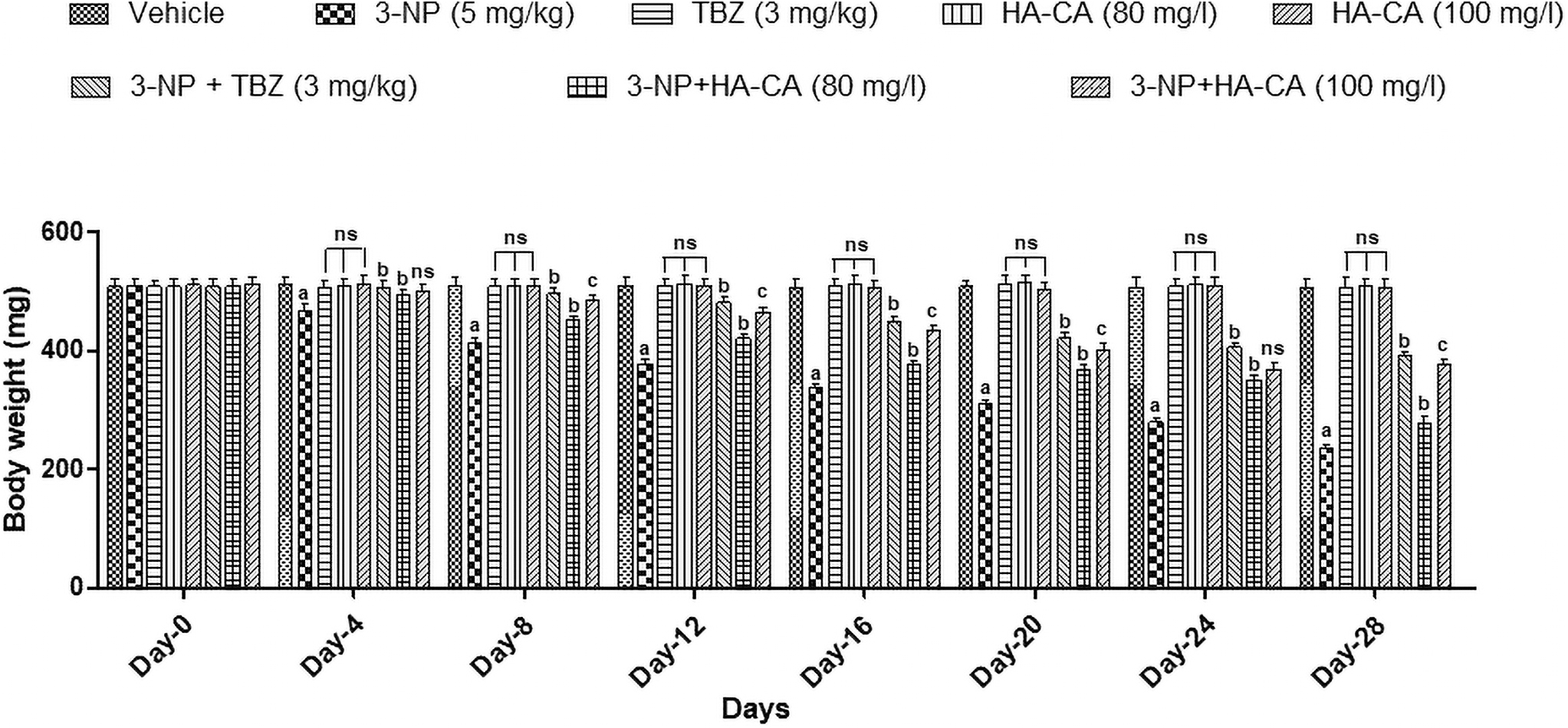
Effect of HA-CA treatment on body weight. Results were expressed as the mean ± SD (n = 6) and analyzed by two-way ANOVA followed by Bonferroni's post hoc test. a p < 0.001 versus vehicle-treated group, ns p > 0.05 versus vehicle treated group b p < 0.001 versus 3-NP-treated group, c p < 0.001 versus 3-NP+HA-CA (80 mg/L) treated group. ANOVA, analysis of variance; SD, standard deviation; TBZ, tetrabenazine.
Behavioral analysis
Effect of HA-CA on LDT in 3-NP-induced HD model of zebrafish
Administration of 3-NP on every fourth day till 28 days significantly (p < 0.001) decreased TSLZ (Fig. 3a) and TELZ (Fig. 3c), while increasing TSDZ (Fig. 3b) when compared to the vehicle-treated group. Furthermore, the TBZ-treated group (3 mg/kg) significantly (p < 0.001) increased 3-NP-induced decreased TSLZ, TELZ, and increased TSDZ when compared to the 3-NP-treated group. Moreover, the HA-CA-treated group (80 and 100 mg/L) significantly (p < 0.001) rescued 3-NP-induced behavioral alterations when compared to the 3-NP-treated group. There was no statistically significant difference has seen between the per se and vehicle groups.

Effect of HA-CA on LDT in 3-NP-induced HD model of zebrafish.
Effect of HA-CA on NTD in 3-NP-induced HD model of zebrafish
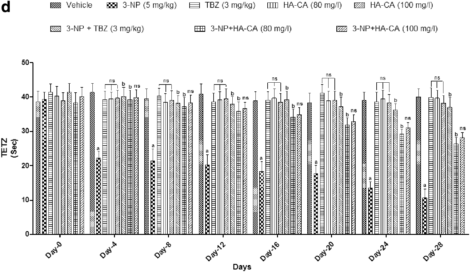
The tracking pattern of different groups are shown (Fig. 4a). Administration of 3-NP showed a significant (p < 0.001) decrease in TSTZ (Fig. 4b) and in TETZ (Fig. 4d), while increasing TSBZ (Fig. 4c) when compared to the vehicle-treated group. Moreover, the TBZ-treated group (3 mg/kg) significantly (p < 0.001) increased TSTZ, TETZ, and decreased TSBZ when compared to the 3-NP-treated group. However, the HA-CA-treated group (80 and 100 mg/L) significantly (p < 0.001) rescued behavioral alterations in NTD when compared to the 3-NP-treated group. There was no statistically significant difference has seen between the per se and vehicle groups.
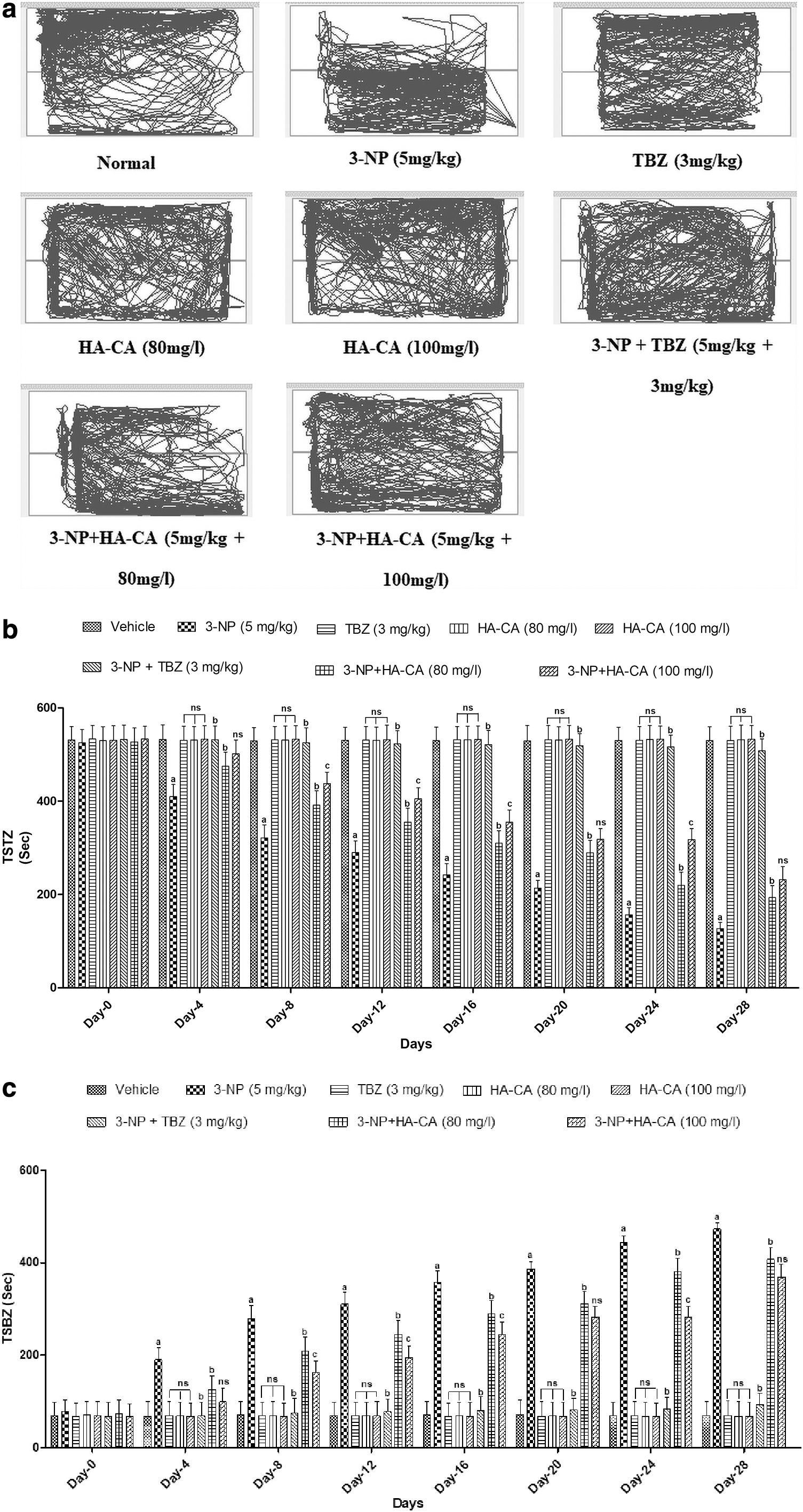
Effect of HA-CA on NTD in 3-NP-induced HD model of zebrafish.
Effect of HA-CA on OFT in 3-NP-induced HD model of zebrafish
The tracking pattern of different groups are shown (Fig. 5a). The locomotor activity in zebrafish altered with the administration of 3-NP, with a significantly (p < 0.001) decreased distance traveled (Fig. 5b) and maximum speed (Fig. 5c) when compared to the vehicle group. The TBZ-treated group significantly (p < 0.001) increased the distance traveled and maximum speed when compared to the 3-NP-treated group. Similarly, the HA-CA (80 and 100 mg/L) treated group significantly (p < 0.001) increased maximum speed and distance traveled in OFT. No statistically significant difference was seen between the per se and vehicle groups.
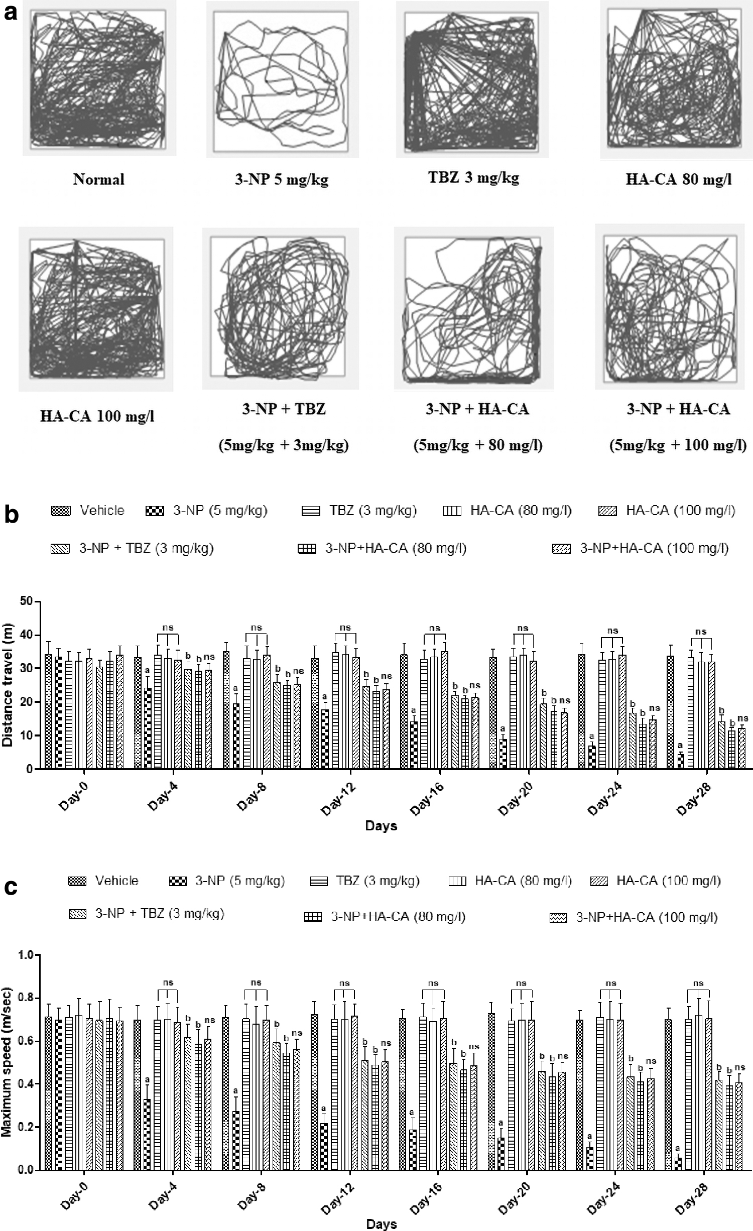
Effect of HA-CA on OFT in 3-NP-induced HD model of zebrafish.
Biochemical analysis
Effect of HA-CA on LPO, nitrite, AChEs, and GSH level in 3-NP-induced HD model of zebrafish
Administration of 3-NP significantly (p < 0.001) increases the levels of LPO (Fig. 6a), nitrite (Fig. 6b), and AchEs (Fig. 6c) while decreasing the level of GSH (Fig. 6d) as compared to the vehicle-treated group. When compared to the vehicle group, the TBZ-treated group significantly (p < 0.001) rescued 3-NP-induced increases in LPO, nitrite, and AchEs and decreased levels of GSH. Similarly, the HA-CA (80 and 100 mg/L) treated group significantly (p < 0.001) reduces the level of LPO, nitrite, and AchEs while increasing GSH levels when compared to the 3-NP treated group. There was no statistically significant difference has seen between the per se and vehicle groups.
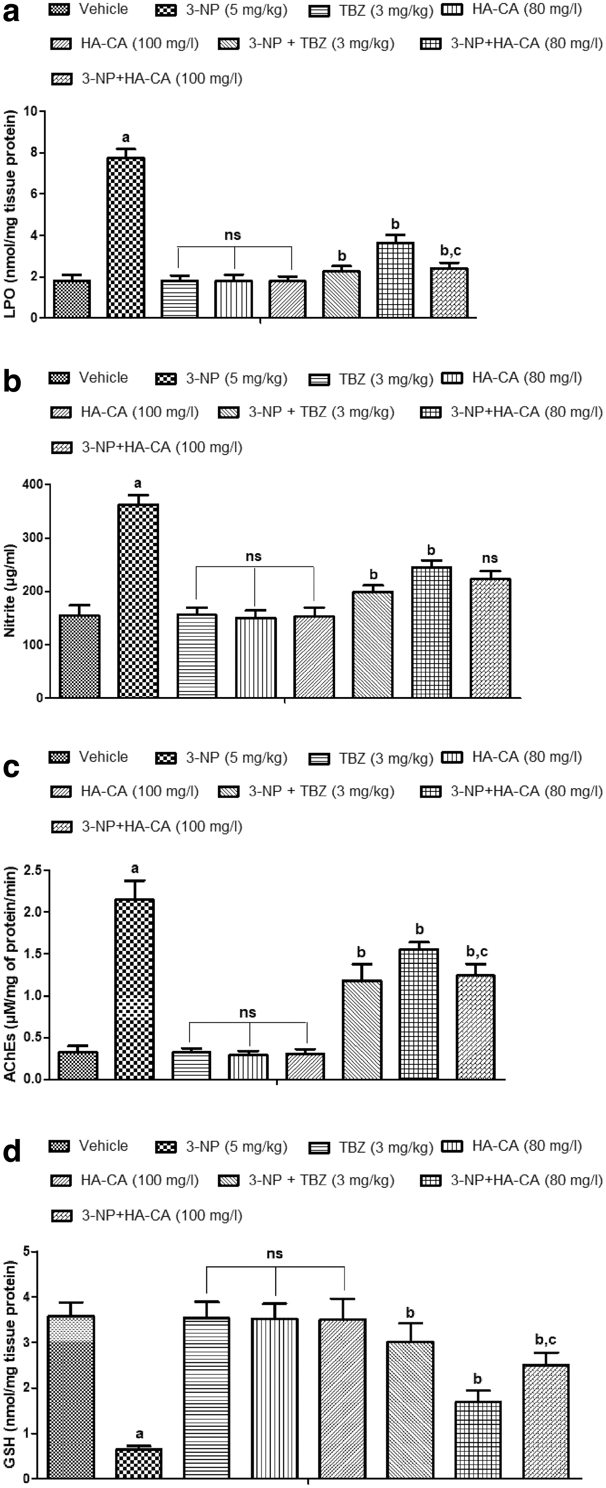
Effect of HA-CA on LPO, nitrite, AchEs, and GSH level in 3-NP-induced HD model of zebrafish.
Neuroinflammatory markers
Effect of HA-CA on the level of TNF-α and IL-1β in 3-NP-induced HD model of zebrafish
TNF-α (Fig. 7a) and IL-1β (Fig. 7b) levels in the 3-NP-treated group were significantly (p < 0.001) higher than in the vehicle group. TNF-α and IL-1β levels were significantly lower (p < 0.01) in the TBZ-treated group than in the 3-NP-treated group. Furthermore, when compared to the 3-NP treated group, the HA-CA (80 and 100 mg/L) treated groups reduced TNF-α and IL-1β levels significantly (p < 0.01, p < 0.001, respectively). No statistically significant difference seen between the per se and vehicle groups.
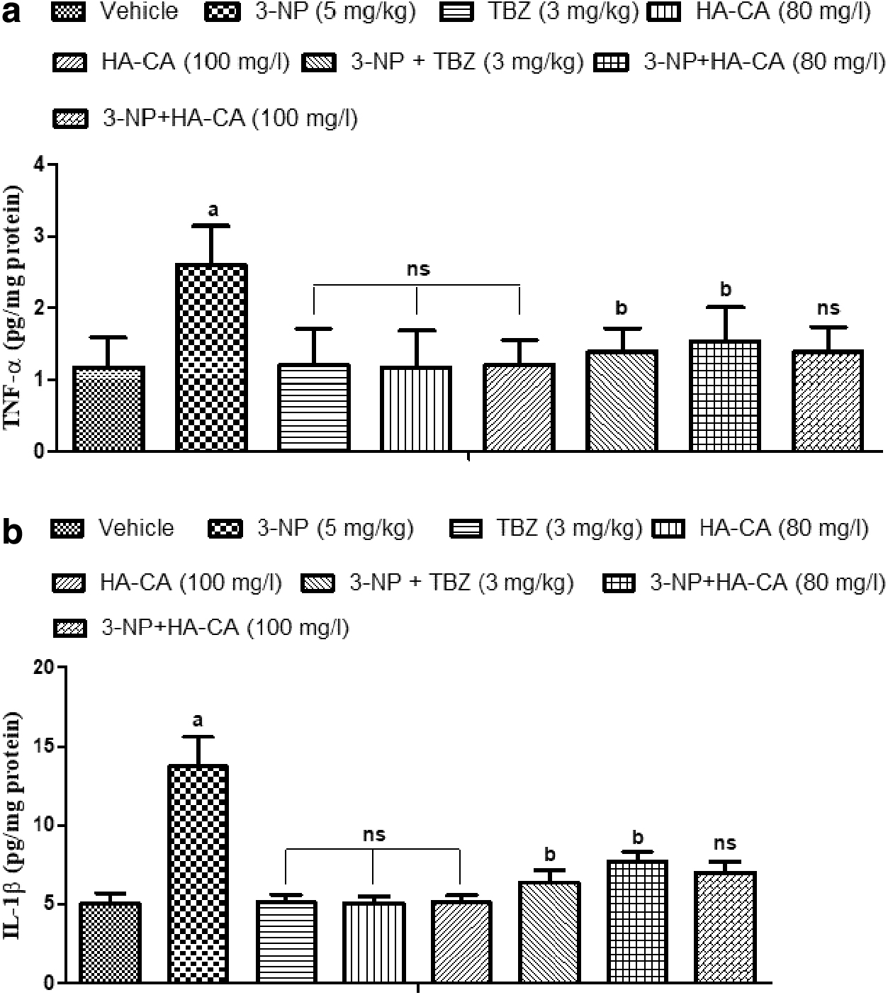
Effect of HA-CA on the level of pro-inflammatory cytokine (TNF-α and IL-1β) in 3-NP-induced HD model of zebrafish.
Histological examination
Histopathological evaluation of brain tissue was conducted by light microscopy; neuronal loss was examined by H&E staining. The 3-NP-treated group shows a greater number of evasive cells when compared to the vehicle group, which indicates neuronal loss in the zebrafish brain. The 3-NP+TBZ-treated group prevented neuronal loss in the striatum when compared to the 3-NP-treated group. However, the 3-NP+HA-CA-treated groups (80 and 100 mg/L) declined neuronal loss in the zebrafish brain, as shown in Figure 8.
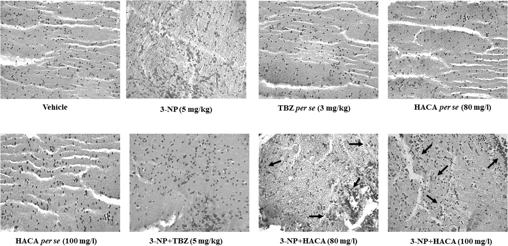
HA-CA reverse 3-NP-induced neuronal loss in adult zebrafish brain. Histopathological evaluation of brain tissue was carried on under light microscopy. The histopathological analysis of vehicle-treated group, TBZ (3 mg/kg) alone treated group, HA-CA (80, and 100 mg/L) alone treated groups showed undamaged neuronal cells. However, 3-NP (3 mg/kg) treated group showed disarrangement of various cell layers and pyramidal neuronal cell loss, which was found significant when compared to vehicle-treated group. However, HA-CA (80 and 100 mg/L) treated groups attenuates the loss of neuronal cell density when compared to 3-NP-treated group.
Discussion
In current study, we verified our hypothesis that oral administration of HA-CA prevents neurotoxicity and neurobehavioral deficits in 3-NP-induced HD in adult zebrafish. 3-NP is being widely used and considered as suitable animal model system for HD. 47 3-NP could induce oxidative stress via increasing oxygen flux through electron transport chain, enhance inflammatory responses and potentiation of excitotoxicity. 48 It is well known as an irreversible inhibitor of mitochondrial complex II. The principal mechanism of 3-NP associated toxicity may occur via the inhibition of Krebs cycle. 49 3-NP is well reported to cause oxidative stress. 48 3-NP is a mycotoxin obtained from plant and fungal species that activates various deleterious pathways such as oxidative stress and neuroinflammation, resulting in neuronal death. Striatum is a part of the basal ganglia, which control motor function; therefore, degeneration of striatal neurons could affect movements. 50
Previous study revealed that HA-CA has antidepressant and anti-anxiety activity. 51 HA-CA possesses nootropic effect that prevents the brain from oxidative damage, and also prevents tissue damage by capturing free radicals. 52 3-NP administration significantly reduced body weight in rat model of HD. It is assumed that 3-NP-induced loss of body weight was not a direct consequence of complex II inhibition, but may have resulted from secondary metabolite. 53 In the current study, we found that HA-CA treatment altered behavior, reduced body weight and GSH levels while increasing LPO and nitrite levels, TNF- and IL-1 levels, and neuronal damage to 3-NP. Through the literature survey, it is interpreted that NTD and LDT are employed to screen for anxiety and depression, while OFT is used to screen locomotor activity in zebrafish.
NTD is one of the methods used to determine anxiety and depression-like behavior in zebrafish. 35,54 It is well reported that depression could affect quality of life in HD associated individuals. Moreover, depression was observed during specific period of illness, especially in early stage of HD. 55 When normal zebrafish are subjected to novel environment, they initially dive to bottom site dwelling and gradually explored the top compartment of NTD and further increased TSTZ, and TETZ. 55 In our study, we have observed significant results. There was no significant difference seen in the per se group; it showed a similar preference toward the top zone and the bottom zone along with TELZ as seen in the vehicle-treated group.
The 3-NP-treated group moved in both zones, but preferred the bottom zone more; thus, animals may experience HD-associated depression and anxiety, whereas the TBZ-treated group showed an increase in TSTZ, TETZ, and a decrease in TSBZ when compared to the 3-NP-treated group. However, the HA-CA-treated group explored both zones, showing more preference for the top zone, thus significantly increasing TSTZ, TETZ, and decreasing TSBZ. So, we thought that the HA-CA-treated group's return to normal exploratory behavior compared to the 3-NP-treated group was a sign of less behavior sickness (anxiety and depression), which meant that depression was getting better.
LDT is the measure of anxiolytic, and memory functions. 56,57 Memory functions were measure by estimating the TSLZ, TSDZ, and TELZ. Usually, the fish chooses darker side for a few seconds when initially put into the new chamber (this possibly is the terror of the new environment); but it shifts toward the light zone within seconds. 58 In the present study, we evaluated memory functions by using LDT, and we observed significant results. We found no significant differences between per se and in the vehicle-treated group when we explored both the light and dark zones, whereas in the 3-NP-treated group, we found decreases in TSLZ and TELZ and an increase in TSDZ, and the animals preferred the dark zone over the light zone when compared to the vehicle-treated group, indicating memory impairment. However, the TBZ and HA-CA-treated group promotes TSLZ and TELZ as compared to the 3-NP-treated group. So, we think that HA-CA could be a better way to help people with HD keep their memories from getting worse.
OFT is commonly used to study animal exploration and emotionality in various species, including rodents, 37 and fish. 38,59 OFT has recently applied to zebrafish to assess their swimming behavior and locomotion, 39,60 also revealing some similarities in strategies of novelty exploration between rodents and zebrafish. 61 3-NP-induced locomotor abnormalities (hyperactivity in the initial stage and hypoactivity in the later stage) in experimental animals. Moreover, for the assessment of locomotor activity, we have performed OFT and measured distance traveled and maximum speed by using ANY-Maze video tracking software (Stoelting Co.). In this study, there was no significant difference between the per se group and the vehicle group. However, the 3-NP-treated group showed that the experimental animals traveled less and went faster than the vehicle group. Moreover, we have observed that HA-CA treated group showed improvement in locomotor activity in response to increase distance travel, and maximum speed against 3-NP-treated group.
Oxidative stress and neuroinflammation have been implicated in the pathophysiology of HD. 62,63 The mutant Huntingtin (mHtt) is known to trigger neuronal damage through elevation of ROS, reactive nitrogen species (RNS), and the level of proinflammatory cytokines (TNF-α and IL-1β). 64 AchEs activity was altered in the 3-NP-treated group, indicating learning and memory dysfunctions. 65,66 In the present study, there were no significant differences seen in the per se group when compared to the vehicle-treated group, whereas 3-NP infusion caused significant elevations in LPO, nitrite (indicating nitrosative stress), AchEs, and decreased levels of GSH (indicating the development of oxidative stress) when compared to the vehicle-treated group.
However, the TBZ and HA-CA-treated groups showed a significant decrease in the level of LPO, nitrite, and AchEs while increasing the level of GSH when compared to the 3-NP-treated group. In the per se group, there was no significant difference in TNF-α and IL-1β levels, but in the 3-NP-treated group, TNF-α and IL-1β levels were higher than in the vehicle-treated group. TNF-α and IL-1β levels in the HA-CA group were lower than in the 3-NP group (Fig. 9).

Mechanistic representation of HA-CA mediated reversal of 3-NP-induced mitochondrial dysfunction.
Kumar and Kumar showed that giving antioxidants to animals with 3-NP changes their behavior and brings back their antioxidative defences. 67 This suggests that HA-CA protects adult zebrafish with 3-NP-induced HD-like symptoms by lowering the level of LPO, nitrite, and ROS, raising the level of GSH, stopping neuroinflammation, and preventing neuronal damage.
The results of histopathological analysis showed that 3-NP treatment causes disorganization of cellular layer when compared to vehicle-treated group. However, treatment HA-CA significantly rescued neuronal damage in histopathological analysis that was found significant compared to 3-NP-treated group.
The current study demonstrated that treatment with HA-CA significantly rescued 3-NP-induced behavioral and biochemical alterations and the level of inflammatory markers. The neuroprotective effect of HA-CA revitalizes its antioxidant and anti-inflammatory effect. HA-CA administration may prevent neuronal damage in HD-associated individuals. So, we can say that HA-CA could be a promising treatment for HD. There are different mechanisms that may involve neurodegeneration in HD, such as oxidative stress, excitotoxicity, mHTT aggregation, and mitochondrial dysfunctions. However, more studies are needed to explore the cellular mechanism associated with HD.
Conclusion
The current study highlighted the neuroprotective potential of HA-CA via the reduction of LPO, nitrite, ROS, neuroinflammation, and neuronal damage and through potentiation of GSH levels in 3-NP-induced HD in adult zebrafish. Administration of 3-NP for up to 28 days on every fourth day results in HD-like phenotypes, memory impairment, neuronal damage, neuroinflammation, and behavioral alterations due to the upregulation of oxidative stress, RNS, and pro-inflammatory markers. HA-CA administration prevents 3-NP-induced behavioral, biochemical, and neuronal damage through its well-known antioxidant mechanism. Hence, it can be concluded that HA-CA could be a promising therapeutic intervention against HD. There are different mechanisms that may play a role in the neurodegeneration associated with HD, such as oxidative stress, excitotoxicity, mHTT aggregation, and mitochondrial dysfunctions. However, more studies are needed to explore the cellular mechanism of HD.
Footnotes
Acknowledgment
Authors thank the institute for providing support to complete this study.
Authors' Contributions
V.K.: Writing—original draft, Methodology; C.S.: Writing—review and editing A.S.: Conceptualization, Methodology, Resources, Supervision.
Author Disclosure Statement
No competing financial interests exist.
Funding Information
No funding was received for this article.
Supplementary Material
Supplementary Figure S1
References
Supplementary Material
Please find the following supplemental material available below.
For Open Access articles published under a Creative Commons License, all supplemental material carries the same license as the article it is associated with.
For non-Open Access articles published, all supplemental material carries a non-exclusive license, and permission requests for re-use of supplemental material or any part of supplemental material shall be sent directly to the copyright owner as specified in the copyright notice associated with the article.
