
Abstract
Cerebral ischemia-reperfusion (CIR) injury occurs as a secondary injury during the treatment of ischemic stroke (IS). There is a high death rate and morbidity due to IS throughout the world. Even though Naoxintong Capsule (NXT) is effective in the treatment of CIR, its mechanisms of action are unclear. The study aims to explore the clear mechanism associated with NXT therapy for CIR. We established the model of middle cerebral artery occlusion to evaluate the neurological function and assess the infarct size. Brain tissue metabolomics was used to identify different metabolites, and metabolic profiling systems enriched metabolic pathways. Then, the potential targets of NXT in the treatment of CIR were explored by proteomic, transcriptomic, and metabolomic methods. NXT improves CIR symptoms. We found potential 11 proteins and corresponding metabolites involved in NXT treatment of CIR. Most of these metabolites are regulated to restore after treatment. According to network pharmacology, we found 6 hub genes, including Glb1, Gmps, Pfas, Atic, Gaa, and Acox1, and their associated core metabolites and pathways. This study reveals the complex mechanism of NXT in treating CIR, and provides a new strategy for future researchers to screen related targets and pathways.
Introduction
Ischemia-reperfusion is a critically pathophysiological change of ischemia stroke 1 and cerebral ischemia-reperfusion (CIR) injury occurs as a secondary injury during treatment of ischemic stroke (IS). 2 IS accounted for 70%–90% of cerebral stroke cases with a high morbidity and mortality rate. 3 Therefore, timely detection and treatment are crucial to the treatment of IS. Currently, the accepted treatment strategy is vascular recanalization, which includes thrombolytic therapy (using drugs such as tissue plasminogen activators), mechanical thrombectomy, and a combination of these. 4
However, a critical problem in both thrombolysis and mechanical thrombectomy is that they only stop ischemia and do not limit additional damage caused by inflammation during reperfusion. They also do not induce the regeneration of any lost neurons. 5 The treatment of IS still has many unanswered questions, such as the short time window, contraindications, and other reasons. 6 Therefore, finding ways to avoid or reduce CIR injury is crucial for improving the treatment of IS. Adjuvant therapy with effective drugs may be a new strategy to improve IS treatment.
Naoxintong Capsule (NXT) is derived from the classic prescription Buyang Huanwu Decoction, which consists of astragalus, salvia, red peony root, chuanxiong, angelica, frankincense, safflower, peach kernel, achyranthes, myrrh, mulberry twig, osmanthus twig, chicken blood vine, dilong, and leech, whole scorpion, clinically used for the treatment of acute and chronic cardiovascular and cerebrovascular diseases for >20 years. 7
In recent years, further studies have confirmed that NXT has neuroprotective effects on IS by improving brain function, reducing serum inflammatory markers and normalizing peripheral blood flow dynamics. 8 Liu et al. demonstrated that NXT could treat cerebral ischemia by interfering with glutamine metabolism and lipid metabolism through ultra-high performance liquid chromatography-quadrupole-time-of-fight mass spectrometry (UHPLC/TOF-MS)-based metabolomic studies. 9 Wang and colleagues used proteomic technology to discover 39 important candidates, key signaling pathways, and core markers of NXT for cerebral ischemic injury and repair. 10 However, the target of NXT for treating IS is still to be further studied.
With the continuous emergence of new omics technologies, the integrated analysis of multiomics data has become a new direction for researchers to explore the mechanism of disease occurrence. Integrating metabolomic and proteomic analyses could allow researchers for understanding the disease-related metabolic and physiological changes. 11 In addition, multiomics combined data can also be applied to personalized medicine, clinical assessment prediction, and risk prediction and clinical outcomes. 12 In this study, we hope to use proteomics, transcriptomics, and metabolomics; then, multiomics analysis systematically explores the molecular mechanism and clarifies the targets of NXT therapy for IS and provides a potential measure of IS treatment. The experimental flow chart is shown in Figure 1.
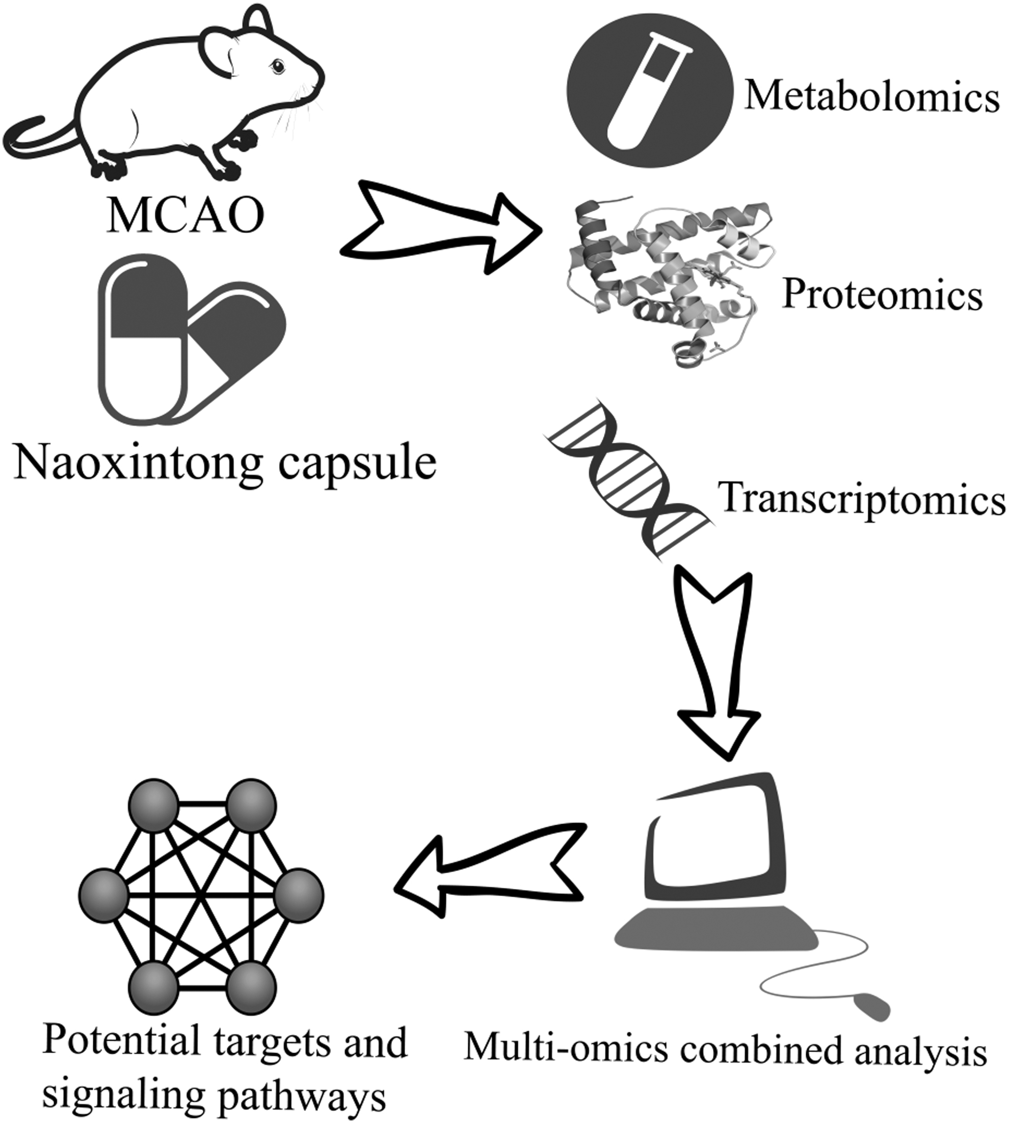
Schematic diagram of the experimental process.
Materials and Methods
NXT preparation
Thanks to Buchang Pharmaceutical Co. Ltd., for providing NXT with a national medicine approval word Z20025001 and mainly contains 16 medicinal materials, which are composed of Buyang Huanwu Decoction and insects to remove blood stasis and collaterals.
CIR model preparation
The animals used in this study were Specific Pathogen Free Sprague Dawley (SD) male rats aged 3–4 months, with a body weight of 280 ± 20 g, obtained from Shanghai Slyke Experimental Animal Co. Ltd., SYXK (Zhejiang) 2018-0012. All the animals were kept in a clean environment in the Animal Experiment Center of Zhejiang University of Chinese Medicine, with a temperature range of 24°C–26°C and free access to drinking water. Adaptive feeding was performed after 7 days. We obtained approval from the Animal Ethics Committee of Zhejiang Chinese Medicine University for all procedures (Approval No. IACUC-20180514-04).
All the rats were anesthetized with 3% pentobarbital sodium (0.15 mL/100 g) and fixed on the operating table in supine position. Rats' skin of the neck was exposed and disinfected with a shaver and surgical scissors, and a mid-neck incision was taken. Blunt dissection found the right common carotid artery (CCA), and upward found the external carotid artery (ECA) and the internal carotid artery (ICA). The proximal CCA and ECA distal ends were ligated, the proximal bifurcation of ICA distal end was attached with a slip-knot, and the proximal bifurcation of ICA was clipped with an arterial clamp. A small incision was made 10 mm away from the bifurcation of CCA. The 4-0 surgical monofilaments nylon suture was inserted from the CCA to the ICA, and the arterial clamp on ICA was slowly loosened. The label 20 mm from the head end of the tether was pushed into the bifurcation, and the tether was slowly pulled out for reperfusion 60 minutes later. Finally, the incision was sutured and disinfected.
Groups
SD rats were randomly distributed into three groups, the sham group, the middle cerebral artery occlusion (MCAO) model group and the NXT group of 13 each. The daily dose of NXT was 0.50 g/(kg·day) by intragastric administration (The dose was selected according to clinical medication conversion and literature references 13,14 ). The dose was given once a day with the volume of each intragastric fluid 1.5 mL for 5 days. The sham group and model group were given the same amount of aseptic distilled water by intragastric administration. Animals in each group were fed under the same conditions and analyzed 5 days after modeling.
Neurological function evaluation
Neurological function evaluations of each rat were scored 1 and 5 days after CIR. Referring to the Longa scoring method, the criteria were as follows: 0 was normal, no neurological impairment; 1 indicated that the left forepaw cannot be fully extended, indicating mild neurological impairment; 2 indicated that the rat turns to the left side (the paralyzed side) while walking, indicating moderate neurological impairment; 3 was when the rat dumps to the left side (paralyzed side) during walking, indicating severe neurological impairment; 4 showed inability to walk spontaneously and loss of consciousness; 5 was death. The higher the score, the more severe the neurological impairment. Each group selected six successful molded rats for scoring.
Assessment of infarct size
After molding 5 days, the rats were sterilized after anesthesia with 3% pentobarbital sodium, the back of the brain was placed in the brain groove, and after freezing at −20°C for 30 minutes, it was cut into coronary artery sections with a thickness of 2 mm, then placed in 2% 2,3,5-triphenyltetrazolium chloride (TTC) solution incubated, and photographed and recorded after 30 minutes of light staining at 37°C. After TTC staining, the infarction part is white and the non-infarction part is red. The area of cerebral infarction is calculated by Image J as follows: White area/total area of the whole brain × 100%.
Transcriptomic research
After 1 hour of the last administration on day 5, each group of rats was anesthetized with 3% pentobarbital sodium (0.15 mL/100 g), the brain was quickly taken, and the ischemic hemilateral brain tissue was quickly separated on ice. Extraction of total RNA from brain tissue. After extracting purified total RNA from brain tissue, Agilent 2100 Bioanalyzer measured total RNA concentration, 28S/18S, RNA integrity number value, and fragment size. After RNA concentration and purity quality control are passed, double-stranded cDNA is synthesized and the final cDNA library is obtained by PCR experiment enrichment. Finally, a cDNA library was added to each channel of the flow tank, and the sequencing analysis using Illumina HiSeq sequencing technology was used to calculate the mass score.
Benjamini and Hochberg's method controls the false discovery rate by adjusting the resulting p value. The raw data of transcriptase sequencing are filtered to obtain high-quality data information, and the threshold is set at the significance level adjusted p < 0.05 and the different fold|log2Fold change 2, and the mRNA of the three samples was screened for differential genes. Using the whole rat genome as the background and reference, with p < 0.05 as the standard, the KEGG database, Cytoscape v3.7.1, and ClueGO v2.5.7 were used to analyze the enrichment pathways involving differentially expressed genes (DEGs) to obtain key pathway information.
Proteomics research
The method of taking materials is the same as aforementioned. Weigh the tissue sample and add liquid nitrogen to a precooled mortar of liquid nitrogen and grind it to the powder. Four times the volume of lysis buffer containing 1% protease inhibitors, 8 M urea, and 2 mM EDTA was added to each sample. After ultrasonic lysis, the samples were centrifuged at 12,000 g for 10 minutes at low temperature. The supernatant was taken and the protein concentration was determined by BCA kit. Dithiothreitol was then added to the protein solution so that the final concentration was 5 mM, and the reduction is 30 minutes at 56°C. Iodide acetamide was added to make the final concentration of 11 mM and incubated for 15 minutes at room temperature under light. Finally, the sample was diluted so that the urea concentration was <2 M.
Incubated overnight at 37°C with 1:50 trypsin-to-protein mass ratios. Then add trypsin to protein 1:100 mass ratio of pancreatic enzyme 4 hours digestion. Peptides are graded with high pH reverse high performance liquid chromatography, the components are separated and combined, the appropriate amount of iRT reagent is added to the combined components according to the iRT reagent instructions, vacuum freeze-dried for follow-up operations, and the peptides are dissolved with liquid chromatography mobile phase A phase (0.1% (v/v) aqueous solution of formic acid) and separated using the EASY-nLC 1000 ultrahigh-performance liquid phase system, and then injected into the nanoelectrospray ionization ion source for ionization, and then using Q Exactive™ HF-X mass spectrometry for data acquisition. Using a high-resolution Orbitrap detector, the parent peptide ions and their secondary fragments were detected and analyzed.
Metabolomics research
The method of taking materials is the same as aforementioned. Weigh 25 mg of the sample and place it in an Eppendorf tube; add 800 μL of precooled dichloromethane/methanol (3:1) precipitant and two small steel balls; grind using a tissue grinder (power set to 50 Hz, time 5 minutes), place the ground sample in a −20°C refrigerator to pellet for 2 hours; centrifuge at 25,000 g for 15 minutes at 4°C, take 550 μL of supernatant, and remove the small steel balls and place them in a beaker; after draining the supernatant into a refrigerated centrifuge, redissolve with 600 μL of lipid compound solution (isopropanol:acetonitrile:water = 2:1:1) and vortex for 1 minute; centrifuge at 25,000 g for 20 minutes at 4°C and 550 μL supernatant will be collected; take 50 μL of each sample mixed into a quality control sample QC and load into liquid chromatograph mass spectrometer injection vials; and all samples were divided into 96-well plates in the order in which they were loaded, with 60 μL per well.
Subsequently, the ACQUITY UPLC CSH C18 column (100 × 2.1 mm, 1.7 μm; Waters, United Kingdom) column was separated, and the small molecules eluted on the column were collected in positive and negative ion mode with high-resolution tandem mass spectrometry Xevo G2-XS QTOF (Waters). Collect centroid data using MSE mode.
Combined analysis of proteome, transcriptome, and metabolome
Bioinformatics analysis of common targets and differential metabolites in CIR
When the sham group was compared with the model group, the NXT group was able to upregulate or downregulate all the differential genes, and the proteome and the transcription group were screened for up- or downregulated genes.
The model group was compared with the NXT group for the screening and de-redundancy of the identified metabolome data. Filters: (1) variable importance in the projection ≥1; (2) fold-change ≥1.2 or ≤0.8333; (3) q-value, three intersect to obtain a common ion. Take the intersection of the three to get the common ions. Match the differential ions with the substance molecules in the metabolite database HMDB, combine the information in the database KEGG, identify the differential metabolites, and obtain differential metabolites.
The identified common targets and differential metabolites were subjected to pathway enrichment analysis through the Joint-Pathway Analysis of the MetaboAnalyst 5.0 database, and the metabolic pathway enrichment map was obtained.
Identify key metabolites and proteins
First: To visualize the interactions between metabolites, pathways, enzymes, and genes, differential metabolites identified in metabolomics are introduced into Cytoscape equipped with Metscape to obtain a network of compound-reaction-enzyme-gene.
Second: Using the multiple proteins module in STRING Version 11.5, a CIR protein–protein interaction (PPI) network was constructed. CytoHubba plug-in for Cytoscape was used to obtain the TOP 20 hub genes by the MCC algorithm.
Third: The identified common targets and differential metabolites were analyzed for KEGG pathway and Gene Ontology (GO) enrichment of potential targets by the ClueGO plugin of Cytoscape 3.7.1. where KEGG pathway analysis was set to pV ≤0.05.
Fourth: Combine compound-reaction-enzyme-gene networks with hub genes and metabolic pathways to identify key metabolites and proteins.
Statistical analysis
The results were expressed as mean ± standard deviation and analyzed by one-way analysis of variance followed by Tukey's post hoc test or Kruskal–Wallis test with SPSS 25.0 software (SPSS, Inc.). A value of p < 0.05 was considered to be statistically significant.
Results
Effect of NXT on neurological function in rats
Based on the neurological function score test, we found that the model group showed significantly improved scores after MCAO when compared with the sham group, whereas the NXT group showed significantly decreased scores after 5 days drug treatment when compared with the MACO group, as shown in Figure 2.
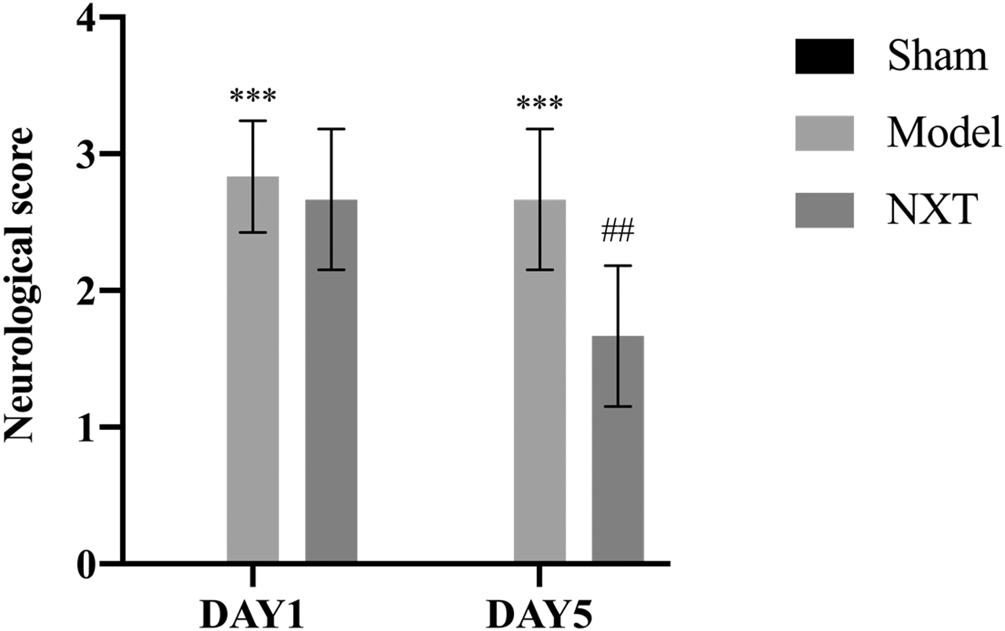
The effect of NXT on 1 and 5 days neurological scores of MCAO rats. Values are expressed as mean ± SD (n = 6). ***p < 0.001 as compared with the sham group. ## p < 0.01 as compared with the model group. SD, standard deviation; MCAO, middle cerebral artery occlusion; NXT, Naoxintong Capsule.
Effect of NXT on cerebral infarct volume
As shown in Figure 3, viable tissues were stained in deep red by TTC staining, whereas the area of infarction in the right cerebral hemisphere is white. In comparison with the Sham group, the model group had a significant increase in infarction volume. And as compared with the model group, the NXT group did significantly reduce infarct volume.
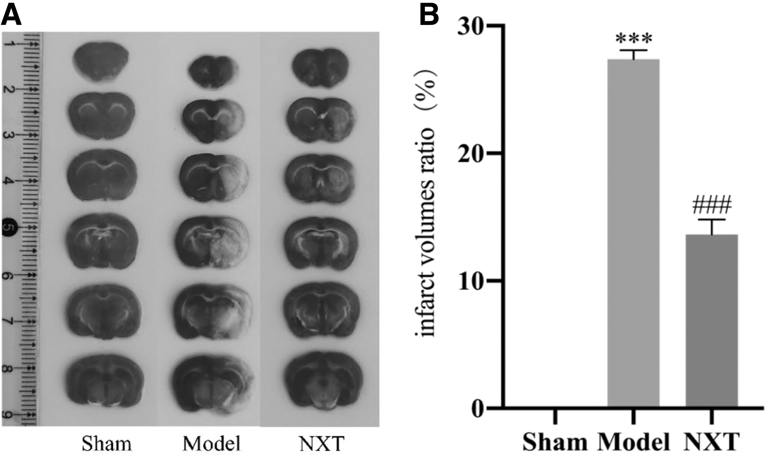
The effect of NXT on cerebral infarct volume in the MCAO rats for 1 hour and reperfusion for 5 days. Representative photographs of TTC stained coronal brain sections of the sham group, model group and NXT-treated group
Combined proteomics and transcriptomics analysis
The sham group, model group, and NXT group were analyzed by metabonomics, proteomics, and transcriptomics, and Figure 4 was obtained.
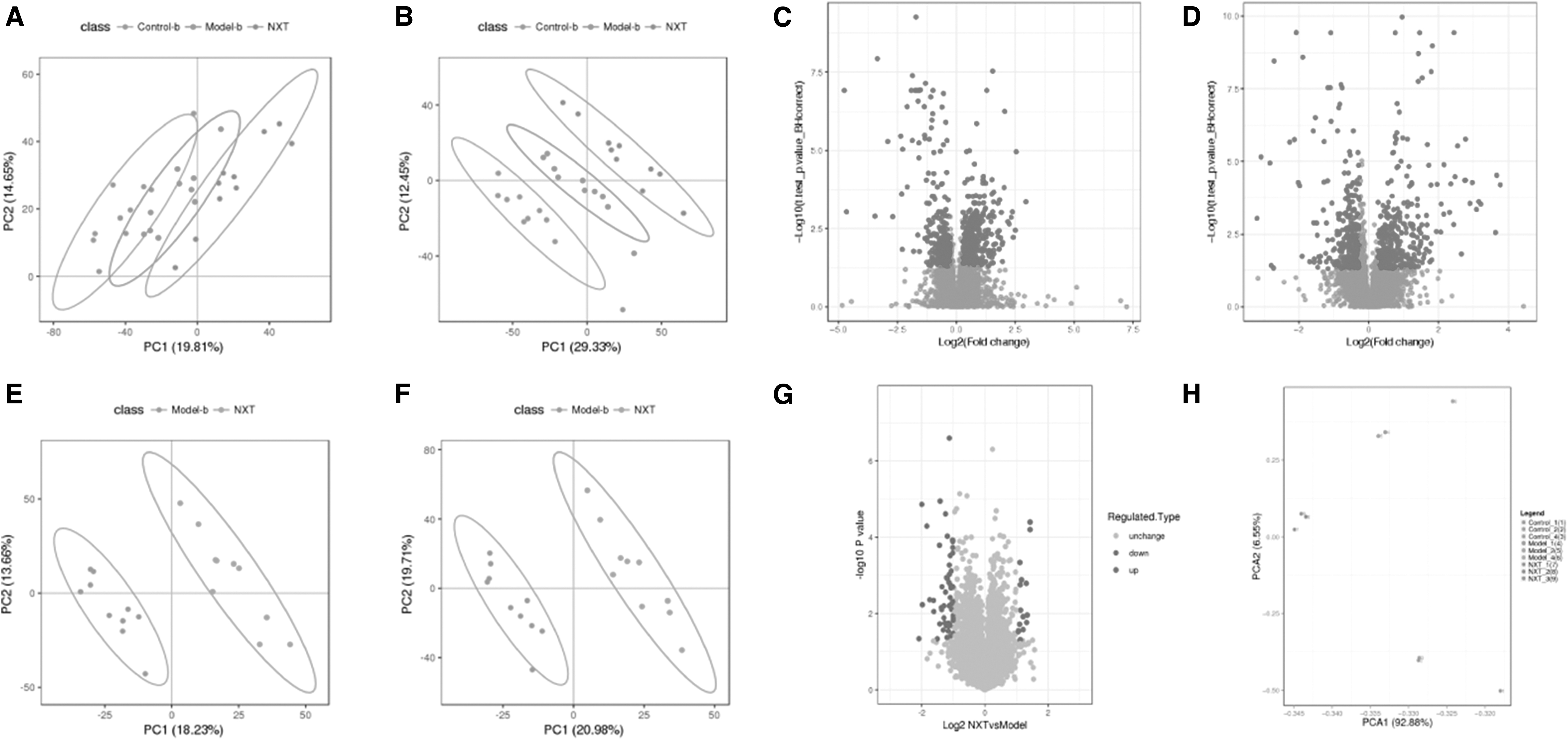
Multiomics analysis of sham (control) group, model group, and NXT group. Metabolomics:
Then three groups were compared and discriminated, and the genes with consistent up- or downregulation in proteomics and transcriptomics were screened. A total of 342 targets (shared targets) were identified, as shown in Figure 5. Among them, 101 were reversed upregulated and 241 were reversed downregulated.
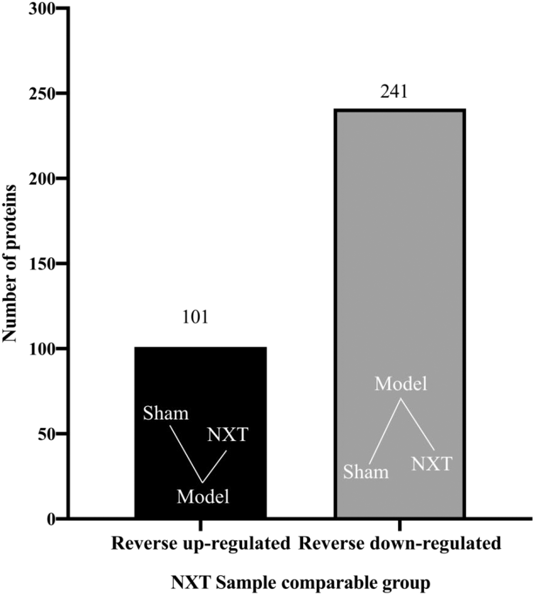
Histogram of shared targets in NXT group. The reversed upregulation represents a “v”-shaped gene expression in the sham, model and NXT groups, whereas the reversed downregulation represents a “∧”-shaped.
Combined analysis of proteomics, transcriptomics, and metabolomics
Bioinformatics analysis of common targets and differential metabolites in CIR
Screening and de-redundancy of the metabolome data identified by comparing the model group with the NXT group were screened according to the screening conditions in the method to obtain 527 total ion identifications in positive ion mode and 390 total ion identifications in negative ion mode. A total of 67 differential metabolites were identified by matching the differential ions with the material molecules in the metabolite database HMDB and combining with the information in the database KEGG.
To explore the metabolic pathways of NXT in CIR rats, we performed pathway enrichment analysis on the identified common targets and differential metabolites through Joint-Pathway Analysis in the MetaboAnalyst 5.0 database, and finally obtained the metabolic pathway enrichment map. and marked the pathway effect >0.2, p < 0.05 on the pathway map, as shown in Figure 6.
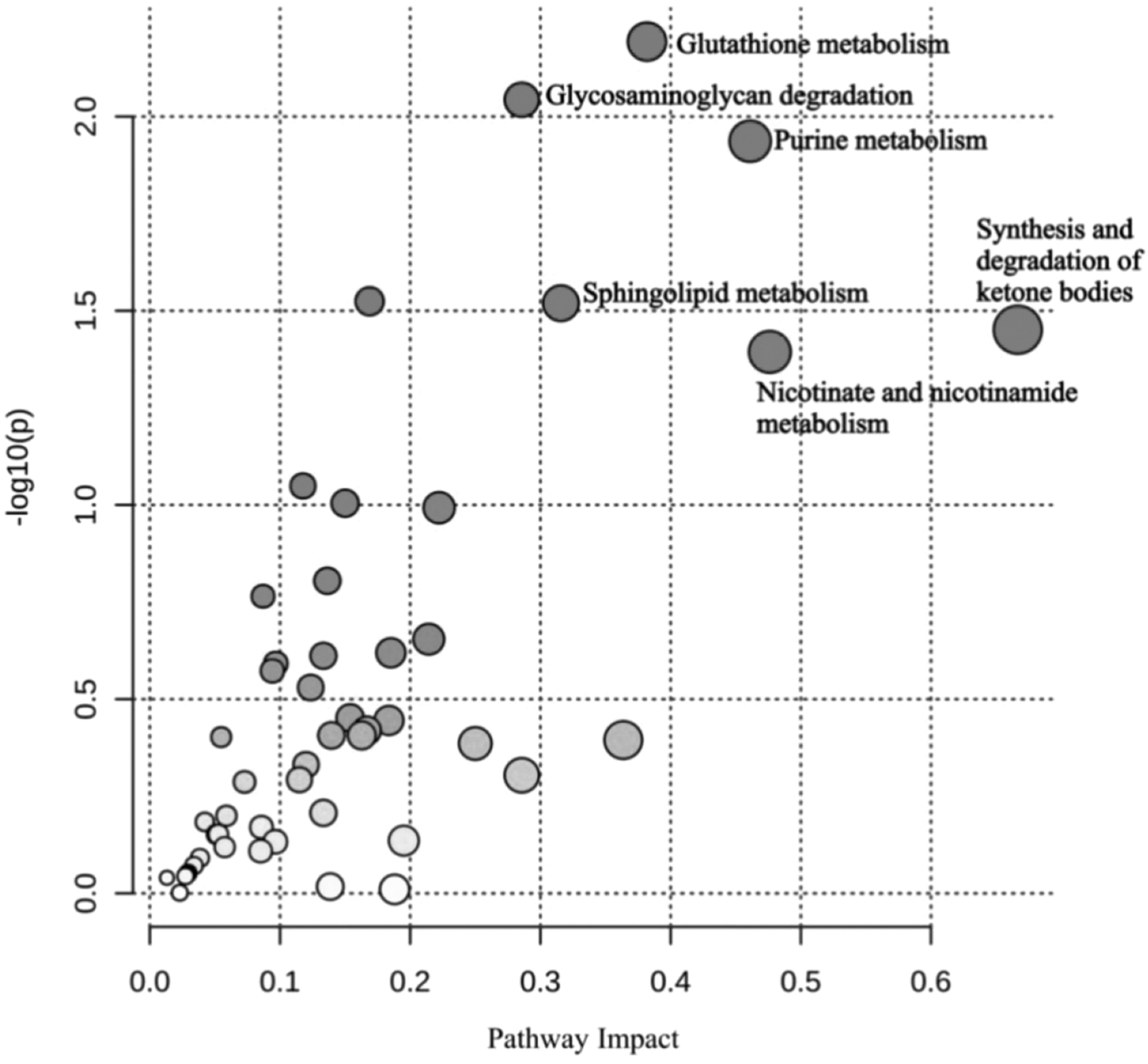
Signal pathway enrichment distribution bubble chart. Node size is based on impact values, node color is based on −log10(p) values.
According to the results of signal pathway enrichment analysis, the main enriched signal pathways of CIR differential metabolites include glutathione metabolism, glycosaminoglycan degradation, purine metabolism, sphingolipid metabolism, synthesis and degradation of ketone bodies, Niacin and niacinamide metabolism. Metabolites associated with these pathways are hypoxanthine, 5-hydroxyisouric acid, inosine, sphingosine, and 3-dehydrotrimethine.
Cytoscape 3.7.1 Database
To identify the hub genes of NXT against CIR, we built a PPI network using STRING and visualized it by Cytoscape. Hub genes were abstracted through CytoHubba. By the MCC algorithm, the top 20 genes were considered as hub genes (Glb1, Hexb, Lap3, Mpi, Gmps, Pfas, Hexa, Me2, Atic, Prps2, Anpep, Gaa, Idh2, Pgm2, Hmgcl, Ilk, Cd38, Renbp, Hmgcs1, and Acox1). A detailed explanation is given in Figure 7.
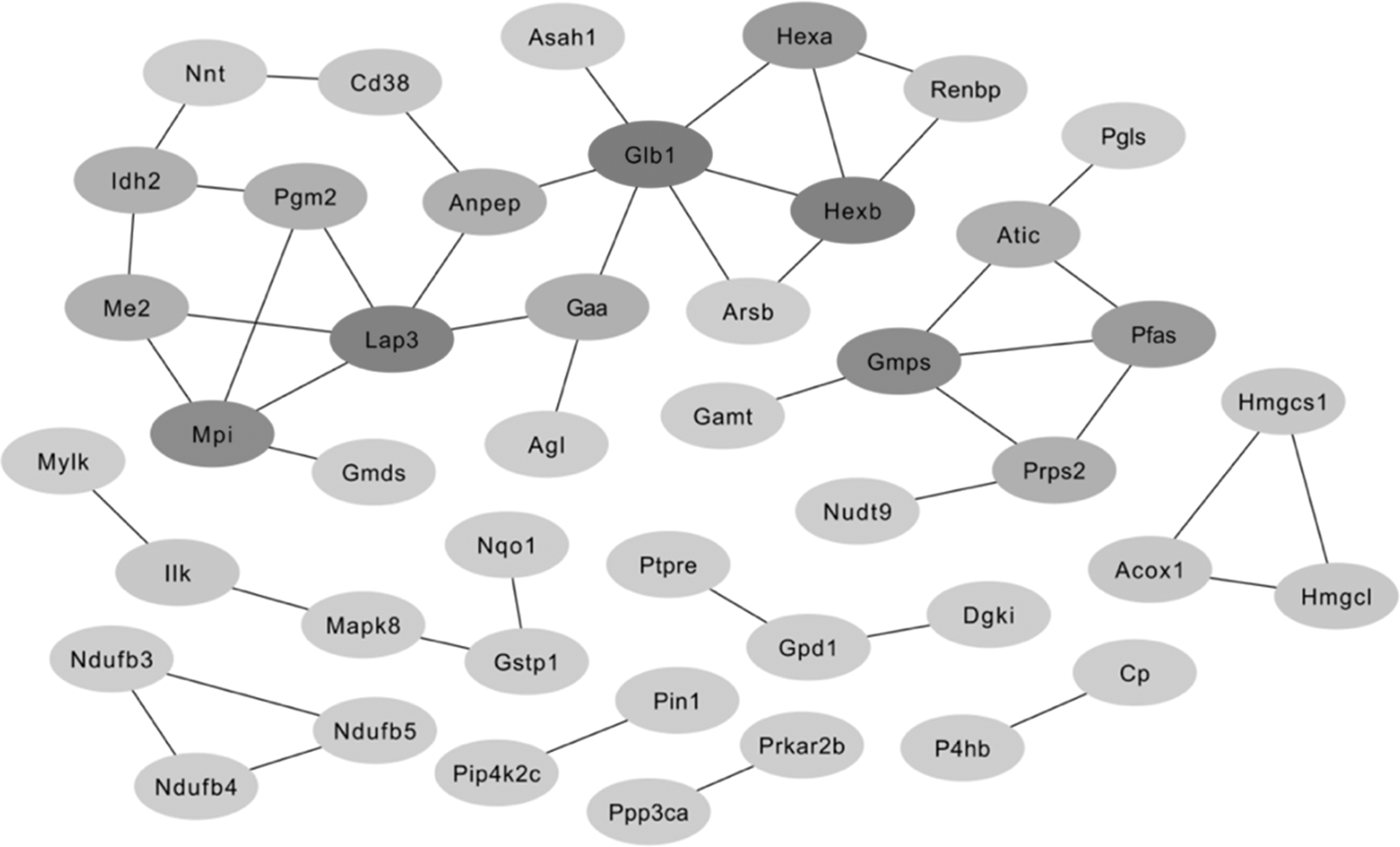
The PPI network of NXT treatment on CIR. Node color reflects its degree. Hub genes are marked in dark grey, and non-hub genes are marked in light grey. CIR, cerebral ischemia-reperfusion; PPI, protein–protein interaction.
To decipher the Anti-CIR function of potential targets, we conducted GO and KEGG pathway enrichment analyses (Figs. 8 and 9). The top terms in the GO analysis were purine ribonucleoside monophosphate metabolism (GO: 0009167), hydrolase activity acting on glycosyl bonds (GO: 0016798), antibiotic catabolism (GO: 0017001), NAD binding (GO: 0051287), and response to vitamin E (GO: 0033197). According to KEGG enrichment analysis, the significantly affected pathways were glycosaminoglycan degradation, arginine and proline metabolism, glutathione metabolism, purine metabolism, and peroxisomes.
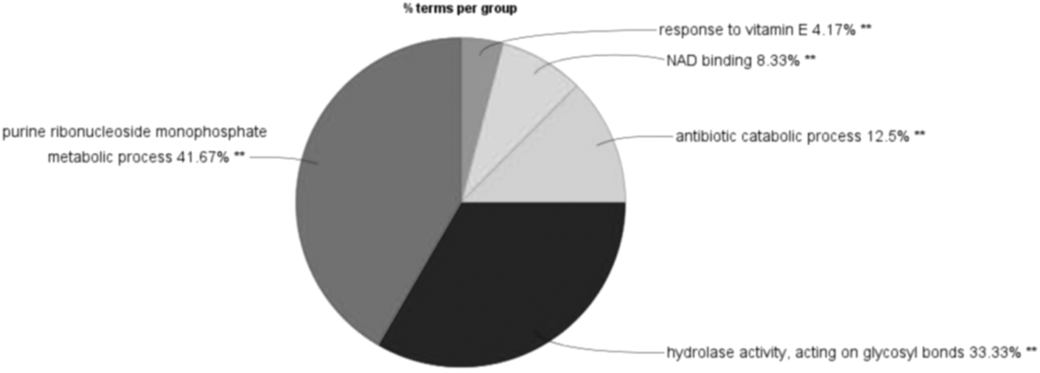
GO enrichment analysis of potential targets by ClueGO. GO, Gene Ontology.
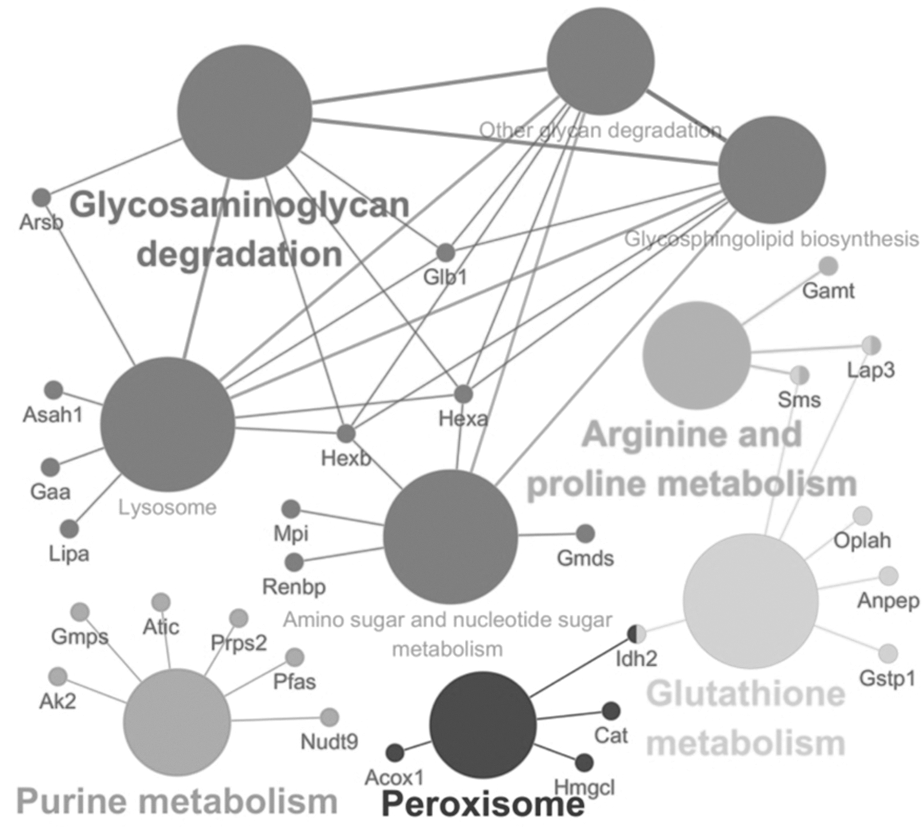
KEGG pathway enrichment analysis of ClueGO. The p-value of all pathways was <0.05.
To comprehensively understand the mechanism of NXT against CIR, we constructed an interaction network based on metabolomics and network pharmacology (Fig. 10). The differential metabolites were imported into the MetScape plugin in the Cytoscape 3.7.2 database to collect compound-reaction-enzyme-gene networks. By matching potential targets identified by transcriptomics and proteomics with genes in the MetScape analysis, 11 key targets have been identified, including Glb1, Dgki, Gpd1, lipa, Gmps, Pfas, Atic, ATP2B2, AK2, Gaa, and Acox1 (Table 1), of which Glb1, Gmps, Pfas, Atic, Gaa, and Acox1 are hub genes. The relevant key metabolites are cholesterol, inosine, hypoxanthine, 5-hydroxyisourate, N-acetyllactosamine, 5,6-epoxyeicosatrienoic acid, and 2-arachidonic acid glycerol. The pathways affected are glycerophospholipid metabolism, purine metabolism, galactose metabolism, and arachidonic acid metabolism. They may play an important role in the therapeutic effect of NXT on CIR injury.
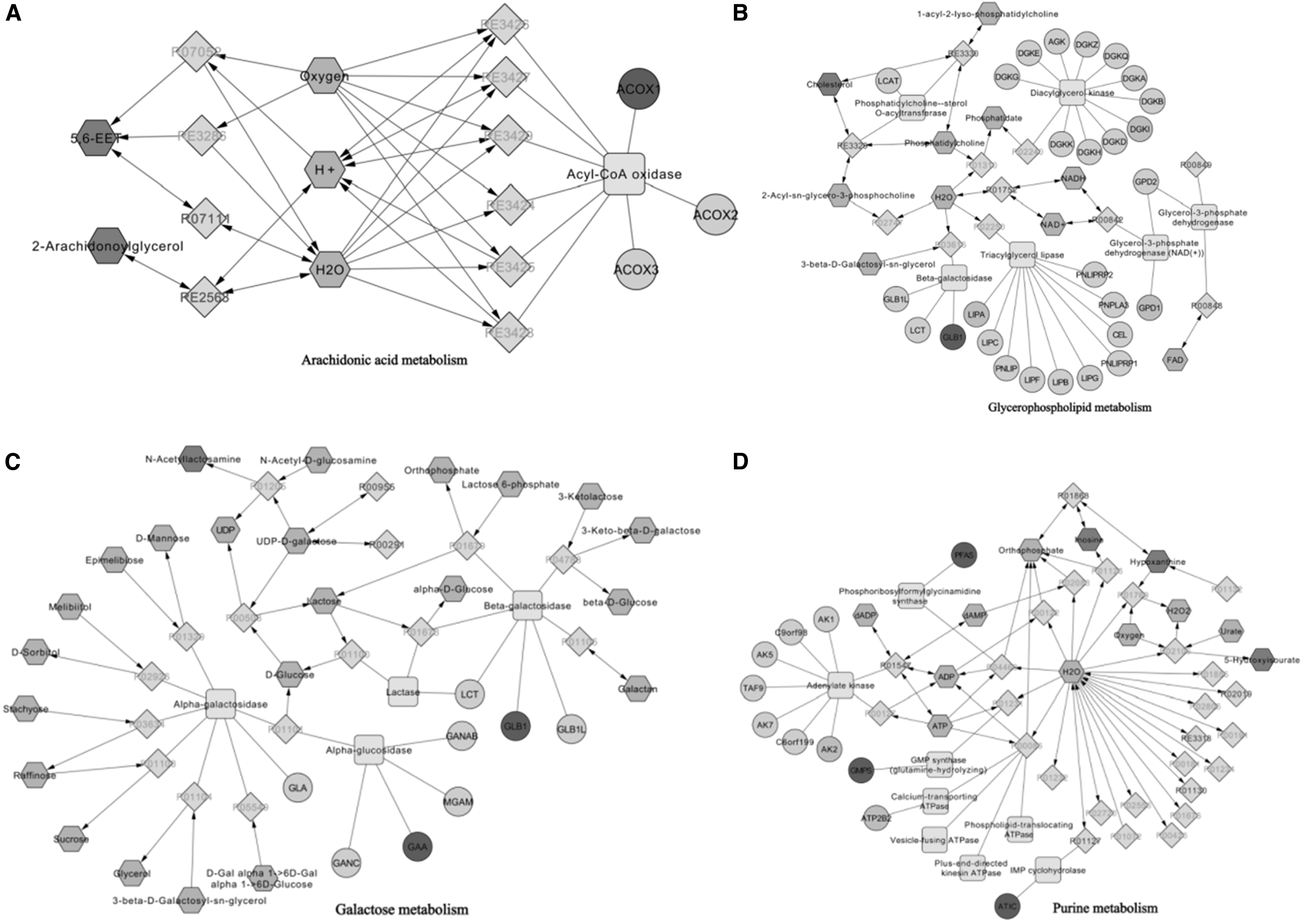
Compound-reaction-enzyme-gene network of NXT.
Information on Key Targets, Metabolites, and Pathways
Glb1, Gmps, Pfas, Atic, Gaa, and Acox1 are the hub targets.
Discussion
Using metabolomics, Liu et al. found that NXT plays a therapeutic role 5 days after prophylactic administration and 12 hours after MCAO rat model by regulating the pathway of glutamine metabolism and lipid metabolism. 9 Wang and colleagues found 39 important candidates, key signaling pathways, and core markers for NXT to prevent cerebral ischemic injury by using transcriptome and proteomics methods. 10 Yin et al. used transcriptome to find that intragastric administration of NXT for 5 days can treat MCAO rats through the “Rpl18-JAK2/STAT3 internal homeostasis,” “Eif3c Shh international homeostasis,” and “Rhoc inflammatory cell adhesion” pathways. 15 Existing single omics studies of NXT have found that transcriptional, protein, and metabolic levels behave differently at different times and at different doses of NXT.
In this study, the dosage and time of NXT were calculated according to clinical dose conversion, which was based on significant improvement in neurological function. To further explore the therapeutic mechanism of NXT on CIR injury in rats, differential expression genes and differential metabolites were first analyzed by three omics separately in this study. Second, transcriptomics and proteomics reversed DEGs and related metabolic pathways were analyzed and discussed. Finally, the association analysis between differential metabolites and DEGs was carried out in depth.
Through data processing and joint analysis, key metabolites and key targets were obtained, and related metabolic pathways were focused. In this study, we found that the regulation levels of 11 proteins and corresponding metabolites in ischemic side brain tissue were affected, and these could be significantly improved by NXT treatment. Including Glb1, Dgki, Gpd1, LIPA, Gmps, Pfas, Atic, ATP2B2, AK2, Gaa, and Acox1 (Table 1), among them Glb1, Gmps, Pfas, Atic, Gaa, and Acox1 are hub genes. The affected pathways are glycerol phospholipid metabolism, purine metabolism, galactose metabolism, and arachidonic acid metabolism.
Val-Gly-Val-Ala-Pro-Gly (VGVAPG) is a repeating hexapeptide in the elastin molecule. Szychowski et al. demonstrated that silencing of the Glb1 gene could reverse the increase in ROS and decrease in NO caused by the VGVAPG peptide. 16 An adequate supply of purines is essential for many life processes, and guanine monophosphate synthase is a glutamine-dependent amidotransferase. Nonsynonymous polymorphisms in guanine monophosphate synthase have been reported as risk factors for poor thiopurine metabolite ratios in patients with inflammatory bowel disease.
Phosphoriboseformylglycamidine synthase (PFAS) is an essential enzyme in the de novo synthesis of purines. PFAS has also been reported to play a role in the activation of innate immunity. Viral pseudoenzyme can hijack RIG-I through PFAS-induced deamidation to reduce proinflammatory cytokine production. 17 5-aminoimidazole-4-carboxamide ribonucleotide formyltransferase/inosine monophosphate cyclohydrolase (ATIC) is the only enzyme involved in de novo purine nucleotide synthesis. Studies have found that ATIC has a signaling mechanism that senses adenylate synthesis, ATP levels, and insulin receptor (IR) activation status and plays a role in regulating IR autophosphorylation and endocytosis. 18
Today, researchers are increasingly emphasizing the importance of blood sugar control in reducing stroke risk. Serum Gaa inhibitors delay carbohydrate absorption and reduce postprandial hyperglycemia, making them therapeutic agents for type 2 diabetes. 19 Acyl-CoA oxidase 1 (ACOX1) is the rate-limiting enzyme in the peroxisomal β-oxidation pathway, producing precursors that can be used to synthesize lipid mediators directly involved in the resolution of inflammatory processes, and have been shown to be used by PGC1-decreased alpha activity drives inflammation in aging. 20
Glycerophospholipids are the most abundant phospholipids in the body, which can be divided into phosphatidylcholine and phosphatidylethanolamine. Dang et al. identified significantly altered metabolites between atherosclerotic-prone and age-matched anti-atherosclerotic models, revealing altered pathways involved in glycerophospholipid and sphingolipid metabolism. 21 The study by Su et al. showed that the ethanolic extract of papaya may inhibit vascular smooth muscle cell apoptosis by reversing lysophosphatidylcholine in the glycerophospholipid metabolic pathway and activating GPR4 to regulate the expression of Bax and Bcl-2, proliferation and migration, thereby inhibiting the production and development of atherosclerosis. 22
Arachidonic acid is one of the most abundant polyunsaturated fatty acids in the human body. Among arachidonic acid metabolites are prostaglandins, prostacyclins, thromboxanes, leukotrienes, and lipoxins, which play an important role in the regulation of vascular tone and cardiovascular complications, including atherosclerosis, hypertension, and myocardial infarction effect. 23 At the same time, arachidonic acid metabolites are also involved in endocytosis, especially the clearance of polymorphonuclear neutrophils and foam cells, which is important for the resolution of inflammation. Impairment of this process can lead to atherosclerotic lesions. 24
Uric acid (UA) is the end product of purine metabolism. The inflammatory mechanisms associated with UA metabolism are mainly as follows: xanthine oxidase generates ROS oxidative stress when it converts hypoxanthine to xanthine and then to UA, and NO is inactivated, leading to endothelial dysfunction. 25 Endothelial novel connexin (Panx1) isoforms are present in endothelial cells throughout the arterial tree, as well as in smooth muscle cells in arteries and arterioles <300 μm in diameter. Deletion of endothelial Panx1 attenuates cerebral ischemic injury by reducing inflammation and the development of myogenic tone, suggesting that endothelial Panx1 may be a new target for therapeutic intervention in IS. 26
Galactose is essential for human metabolism and plays an established role in energy transfer and galactosylation of complex molecules. 27 New research suggests that loss of galactose and sialic acid and IgG bisecting GlcNAc may be involved in the molecular mechanism of inflammation in the development of IS. 28
Conclusions
In conclusion, this study combined multiomics to systematically explore the molecular mechanism of NXT in the treatment of CIR injury. The results showed that the protective effect of NXT on CIR injury in rats was related to glycerophospholipid metabolism, purine metabolism, galactose metabolism, and arachidonic acid metabolism, involving 11 key targets, reflecting the multiple pathways and multiple targets of NXT in the treatment of CIR injury. These results suggest that the selected target can be used as a potential drug target for the diagnosis or treatment of CIR injury, but its clearer mechanism needs further study.
Footnotes
Authors' Contributions
Data curation and analysis were performed by N.F., P. Z., X.W., B. L. and W. F.; drafting of the manuscript was performed by J.Y. and H.Z.; and concept and design of the study were performed by Z.D. and H.W.
Acknowledgments
We gratefully acknowledge the Key Laboratory of TCM Encephalopathy of Zhejiang Province, Zhejiang Chinese Medical University for being awarded a Research Starter Grant and support.
Author Disclosure Statement
No competing financial interests exist.
Funding Information
This study was supported by National Key R&D Projects of China (grant nos. 2019YFC1708600 and 2019YFC1708604); National Natural Science Foundation of China (grant no. 81630105); Key Laboratory of TCM Encephalopathy of Zhejiang Province (grant no. 2020E10012).