




Abstract
To observe the effects of moxibustion on T cells and T cell immunoglobulin and mucin-domain-containing molecule-3/galectin-9 (Tim-3/Gal-9) pathway in rats with rheumatoid arthritis (RA). To further explore the possible anti-inflammatory mechanism of moxibustion in the treatment of RA. Thirty Sprague Dawley rats were randomly divided into three groups, including a control group, an RA model group, and a moxibustion group. An RA model was created through the injection of Freund’s complete adjuvant. In the moxibustion group, rats were treated with moxibustion at acupoints of “Shenshu” and “Zusanli.” A total of three courses of treatment were conducted. Then the thickness of foot pad was measured, joint pathological changes were observed by hematoxylin–eosin (HE) staining, the proportion of CD4+T and CD8+T in peripheral blood was detected by flow cytometry, the expression levels of Tim-3 and Gal-9 in synovium were detected by polymerase chain reaction (PCR), and the expressions of CD4+T and CD8+T in synovium were detected by immunofluorescence. HE staining showed that the synovial tissue of the control group was smooth and neatly arranged without inflammatory cell infiltration. In the model group, the joint space was narrowed, the synovial tissue had congestion and edema, and a large number of inflammatory cells infiltrated. Compared with the model group, in the moxibustion group, the joint space narrowed with synovium hyperemia and edema, and the level of inflammatory cell infiltration decreased. Flow cytometry showed that compared with the model group, CD4+T expression in the moxibustion group was downregulated, while CD8+T expression was upregulated. PCR results showed that compared with the model group, the expressions of Tim-3 and Gal-9 in the moxibustion group were upregulated. Immunofluorescence results showed that compared with the model group, CD4+T expression in the moxibustion group was decreased, while CD8+T expression was increased. The results demonstrate that moxibustion not only suppressed the expression of CD4+T but also promoted the expression of CD8+T. The anti-inflammatory effect of moxibustion may be related to the regulation of T cell expression through the Tim-3/Gal-9 signaling pathway.
Introduction
Rheumatoid arthritis (RA) is an autoimmune disease that mainly damages the synovium. Continuous inflammatory stimulation can lead to the destruction of the bone and cartilage, limiting the motion of the joint and thereby reducing the quality of life of the patient. Studies have shown that about 0.5%–1% of the world’s population suffers from this disease, and women are more common than men. 1 Although the pathogenesis of RA is complex, it is generally believed that abnormal activation of T cells is the initial stage of RA pathogenesis. 2 However, costimulatory molecules can provide key stimulatory signals for T cell activation and play an important role in the occurrence and development of autoimmune diseases. 3
T cell immunoglobulin and mucin-domain-containing molecule-3(Tim-3) is a key member of the costimulatory family and plays an important role in maintaining autoimmune tolerance by binding to its ligand galectin-9 (Gal-9) to inhibit T cell activation. 4 As immune cells, T lymphocytes mainly include CD4+T and CD8+T subsets, and the ratio of CD4+T/CD8+T is the reflection of the state of immune balance. It was found that the expression of Tim-3 on CD4+T and CD8+T cells in synovial fluid and peripheral blood of RA patients was significantly higher than that of the control group, and the level of Tim-3 was negatively correlated with the severity of the disease. 5,6 In the pathological process of RA, Tim-3 acts on CD4+T and CD8+T cells through its negative regulation, inhibits the production of proinflammatory cytokines, and promotes the secretion of anti-inflammatory cytokines by T cells, thus playing an important role in the activation and proliferation of T cells. 7 Therefore, the abnormality of the Tim-3/Gal-9 signaling pathway is an important link in the occurrence and development of RA, and correcting its imbalance can regulate the activation of T cells, inhibiting the inflammatory response of joints, which is beneficial to the treatment of the disease.
As an important part of traditional Chinese medicine, moxibustion can not only relieve joint pain and swelling caused by RA, but can also reduce the level of rheumatoid factor, which plays an important role in the treatment of RA. 8,9 However, whether the anti-inflammatory effect of moxibustion is related to the regulation of T cell differentiation by the Tim-3/Gal-9 signaling pathway remains to be further studied. Therefore, this study took the Tim-3/Gal-9 signaling pathway as the breakthrough point to observe the effect of moxibustion on the expression of T cells and related inflammatory factors in RA rats, which is of great significance to deepen the scientific connotation of moxibustion in the treatment of RA.
Materials and Methods
Rats
Six-week-old male Sprague Dawley rats (weight, 200 ± 20 g) were purchased from Chengdu Dasuo Animal Science and Technology Co., Ltd (Chengdu, China) (Permit Number: SCXK 2021–030). The rats were randomly divided into a control group (Ctrl), RA model group (RA), and moxibustion group (Moxa), with 10 rats in each group. The rats were fed in a room with a temperature of 22–24°C, a humidity of 20%, and natural light and dark cycles. These animals were free to eat and drink. All animal-related procedures were approved by the Ethics Committee of Chengdu University of Traditional Chinese Medicine (Approval Number: 2020–11).
Establishment of the RA model
On the first day of the experiment, the rats were induced to develop RA with an injection of Freund’s complete adjuvant (FCA) (Sigma Aldrich, St Louis, USA) on a foot pad at 0.5 mL/kg. 10 The thickness of the foot pad, redness, and swelling were the indicators to assess the development of RA.
Moxibustion methods
Moxibustion therapy started on the seventh day of the experiment. The locations of Shenshu (BL23) and Zusanli (ST36) are determined according to the standards of the Experimental Acupuncture. 11 BL23 is located 7 mm laterally below the spinous process of the second lumbar spine. ST36 is below the knee joint and 6 mm beside the anterior tibial muscle. The rat’s fur near the acupoints of BL23 and ST36 was shaved for moxibustion. Lighting moxa granules Ω (diameter: 2 mm; length: 5 mm) were placed on these acupoints. Five moxa granules were burned at each point per day by alternating bilateral acupoints for 3 consecutive weeks. There was a day off on the seventh day every week.
After treatment for 3 weeks, the rats were anesthetized with isoflurane and then sacrificed. In total, 4 mL of blood was extracted from the abdominal aorta and put into a centrifuge tube labeled with serial numbers. After 1 hour, the supernatant was centrifuged and put into an Eppendorf tube stored in a refrigerator with a temperature of −20°C. The sample of the ankle was stored in 4% paraformaldehyde solution. The synovial tissue was put into a cryopreservation tube and frozen in liquid nitrogen, and then it was moved to a refrigerator with a temperature of −80°C for preservation.
Histological analysis
The ankle was immersed in the 4% paraformaldehyde solution. According to the standard operating procedures of pathological examination, dehydration, paraffin embedding, the making of 5-µm thick sections, HE staining, and gum sealing were carried out. The pathological changes of joint were observed under a light microscope.
Quantitative real-time polymerase chain reaction
Total ribonucleic acid (RNA) was extracted with the TRIzol method. The purity and the concentration of RNA were determined. The mRNA was converted into complementary deoxyribonucleic acid and detected by real-time quantitative polymerase chain reaction (PCR). The β-actin gene was used as an internal reference. Primer sequences of Tim-3 and Gal-9 were synthesized by Sangon Biotech (Shanghai, China), given in Table 1. 2−
The Primer Sequences of Real-Time Polymerase Chain Reaction
Flow cytometry
In total, 100 µL of peripheral blood was put into flow tubes, and 1µg of CD4 and CD8 flow antibodies was added, respectively. After mixing, the blood was dyed at 4°C for 30 minutes without light. The next step was conducted by adding 1 mL of erythrocyte lysate and incubating it for 10 minutes at room temperature. Then the researcher added 1 mL of phosphate-buffered saline (PBS) at 4°C, mixed it well, and centrifuged it for 5 minutes. After that, the researcher discarded the supernatant, added 2 mL of PBS for washing once, added 400 µL of PBS, and then immediately put it on the machine for testing. Data were analyzed by using Kaluza 2.1 software (Back-man, USA).
Immunofluorescence detection
Antigen retrieval was performed after paraffin sections were deparaffinized in water. Sections were washed three times in PBS every 5 minutes. Then it was sealed with goat serum at room temperature for 1 hour. After sealing, the sections were dried by shaking and a primary antibody (CD4
Statistical analysis
SPSS25.0 was used to analyze the data. First, a normality test is performed on the data. The independent-sample t-test was used for intergroup pairwise comparison, the paired-sample t-test was used for pairwise comparisons within groups, and the one-way analysis of variance (ANOVA) was used for comparisons between multiple groups. Nonparametric test was used for comparison between groups if the distribution was abnormal. Two-way repeated-measures ANOVA was used for the analysis of plantar thickness, and p < 0.05 was considered statistically significant.
Results
Moxibustion reduced the swelling of the foot
RA is characterized by joint swelling and pain. The joint swelling can indirectly reflect the degree of inflammation. In this study, we observed that the feet of rats with FCA injection showed obvious redness and swelling, but after the intervention of moxibustion, the redness and swelling symptoms were significantly improved (Fig. 1A). The result was consistent with our previous report 12 as FCA-injected rats developed more severe inflammatory reaction, with thicker foot pad, compared with the FCA-injected rats given moxibustion treatment.
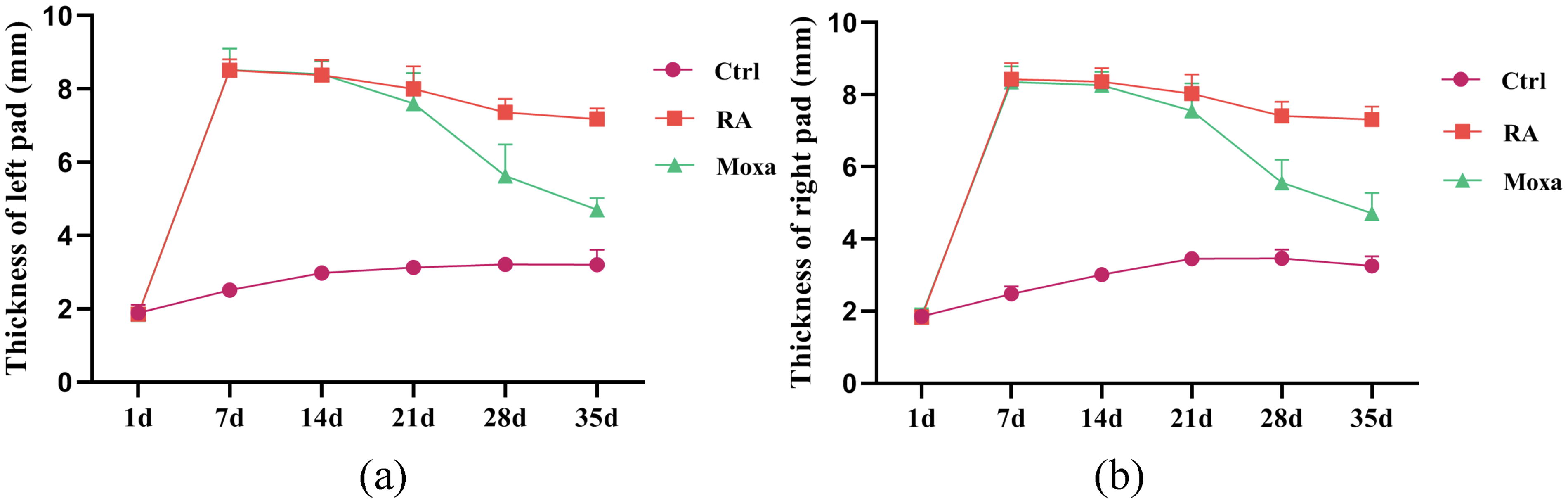
The effect of moxibustion on reducing the swelling of the foot.
Moxibustion improved the pathological changes of joint
The state of joint directly reflects the pathological changes in RA. HE staining showed that the joint structure of the control group was clear, the synovial tissue was smooth and orderly, and there was no infiltration of inflammatory cells. The synovium of the RA group showed significant hyperemia and edema, with a large number of inflammatory cell infiltration and vascular dilation. In the moxibustion group, the degree of synovial hyperemia and edema was improved, and the infiltration of inflammatory cells and vascular dilation were reduced, as shown in Figure 2.

The effect of moxibustion on joint pathology in RA rats. The pathological changes of the joint (scale bar, 100 μm and 50 μm) are shown. Data were expressed as the mean ± SD (n = 8). RA, rheumatoid arthritis.
Moxibustion restored the balance between CD4+T/CD8+T cells
Activation of T lymphocytes in peripheral blood is the first step to trigger RA and cause chronic inflammation. As immune cells, T lymphocytes mainly include CD4
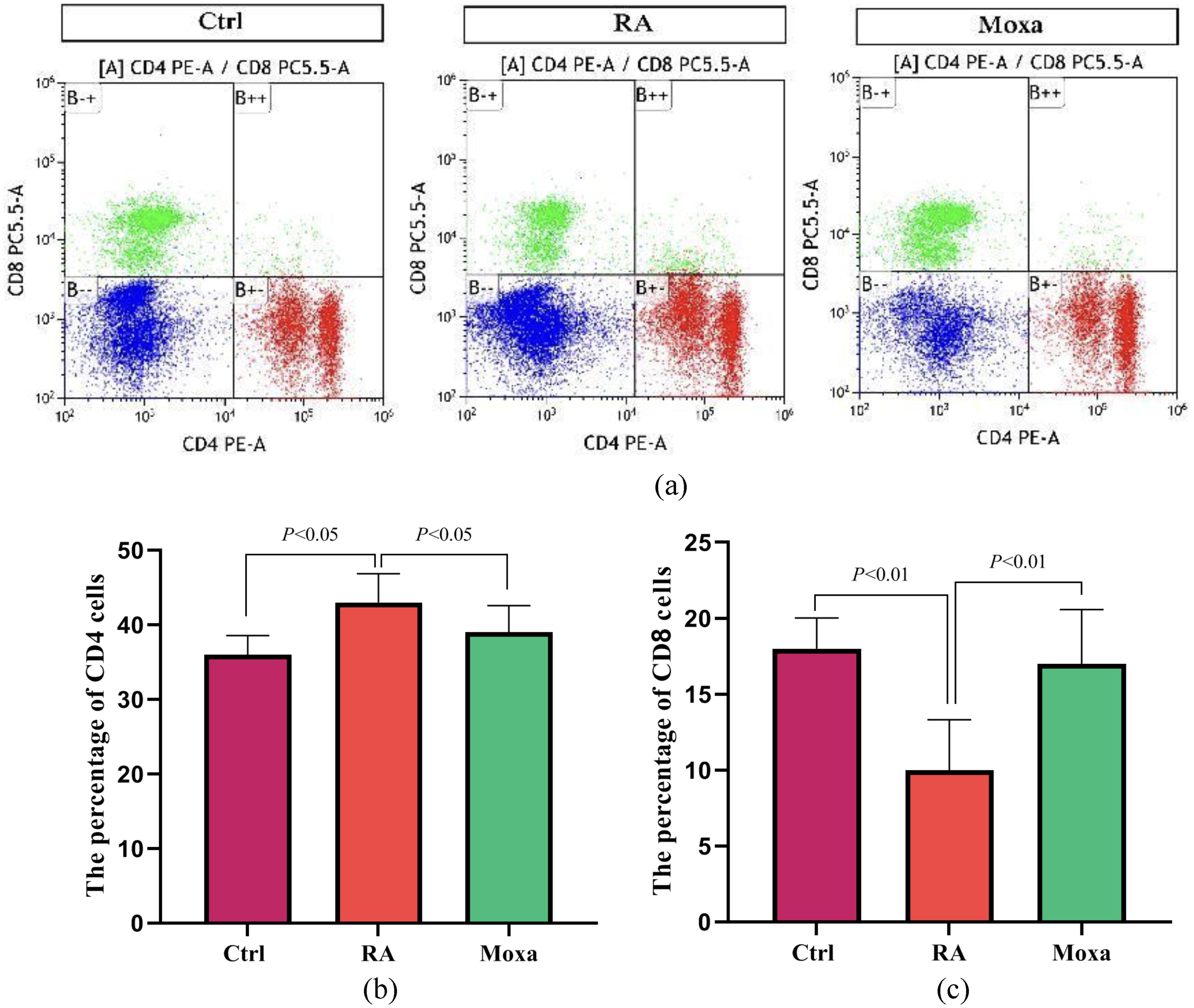
Effect of moxibustion on the expression of Tim-3 and Gal-9 mRNA
Tim-3 is an important member of the immune system’s costimulatory inhibitory receptor family and plays an immunoregulatory role through binding with its ligand Gal-9. The PCR results showed that compared with the control group, the expression levels of Tim-3 and Gal-9 in the synovium of the rats in the RA group were significantly increased. Compared with the RA group, the expressions of Tim-3 and Gal-9 in the moxibustion group were upregulated, as shown in Figure 4.
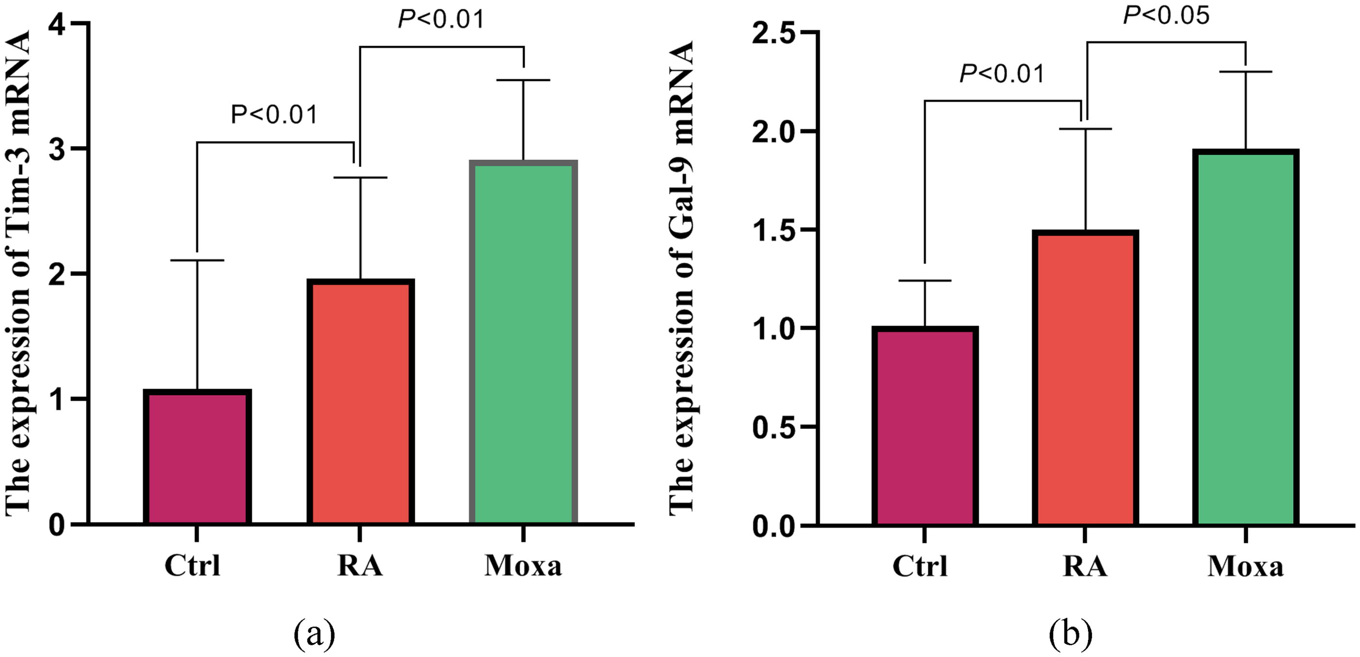
Expression of genes related to Tim-3/Gal-9 pathway. The expression level of mRNA was examined in synovium from RA rats by real-time PCR.
Effect of moxibustion on the expression of CD+T and CD8+T in synovium
The synovium is the target tissue of RA. Immunofluorescence was used to detect the expression of CD4+T and CD8+T in the synovium. It showed that compared with the control group, the expression of CD4+T in the synovium of the RA group increased and the expression of CD8+T decreased; compared with the RA group, the expression of CD4+T in the moxibustion group decreased, and the expression of CD8+T increased, as shown in Figure 5.

Immunofluorescence was used to examine the expression of CD4
Discussion
RA is an autoimmune disease whose pathogenesis is closely related to the overactivation of T cells. Continuous inflammatory stimulation makes the body to be in a state of high response for a long time and therefore disrupts the autoimmune balance, which eventually leads to joint damage. Traditional Chinese Medicine believes that RA belongs to the category of “arthralgia syndrome,” The “Huangdi Neijing” (China’s earliest classical work on medicine) believes that “The combination of the three qi, including wind, cold, and dampness, causes paralysis.” It can be seen that the occurrence of this disease is related to the invasion of wind, cold, and dampness, so the methods of dispelling wind and dispersing cold, dehumidifying, and dredging collaterals are often used in the treatment of RA. Moxibustion has the functions of warming meridians, dispelling cold, nourishing qi, and dredging collaterals, and has a definite curative effect on autoimmune diseases. It is often used as a clinical treatment for RA. Zusanli (ST36) is the Wushu acupoint of the stomach meridian, and Shenshu (BL23) is the back-shu point of the kidney. Modern studies showed that acupuncture at this acupoint involves regulating the inflammatory signaling pathway and autophagy and regulating the secretion of inflammatory factors, which plays an important role in improving immunity and rebuilding autoimmune homeostasis. 12 –14
The abnormal activation of T cells is a key link in the pathogenesis of RA. As an important member of the immune system costimulatory inhibitory receptor family, Tim-3 is mainly expressed on the surface of T cells, macrophages, NK cells, etc., and plays an immunoregulatory role through binding with its ligand Gal-9. 15 Tim-3 can inhibit Th1-mediated autoimmune response, promote immune tolerance, and may affect a series of inflammatory states. 16 Studies showed that the expression of Tim-3 is upregulated in various types of immune cells in RA patients, and the expression of Tim-3 on CD8+T cells and NK cells in RA patients is negatively correlated with disease activity score 28. 17 Meanwhile, the expression of Tim-3 mRNA in the synovial tissue of RA patients was significantly higher than that of osteoarthritis, and the expression of Gal-9 mRNA in peripheral blood mononuclear cells was also higher. The expression of Tim-3 was positively correlated with the expression of regulatory FoxP3. 18 In this study, an RA model was established in FCA rats, and it was observed that the expression of Tim-3 was upregulated in the synovial tissue of rats, which was consistent with the previous research results. Due to the persistent antigen stimulation mechanism in RA pathology, the reactivity of the body inhibits the proliferation of T cells and secreted cytokines, which promotes the enhancement of negative signals in the body, and the expression of Tim-3 also increases correspondingly, but its upregulation is insufficient. To regulate continuous antigen stimulation and provide sufficient negative inhibitory signals for the cellular immune response, the homeostasis of immune function is broken, resulting in immune damage. 19 After moxibustion, the expression of Tim-3 was significantly increased, and the inflammatory response of RA was inhibited, indicating that moxibustion treatment can enhance the negative signal of Tim-3 in RA rats, and exert its immune regulation function and anti-inflammatory effect.
As immune cells, T lymphocytes mainly include CD4
Conclusions
In conclusion, the present study demonstrated that moxibustion of Zusanli (ST36) and Shenshu (BL23) acupoints alleviated the pathological changes of the joints in RA rats and had a definite anti-inflammatory effect. The results also revealed that moxibustion not only suppressed the expression of CD4+T but also promoted the expression of CD8
Footnotes
Acknowledgment
The authors thank the Chengdu University of TCM for providing the research platform for the experiment.
Authors’ Contributions
Y.-M.Z. carried out the data collection, participated in the research coordination, and drafted the article. K.L. participated in the moxibustion therapy. Y.-D.G. carried out the data analysis. X.-H.G. revised the article. X.Y. and H.-Y.Z. contributed to the conception and design of the research. The authors read and approved the final article.
Ethics Approval Statement
The research was approved by the Ethics Committee of Chengdu University of Traditional Chinese Medicine.
Data Availability Statement
Data supporting the results of this study are available from the corresponding authors. The all-original data supporting the findings of this study were supplied by Hai-yan Zhou. Requests for access to these data should be made to Hai-yan Zhou (
Author Disclosure Statement
All authors have no conflicts of interest.
Funding Information
This work was supported by